
Endoluminal medical access device

a medical access device and endoluminal technology, applied in the field of catheter based medical devices, can solve the problems of significant surgical risks, inaccessible target areas in the body, and inapplicable techniques for all organ systems

Inactive Publication Date: 2015-01-08
SMARTWISE SWEDEN
View PDF3 Cites 15 Cited by
  • Summary
  • Abstract
  • Description
  • Claims
  • Application Information

AI Technical Summary

Benefits of technology

Enables safe and minimally invasive delivery and sampling in microvasculature sites, including the CNS and pancreas, by providing a communication channel that seals automatically, reducing the risk of bleeding and allowing for prolonged vessel access without leaving a catheter system in place.

Problems solved by technology

However, these techniques are not applicable for all organ systems, e.g. due to the limited resolution of the imaging modalities and sub-sequential planning of safe route at justifiable patient risk and radiation doses.
Moreover, there are some target areas in the body that are not accessible by known minimally invasive techniques and devices via safe routes.
For such organs with less accessible anatomical location, parenchymal access can be associated with significant surgical risks.
In addition, it appears that the system disclosed in U.S. Pat. No. 6,602,241 does not provide a satisfactory solution to avoid bleeding at the penetration side inside the body after completed treatment when the catheter is retracted.
However, this solution to avoid bleeding at the penetration site is not satisfactory from a clinical point of view as it is difficult to perform and to monitor the success thereof.
In addition, the injection of adhesive or embolization material may induce thrombotic embolies or unintentionally occlude the delivery vessel completely.
Furthermore, the use of adhesives is not feasible in arterial vessels due to the existing higher blood pressure pushing the adhesive material out of the penetration site into the surrounding tissue before the penetration site is closed.
Moreover, the vessel wall penetrating catheter disclosed in U.S. Pat. No. 6,602,241 is of such large size that it cannot navigate into the microvasculature, e.g. into the central nervous system (CNS).
Hence, the vessel penetrating catheter disclosed in U.S. Pat. No. 6,602,241 is not suited for vascular navigation into the CNS or other similar small vessels in the body.
The vessel wall penetrator body is, amongst other things due to the multi lumen design, so large that it would occlude such small vessels, which is highly undesired, and may be fatal to the CNS parenchyma supported by such an artery.
However, this endovascular technique is not suited for the arterial part of the body vascular system.
Furthermore, it is not suited for use in microvessels, but in large vessels.
Moreover, in practice the radiologist usually pushes and retracts the needle several times until the second vein is hit, which implies a risk for bleedings.
The amount of bleeding that can occur can sometimes be life threatening needing costly patient monitoring in intensive care.
A further issue is when vascular walls are penetrated, e.g. by a needle, upon retraction of the needle, a compression of the exit site is needed in order to avoid bleeding.
However, often it is not possible to provide a compression of such an exit site at conventionally difficult accessible target sites in a human or animal body.
A self sealing ability is not sufficient on the arterial side of the vascular system especially in areas where no bleeding is tolerated, e.g. in connection with CNS interventions.
Conventionally difficult accessible target sites in the body may not be reached with the aforementioned devices.
Hence, it is difficult to deliver substances to and / or from conventionally difficult accessible target sites in a human or animal body.
However, a penetration of vessel walls is not anticipated or implementable with this type of microcatheter as the distal tip of the disclosed microcatheter is blunt, and the distal end portion is in addition flexible and has spiral cuts.
Thus, extravascular target sites are not accessible for this kind of microcatheters.
This device appears to be not suited for use in the microvasculature.
An issue needing a novel and inventive solution is thus delivery of substances to and / or from conventionally difficult accessible target sites in a human or animal body, such as the microvasculature, e.g. in the CNS or pancreas.

Method used


Image

  • Endoluminal medical access device
  • Endoluminal medical access device
  • Endoluminal medical access device

Examples


examples

[0142]In Table 1, given below, typical dimensions of some embodiments are given.

TABLE 1Typical dimension ranges and ratios of some embodiments of the extroducerdimensions in micrometers,Length ofapproximalLength ofLength ofhollowmeasuresdistal penetrationdetachmentLumenFlangeonlyportion 102limiterzoneOuter diameterdiameterdiameterMinApprox. 3xa few0, if the140100300outer diametermicrometerscatheter isof extroducer10-20dissolved400-450no zone isneededMaxNo limit if itMust notLimited byLimited by polymer Limited byLimited byis made of aaffect bloodtime / energytube and micro polymerdimensionshighly flexiblestream.amount ofcatheter system, tube andof the vessel.material,Approx 0.3dissolvedand intended microThe wheelsuch astimes vesselmaterial.target sitecatheterdesign isnitinol. Itdiameter,Processsystem andlimited bymust betypicallymust notintendedthe vesselpossible to250 harm tissue.target sitediameter toadvance itmicrometerSmallerapprox.through theis better.1.5x ofvascular treeApprox. ...


PUM


Abstract

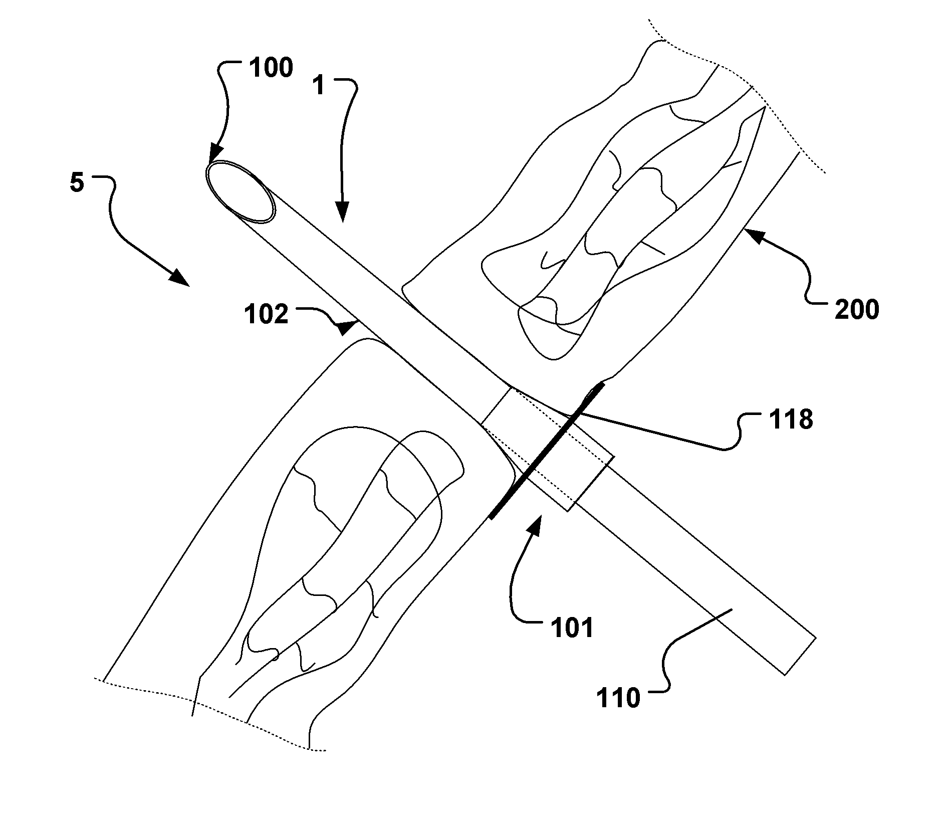
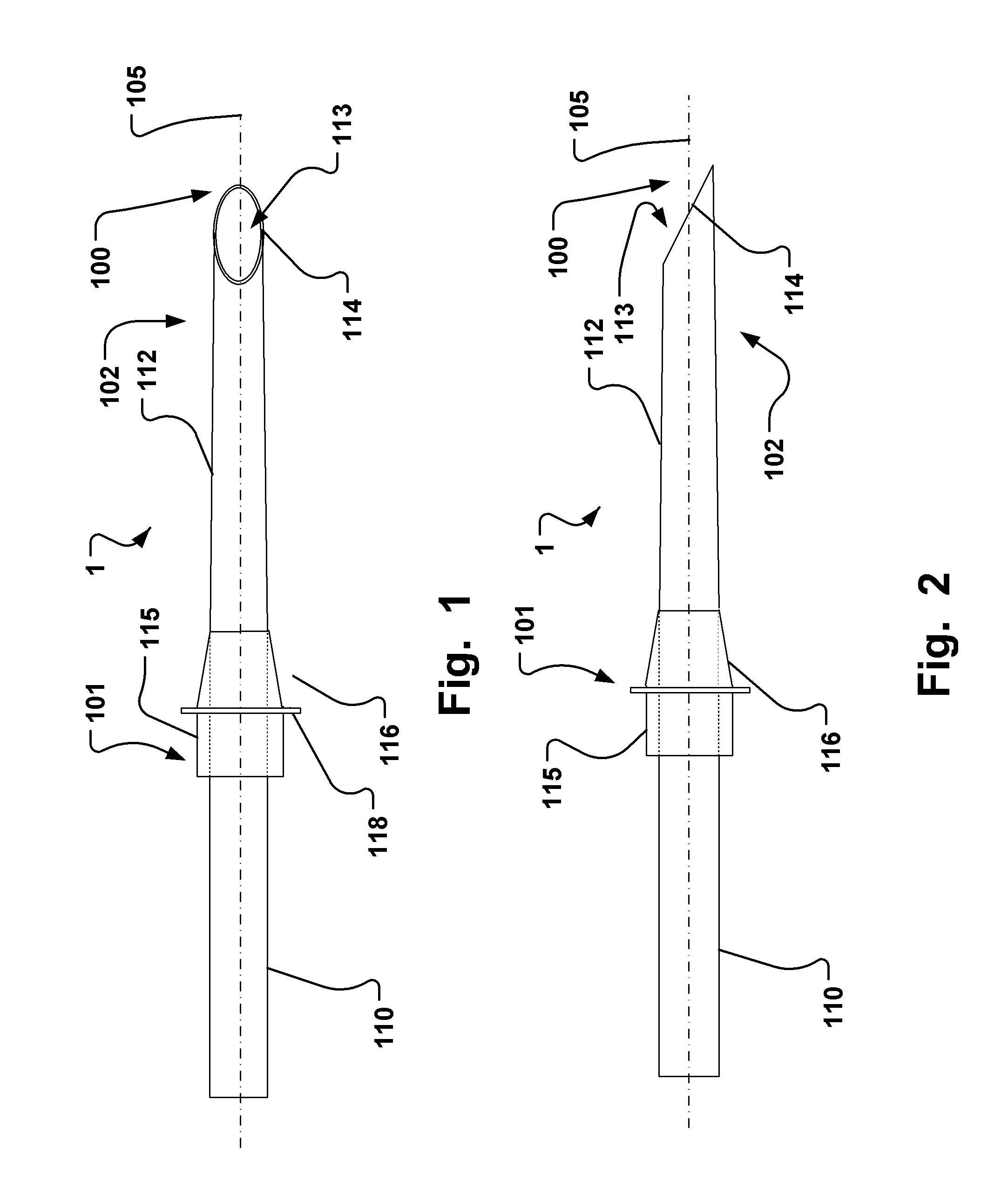
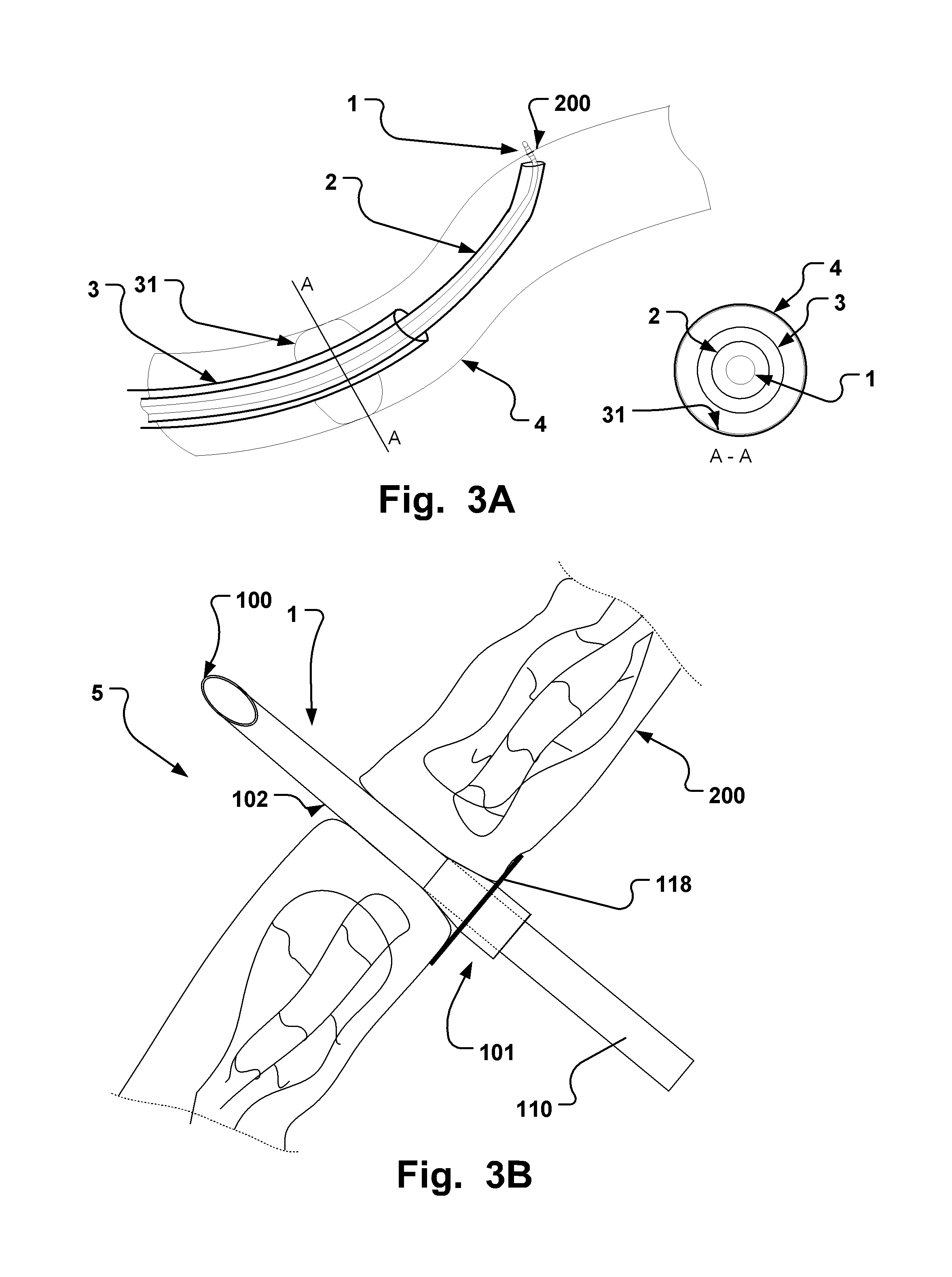
An endoluminal medical access device (1) is disclosed that is devised for endoluminal delivery to an extravascular target site (5) at a vasculature site of a human or animal body vasculature, such as the microvasculature. The device (1) comprises a hollow body (112) arranged around a continuous channel (113) that ends in a distal end (100) and comprises a distal penetration portion (102) that is devised to extend across a tissue wall of said microvasculature said microvasculature site (4) at an extravascular target site in said body to provide communication with said extravascular target site through said channel (113) and devised for at least partly apposition to said tissue wall, and a proximal connection section (101), which proximally adjoins said penetration portion (102), and optionally comprises an intrusion depth limit unit (116, 118) and / or a hollow separation section (115) devised to provide a controllable separation of the penetration portion (102) from a connected proximal portion (110) of the hollow body.

Description

FIELD OF THE INVENTION[0001]This invention pertains to the field of catheter based medical devices. More particularly, the invention relates to hollow probes having a piercing tip. Even more particularly, the invention relates to an endoluminal vascular medical access device for endoluminal delivery of substances to and / or from a vasculature site of a human or animal body and access to an extravascular target site located outside of the lumen of the vasculature at said site. The vascular site may be a microvasculature site.BACKGROUND OF THE INVENTION[0002]There is today a trend towards minimally invasive techniques for administration or sampling of substances or cells to or from various organ systems. Most organs and tissues in the body can be reached by needles with or without ultrasonic or computerized tomography guidance and if this is not possible, open surgery is an option. Stereotactic delivery, assisted by modern imaging techniques, are also existing alternatives.[0003]Howeve...

Claims


Application Information

Patent Timeline
Patent Type & Authority Applications(United States)
IPC IPC(8): A61B17/34A61B10/04
CPCA61B17/3423A61B2017/3425A61B10/04A61M25/0069A61M25/0084A61M25/06A61M2025/0042A61M25/0082A61B2017/3492A61B2018/1892A61M25/0068A61B17/3494A61N1/05
Inventor HOLMIN, STAFFANJONSSON, STEFANLUNDBERG, JOHAN
Owner SMARTWISE SWEDEN
Learn More