Data detection device for use in combination with an MRI apparatus

Active Publication Date: 2015-01-01
KONINKLIJKE PHILIPS ELECTRONICS NV
View PDF7 Cites 20 Cited by
  • Summary
  • Abstract
  • Description
  • Claims
  • Application Information

AI Technical Summary

Benefits of technology

The patent describes a method for detecting magnetic signals generated by a magnetic resonance imaging (MRI) apparatus. This can be done without needing a special interface between the detection device and the MRI apparatus, making the process easier and safer. The detection device uses a coil made of printed circuit board traces, which is efficient and cost-effective. The method can provide a meaningful temporal relation to the acquired MRI data, making it easier to analyze. Overall, the patent describes a simple and efficient way to detect the magnetic signals generated by an MRI machine.

Problems solved by technology

However, the use of CT in combination with PET has its limitations, such as notable irradiation (X-ray) dose and reduced soft tissue contrast compared to magnetic resonance imaging (MRI).

Method used


Image

  • Data detection device for use in combination with an MRI apparatus
  • Data detection device for use in combination with an MRI apparatus
  • Data detection device for use in combination with an MRI apparatus

Examples


Embodiment Construction

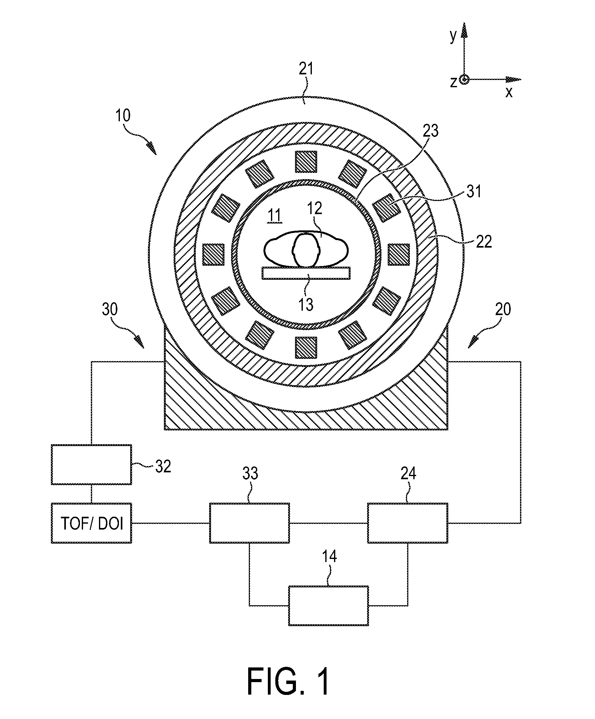
[0043]FIG. 1 shows schematically and exemplarily an embodiment of an imaging apparatus 10 comprising a magnetic resonance imaging (MRI) apparatus 20 and a positron emission tomography (PET) apparatus 30. The imaging apparatus 10 is capable of acquiring both MRI data and PET data, for example, from spatial areas of an imaging region 11 that are at least partially overlapping or spatially adjacent.

[0044]The MRI apparatus 20 comprises a main magnet 21 that serves to generate a strong, uniform, static magnetic field B0 through the imaging region 11 (along the z direction, by convention). The main magnet 21 may be an annular magnet or a bore-type magnet. Moreover, the main magnet 21 may be superconducting or resistive in nature; in the former case, it is typically disposed in a cryogenic devar or other cooling system (not shown). The MRI apparatus 20 also comprises magnetic field gradient coils 22 that serve to superimpose switched magnetic field gradients in the x, y, and z directions o...


PUM


Abstract

The invention relates to a data detection device for use in combination with a magnetic resonance imaging (MRI) apparatus. A magnetic field detection unit (34) serves to detect a temporally varying magnetic field generated by the MRI apparatus, and a timestamping unit (35) generates magnetic field detection timestamps in dependence of the detected temporally varying magnetic field. This allows determining a temporal relation to acquired MRI data.

Description

FIELD OF THE INVENTION[0001]The invention relates to a data detection device for use in combination with a magnetic resonance imaging (MRI) apparatus as well as to a corresponding data detection method and data detection computer program. The invention relates further to an imaging apparatus as well as to a corresponding imaging method and imaging computer program.BACKGROUND OF THE INVENTION[0002]In recent years, there has been a trend towards the combination of different medical imaging modalities for improved diagnosis. For example, the integration of biochemical or metabolic imaging provided by positron emission tomography (PET) and anatomical imaging provided by computed tomography (CT) has proven itself as a standard clinical tool. However, the use of CT in combination with PET has its limitations, such as notable irradiation (X-ray) dose and reduced soft tissue contrast compared to magnetic resonance imaging (MRI). For this reason, MRI is now considered to be an improved alter...

Claims


Application Information

Patent Timeline
IPC IPC(8): G01R33/48
CPCG01R33/481A61B5/055A61B6/5247A61B6/037A61B6/4417A61B5/0035G01R33/48
Inventor WEISSLER, BJOERNZINKE, MANFRED BRUNO
Owner KONINKLIJKE PHILIPS ELECTRONICS NV
Why Eureka
Try Eureka