
Differentiation between transient and persistent high-risk HPV infection by in situ hybridization

a high-risk, transient technology, applied in the field of in situ hybridization assays and cervical sample analysis, can solve the problems of poor clinical specificity, unnecessary follow-up procedures, and insufficient hpv infection alone for oncogenic transformation

Inactive Publication Date: 2014-12-04
ADVANCED CELL DIAGNOSTICS INC
View PDF0 Cites 16 Cited by
  • Summary
  • Abstract
  • Description
  • Claims
  • Application Information

AI Technical Summary

Benefits of technology

The patent is about a new method for categorizing cervical tissue samples based on the presence of HPV DNA or RNA. This is done using an antisense probe that detects both HPV DNA and RNA, allowing for a more accurate and reliable classification of the tissue sample.

Problems solved by technology

However, HPV infection alone is not sufficient for oncogenic transformation; 80-90% of infections are transient and self-cleared by the immune system.
As a result, these tests have poor clinical specificity (40-50%) for high-grade intraepithelial neoplasms (CIN2+), leading to unnecessary follow-up procedures for many women (Kinney et al., Am. J. Clin. Pathol.

Method used


Image

  • Differentiation between transient and persistent high-risk HPV infection by in situ hybridization
  • Differentiation between transient and persistent high-risk HPV infection by in situ hybridization
  • Differentiation between transient and persistent high-risk HPV infection by in situ hybridization

Examples


example i

HPV Detection in Caski Cells

[0134]This example describes detection of HPV nucleic acids in Caski cells.

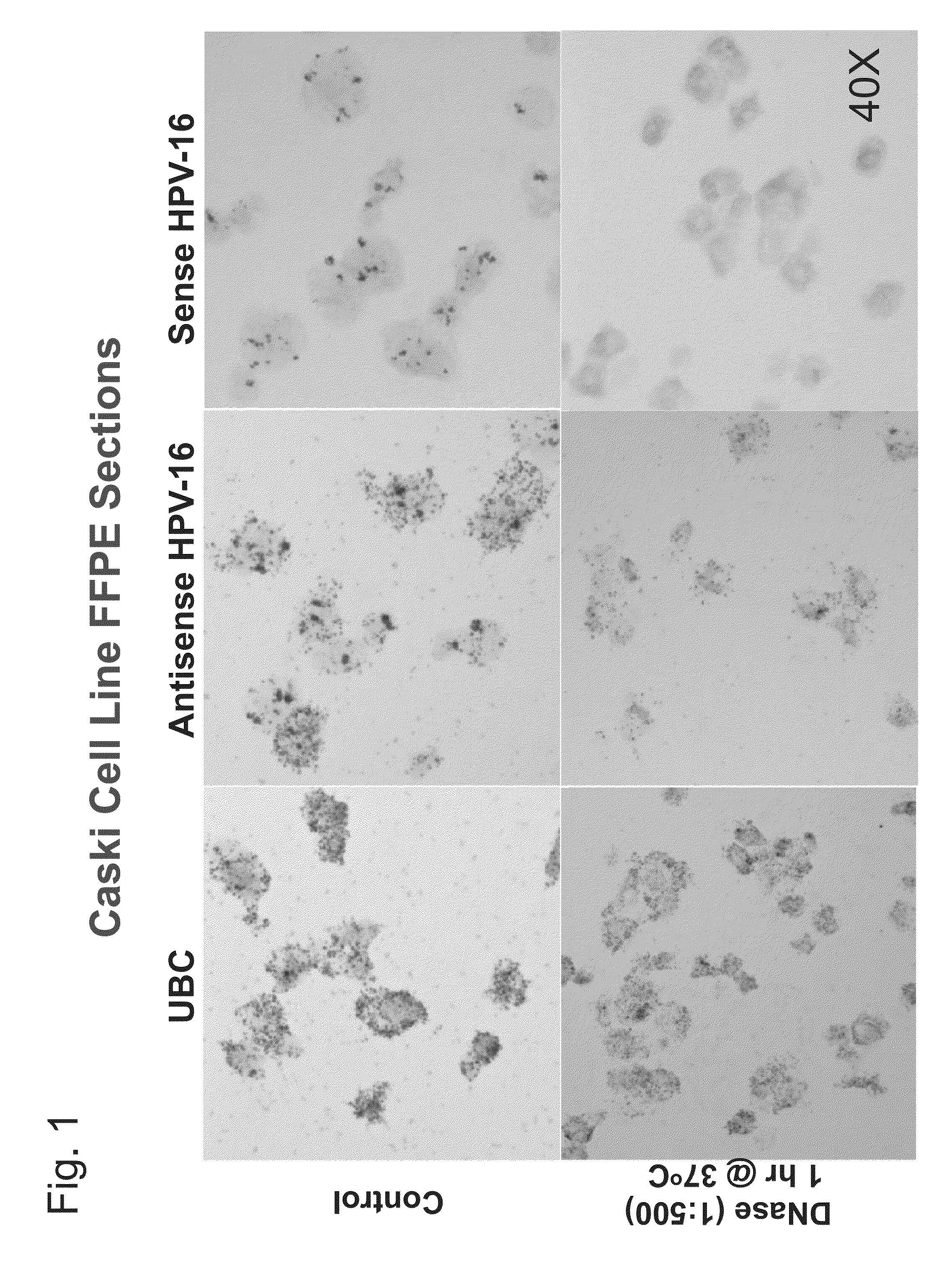
[0135]An RNA in situ hybridization (ISH) assay was developed based on RNAscope® technology (Advanced Cell Diagnostics) to detect the mRNA of E6 and E7 oncogenes of HR-HPVs (Bishop et al., Am. J. Surg. Pathol. 36:1874-1882 (2012); Schache et al., Br. J. Cancer 108:1332-1339 (2013); Ukpo et al., Am. J. Surg. Pathol. 35:1343-1350 (2011); Wang et al., J. Mol. Diagn. 14:22-29 (2012)). In the assay, frequent nuclear staining nuclear staining was observed, which may be DNA in origin, in HPV-infected cells. To investigate this, RNAscope® ISH assays were performed on the cervical cancer cell line Caski known to harbor HPV 16 using probes targeting the E6 and E7 genes. Two types of probes were used: a standard antisense probe complementary to E6 and E7 mRNA, and a sense probe targeting the same regions but in the same sense as the E6 / E7 mRNA (probe sets commercially available from Advanced C...

example ii

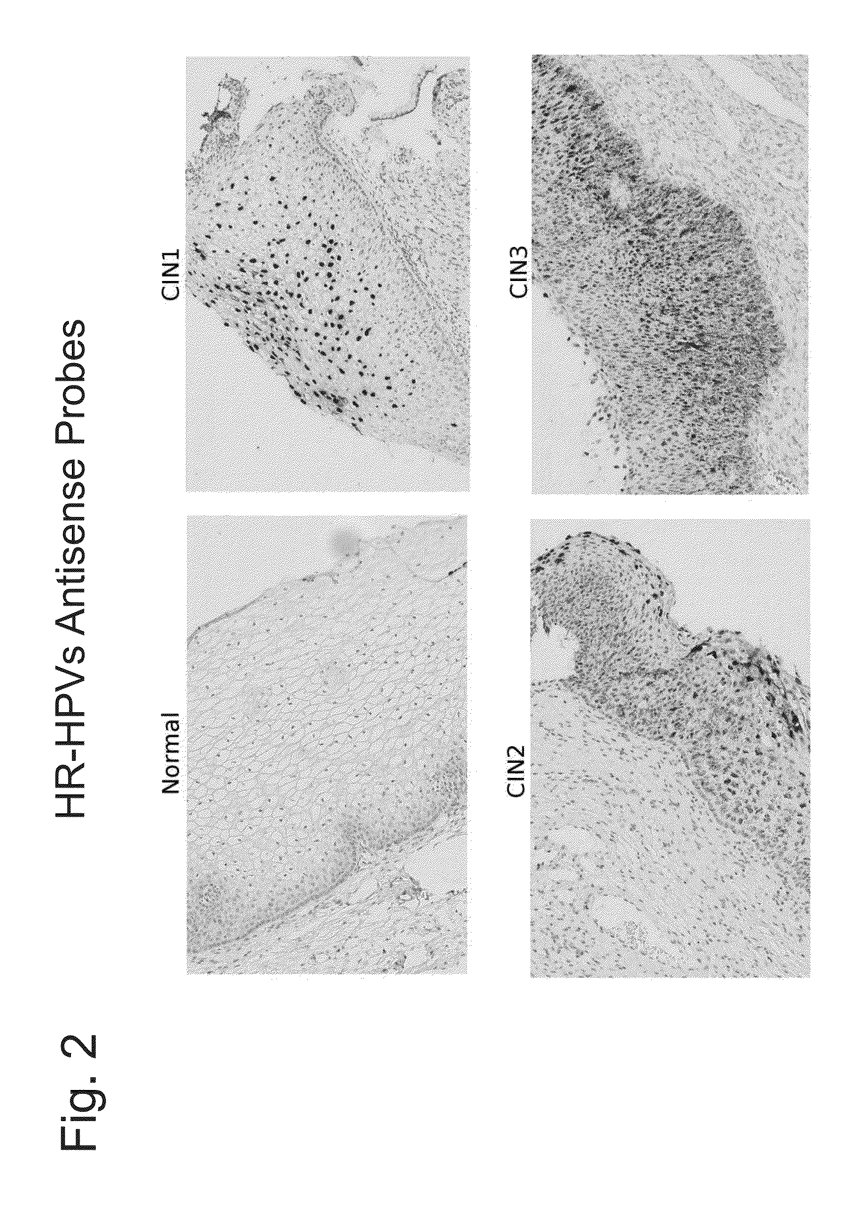
HPV Detection in Cervical Tissue Samples

[0139]This example describes detection of HPV nucleic acids in cervical tissue samples.

[0140]High-risk HPV E6 / E7 mRNA is known to be up-regulated in high grade CIN lesions (CIN2+) but not in low grade lesions (CIN1)(Lie and Kristensen, Expert Rev. Mol. Diagn. 8:405-415 (2008)). An in situ hybridization assay was performed essentially as described in Example I on cervical tissue samples. Briefly, 5 micron thickness slide-mounted sections were de-paraffinized through 100% xylene and ethanol washes. Tissues were then treated serially with commercially available buffers (Advanced Cell Diagnostics): Pre-Treatment 1 solution (endogenous hydrogen peroxidase block with Pretreat 1 solution for 10 minutes at room temperature); Pre-Treatment 2 (100° C., 15 minutes immersion in Pretreat 2 solution); and, Pre-Treatment 3 (protease digestion, 40° C. for 30 minutes); rinses with water were performed after each Pre-Treatment step. Tissues were then hybridized...


PUM

PropertyMeasurementUnit
temperatureaaaaaaaaaa
Tmaaaaaaaaaa
Tmaaaaaaaaaa

Abstract

The invention relates to methods of categorizing a cervical tissue or cytology sample by performing an in situ hybridization assay using an antisense E6 or E7 probe on a cervical tissue sample, wherein the antisense E6 or E7 probe can simultaneously detect HPV DNA and HPV RNA; detecting the presence of HPV nucleic acid; and categorizing the cervical tissue sample based on HPV nucleic acid expression.

Description

[0001]This application claims the benefit of priority of U.S. Provisional application Ser. No. 61 / 806,360, filed Mar. 28, 2013, the entire contents of which are incorporated herein by reference.BACKGROUND OF THE INVENTION[0002]The present invention relates generally to in situ hybridization assays and cervical sample analysis, and more specifically to assays for testing for human papillomavirus (HPV) infection and analysis of cervical tissue samples.[0003]High-risk human papillomavirus (HR-HPV) infection causes essentially all cervical cancer and a subset of head and neck cancer (Zur Hausen, Virology 384:260-265 (2009)). However, HPV infection alone is not sufficient for oncogenic transformation; 80-90% of infections are transient and self-cleared by the immune system. Current HPV testing methods such as Hybrid Capture® 2 (Digene Corporation; Gaithersburg, Md.) and GP5+ / 6+ PCR have excellent analytical sensitivity but do not differentiate transient from persistent HPV infections, on...

Claims


Application Information

Patent Timeline
Patent Type & Authority Applications(United States)
IPC IPC(8): C12Q1/70
CPCC12Q1/708C12Q2600/118C12Q2600/112G01N2333/025
Inventor MA, XIAO-JUNWANG, HONGWEIWU, XINGYONGLUO, YULING
Owner ADVANCED CELL DIAGNOSTICS INC
Learn More