Marker-flange for mri-guided brachytherapy
a magnetic resonance imaging and mark-flange technology, applied in radiation therapy, medical science, therapy, etc., can solve the problems of low radiation dose delivered to surrounding or intervening healthy tissues, ct imaging does not offer the same accuracy in identifying these soft tissues, and three-dimensional reconstruction of brachytherapy, so as to facilitate the identification of relevant points, improve geometric accuracy, and overcome susceptibility artifacts
Inactive Publication Date: 2014-09-18
UNIV OF IOWA RES FOUND
View PDF8 Cites 13 Cited by
- Summary
- Abstract
- Description
- Claims
- Application Information
AI Technical Summary
Benefits of technology
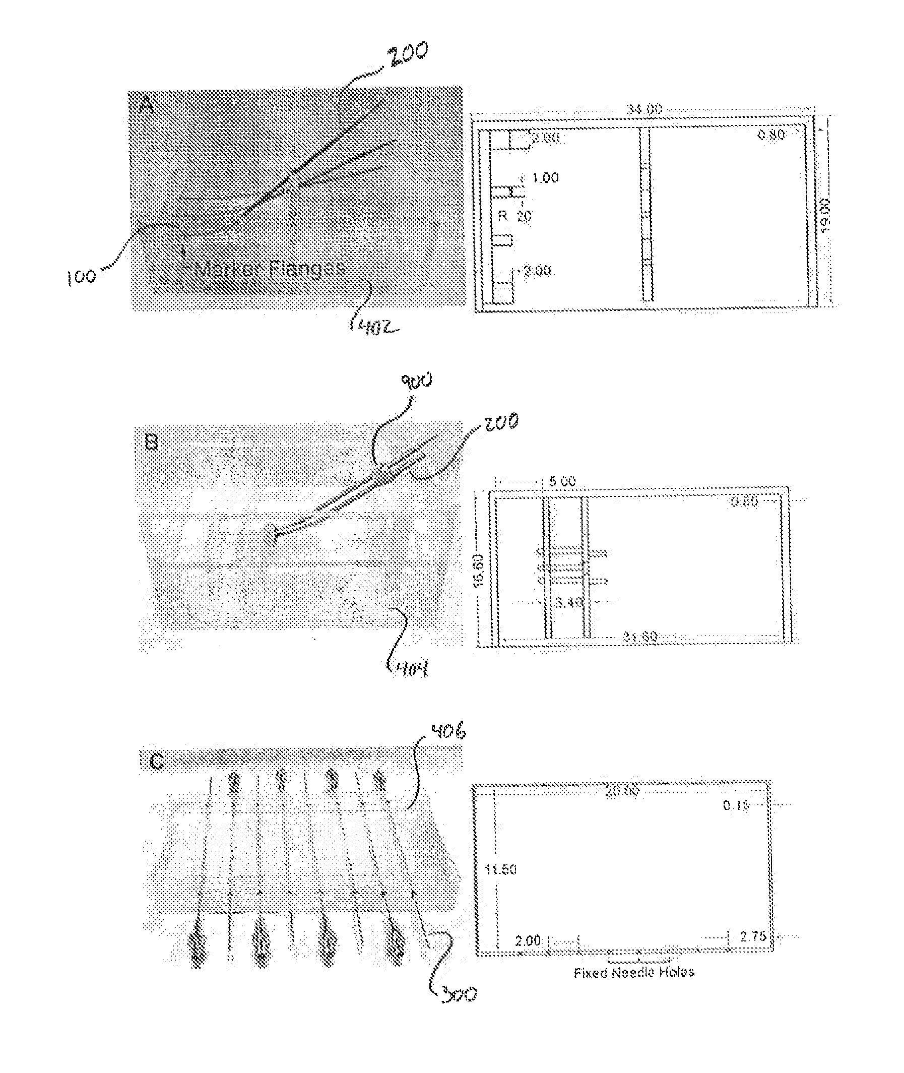
[0015]The marker agent contained within the hollow chamber of the marker-flange is an MR imaging responsive liquid marker material that generates MR image signal intensities sufficient to overcome susceptibility artifacts generated by titanium materials when subjected to MR imaging. In this manner, the combination of the external marker-flange with the marker agent provides clearly defined images with improved geometric accuracies, thereby facilitating the identification of relevant points on a physical brachytherapy applicator, and the registration of those p
Problems solved by technology
However, by placing a radiation source inside or directly adjacent to the target tissue, brachytherapy procedures are capable of delivering a focused dose of radiation to the target tissue with relatively low dosages of radiation being delivered to surrounding or intervening healthy tissues and nearby organs at risk.
CT imaging does not offer the same accuracy in identifying these soft tissues.
One challenge that has arisen with MRI-guided brachytherapy is the three-dimensional reconstruction of brachytherapy applicators.
Applicator reconstruction inaccuracies lead to uncertainties in the delivery of radiation doses to both the target tissue and organs-at-risk.
In particular, a mispositioning of the applicator may result in one or both of an underdosage to the target tissue (leading to the recurrence of the cancerous tissue) and the overdosage of nearby healthy tissue (causing undesired damage of healthy tissues).
However, titanium has been found to generate substantial imaging artifacts when imaged under an MR imaging modality.
These artifacts result in an increased uncertainty when attempting to reconstruct a three-dimensional model of a titanium applicator, in both its geometric dimensions and location within the treatment region.
However, plastic brachytherapy applicators are not ideal.
This increase
Method used
View moreImage
Smart ImageViewing Examples
Examples
 PUM
 Abstract
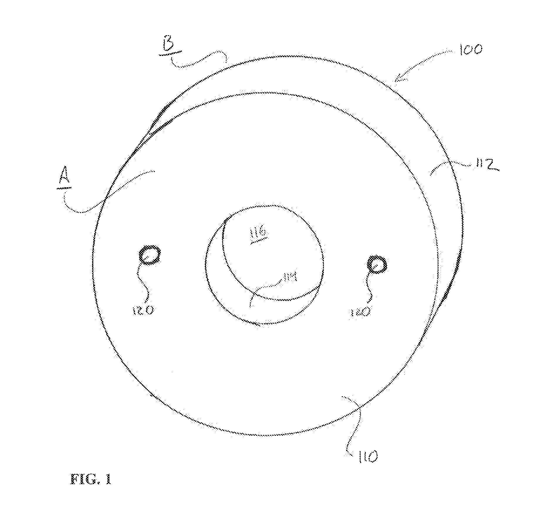
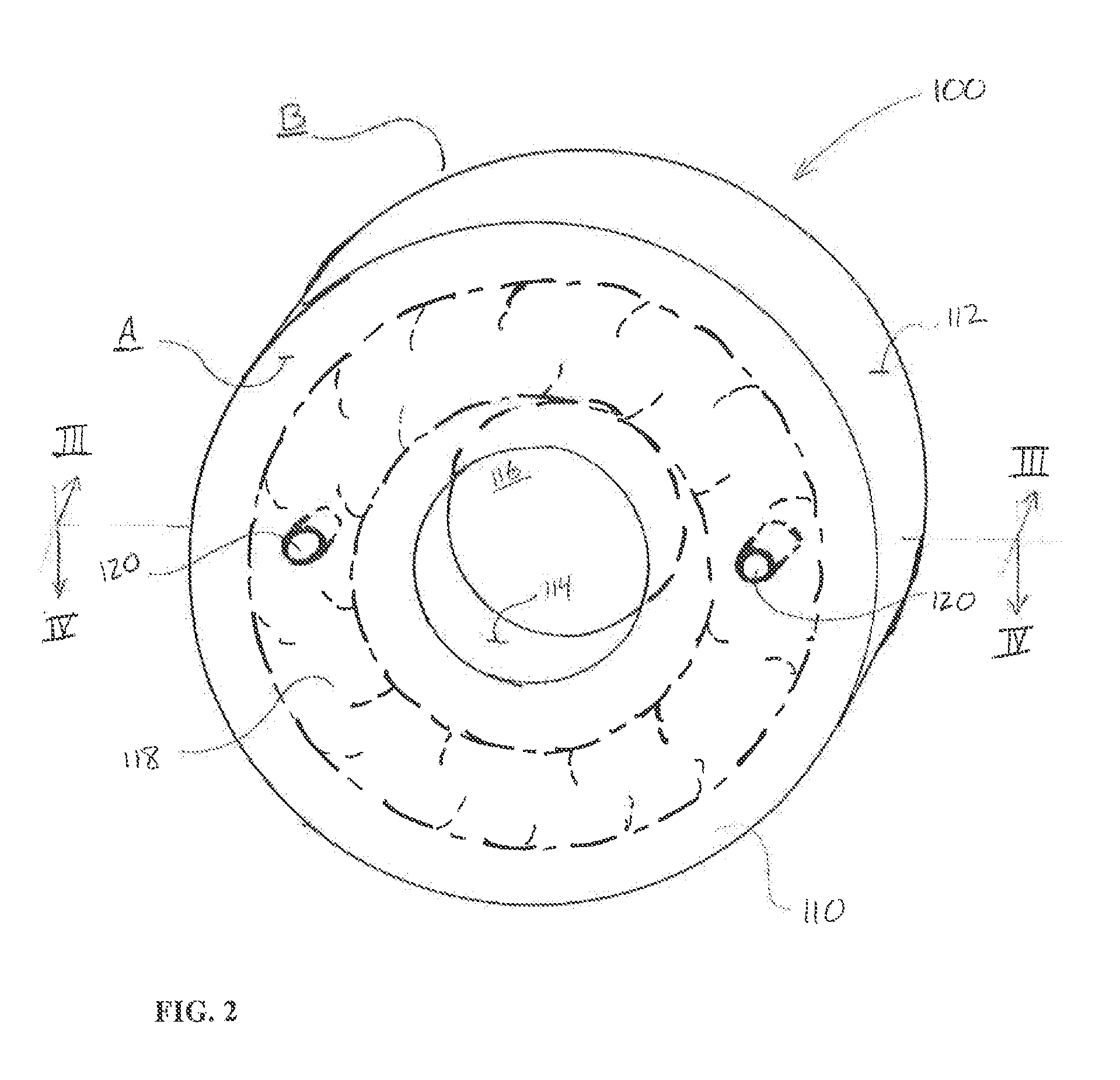
Disclosed herein is a marker flange for use with a brachytherapy applicator, having a flange body with a first face, a second face, and a hollow chamber positioned between the first and second faces. A cavity extending through the flange body is dimensioned to receive a tandem in a brachytherapy applicator in a press-fit connection, so as to affix the flange-marker to an outside surface of the tandem. The hollow chamber includes an MR imaging responsive marker agent that provides strong signal intensities and a high geometric accuracy in MR imaging.
Description
BACKGROUND OF THE INVENTION[0001]1. Field of the Invention[0002]This invention relates to devices and methods for use in therapeutic treatments. In particular, the present invention relates to a marker-flange for use in Magnetic Resonance Imaging (MRI) guided radiation therapy with a brachytherapy applicator.[0003]2. Description of the Related Art[0004]Brachytherapy (also referred to as “short-distance therapy”) is a medical treatment procedure wherein target tissues, such as cancerous tumors, are treated with radiation sources that are placed inside or directly adjacent to the target tissue. Example target tissue includes cervical, vaginal, endometrial, breast, prostate, esophageal, lung, and skin cancers. Example applicators include interstitial needles, intraluminal applicators and intracavitary applicators.[0005]Brachytherapy is a favorable alternative or supplemental treatment to External Beam Radiation Therapy (EBRT). In particular, EBRT procedures are performed by directing a...
Claims
Login to View More Application Information
Patent Timeline
 IPC IPC(8): A61N5/10
CPCA61N5/1007A61B90/39A61B2090/3954A61N5/1016A61N2005/1012
Inventor KIM, YUSUNG
Owner UNIV OF IOWA RES FOUND
Features
- R&D
- Intellectual Property
- Life Sciences
- Materials
- Tech Scout
- Unparalleled Data Quality
- Higher Quality Content
- 60% Fewer Hallucinations
Learn More Browse by: Latest US Patents, China's latest patents, Technical Efficacy Thesaurus, Application Domain, Technology Topic, Popular Technical Reports.
© 2025 PatSnap. All rights reserved.Legal|Privacy policy|Modern Slavery Act Transparency Statement|Sitemap|About US| Contact US: help@patsnap.com


