
Pharmaco-cellular therapeutic method for the treatment of muscular dystrophies

a technology of cellular therapy and muscular dystrophies, applied in the direction of biocide, drug composition, muscular disorder, etc., can solve the problems of affecting the fiber environment, affecting the normal physiology of muscle physiology, and children with this condition

Inactive Publication Date: 2014-09-18
PONTIFISIA UNIVERSIDAD KATOLIKA DE CHILE
View PDF1 Cites 13 Cited by
  • Summary
  • Abstract
  • Description
  • Claims
  • Application Information

AI Technical Summary

Benefits of technology

The patent is about a combination of therapies for treating Muscular Dystrophies, specifically using a botanical drug called Andrographis paniculata and a cell therapy called stem cells. This combination has been found to be more effective than using either therapy alone. The invention is based on the discovery that Andrographis paniculata can reduce fibrosis, which is a common feature of dystrophic diseases and can negatively impact muscle function. By using both therapies together, the invention aims to increase the efficacy of stem cell therapy and reduce the impact of fibrosis on muscle function.

Problems solved by technology

Thus, children with this condition gradually and progressively lose muscle strength, requiring the use of a wheel chair since age 10 and dying in the late second or early third decade of life by to cardio-respiratory arrest due to severe muscle damage in the heart and diaphragm muscles.
One cause of this damage and loss of muscle function is the appearance of fibrosis, which is characterized by excessive accumulation of ECM replacing muscle tissue by connective tissue, dramatically affecting the fibers environment and therefore normal muscle physiology.
The process leads to progressive distortion of tissue architecture with consequent dysfunction and ultimate failure of fibrotic organs (Varga et al., 2005; Wynn, 2008).
Glucocorticoids are the first line therapy in the treatment of DMD, which retards the use of wheelchairs for around 2 to 4 years, but involves troublesome and severe side effects.
Nevertheless, these therapies represent a major challenge, since muscle is the most abundant tissue in the body and more over fibrosis reduce the efficacy of these approaches (Zhou and Lu, 2010).
Therefore, even if current trials are successful, they are unlikely to elicit a significant benefit when extended to people at more advanced stages of the disease.
One problem associated with these therapies is the low tissue colonization efficiency by the stem / progenitors muscle cells by the presence of an important physical barrier formed by the excessive connective tissue (fibrotic) present in the dystrophic muscle, which impedes the efficient migration and colonization of these stem / progenitor cells (Gargioli et al., 2008).
This type of strategy is completely new, since a method or an effective therapy for the treatment of DMD is unavailable.
Although gene therapy and cell therapy may ultimately provide a cure for DMD, currently the disease is devastating, with no effective therapies.
However, it has been so difficult bring these therapies to clinical assays, because dystrophin is a big protein and moreover a large gene, then find good vectors to deliver the gene is a trouble.
The efficacy of these approaches is very low.
The size of the dystrophin gene makes it difficult to work with in gene therapy.
However, in preliminary studies on humans, 90 days after treatment initiation the gene expression was lost (Arechavala-Gomeza et al., 2010; Mendell et al., 2010).
Nonetheless, these approaches only control the symptoms but not the primary cause of DMD dystrophies that is the absence of dystrophin gene expression, therefore the disease is less severe but is not healed.
A disadvantage of this drug is that the effect is only transitory and limited to the time in which this antisense oligonucleotide remains in the tissue.
However, studies on DMD patients remain controversial.
However, there is no evidence of these results in skeletal muscle and even less in muscular dystrophies.
However, they don't show direct experimental evidence of the use of this drug, and more important is that they don't show or propose the use of andrographolide in combination with cell or molecular therapies to treat muscular dystrophies.
A plethora of different strategies have been used to restore the dystrophin expression, nonetheless no one of them has proved fully effective.
There are not previous evidences testing methods that work with anti-fibrotic or anti-inflammatory drugs and gene or molecular therapies working together.

Method used


Image

  • Pharmaco-cellular therapeutic method for the treatment of muscular dystrophies
  • Pharmaco-cellular therapeutic method for the treatment of muscular dystrophies
  • Pharmaco-cellular therapeutic method for the treatment of muscular dystrophies

Examples


example 1

Effect of Andrographolide on CTGF, Fibronectin and Collagen Type III Induction by TGF-β1 In Vitro

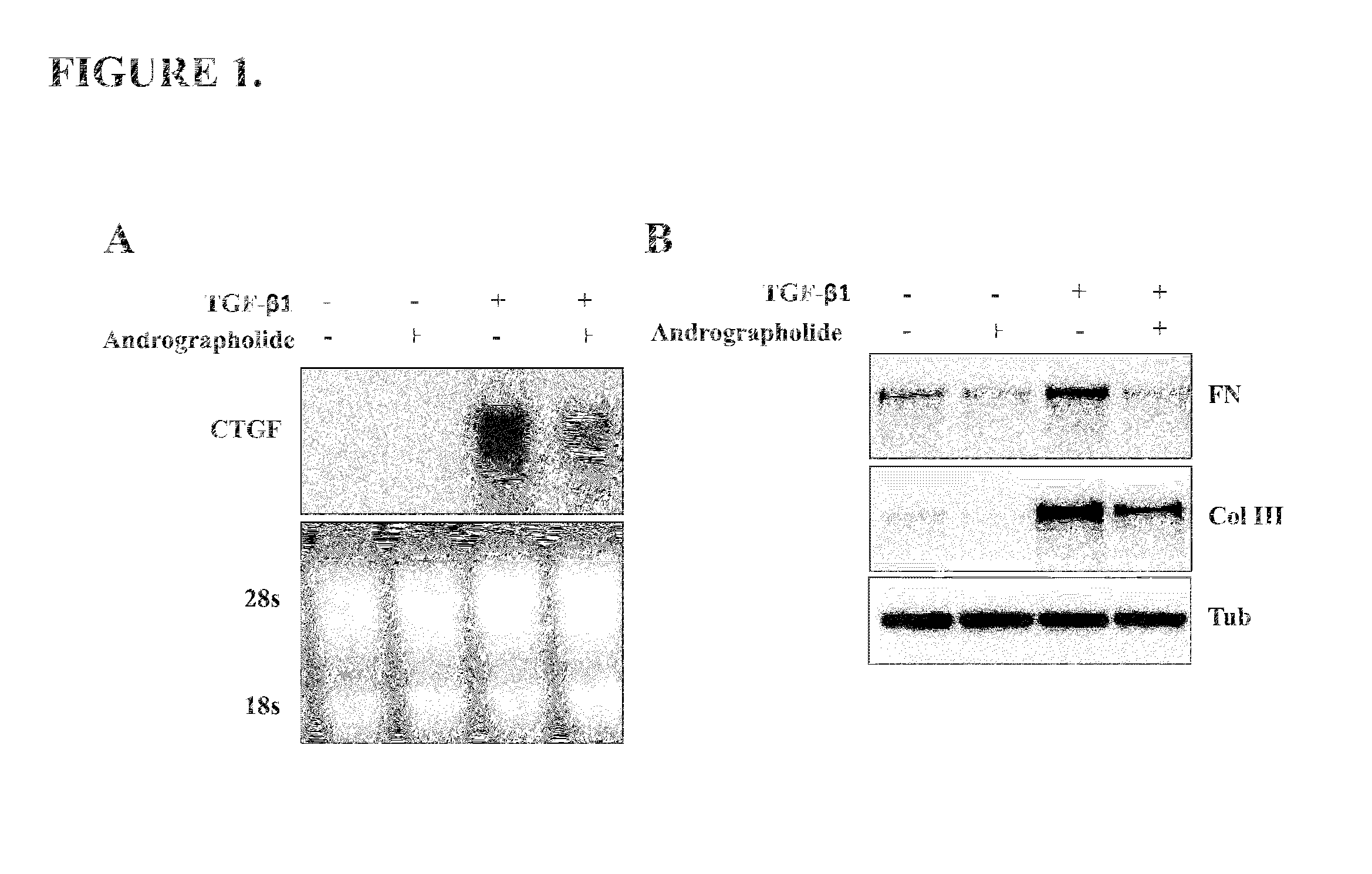
[0047]To evaluate if andrographolide could be an anti-fibrotic factor we determine in vitro the mRNA levels of two known pro-fibrotic factors: Connective tissue growth factor (CTGF) and Transforming growth factor type beta 1 (TGF-β1) (Cabello-Verrugio et al., 2012; Morales et al., 2011). TGF-β1 induces the expression of CTGF in skeletal muscle cells (Vial et al., 2008). The FIG. 1A shows that andrographolide reduced the induction of CTGF expression in response to TGF-β1. A molecular feature of fibrotic diseases is the accumulation ECM molecules such as collagen and fibronectin, both molecules are induced by TGF-β1. FIG. 1B shows that andrographolide decreased both fibronectin and collagen type III protein levels, induced by TGF-β1 in vitro.

example 2

Effect of Andrographolide on TGF-131 in Mdx Mice

[0048]Since we show that andrographolide have anti-fibrotic effects in vitro, we decide to evaluate these results in vivo. We previously showed the antifibrotic effects of andrographolide in vitro, thus we decide to evaluate the andrographolide properties in an animal model of the disease.

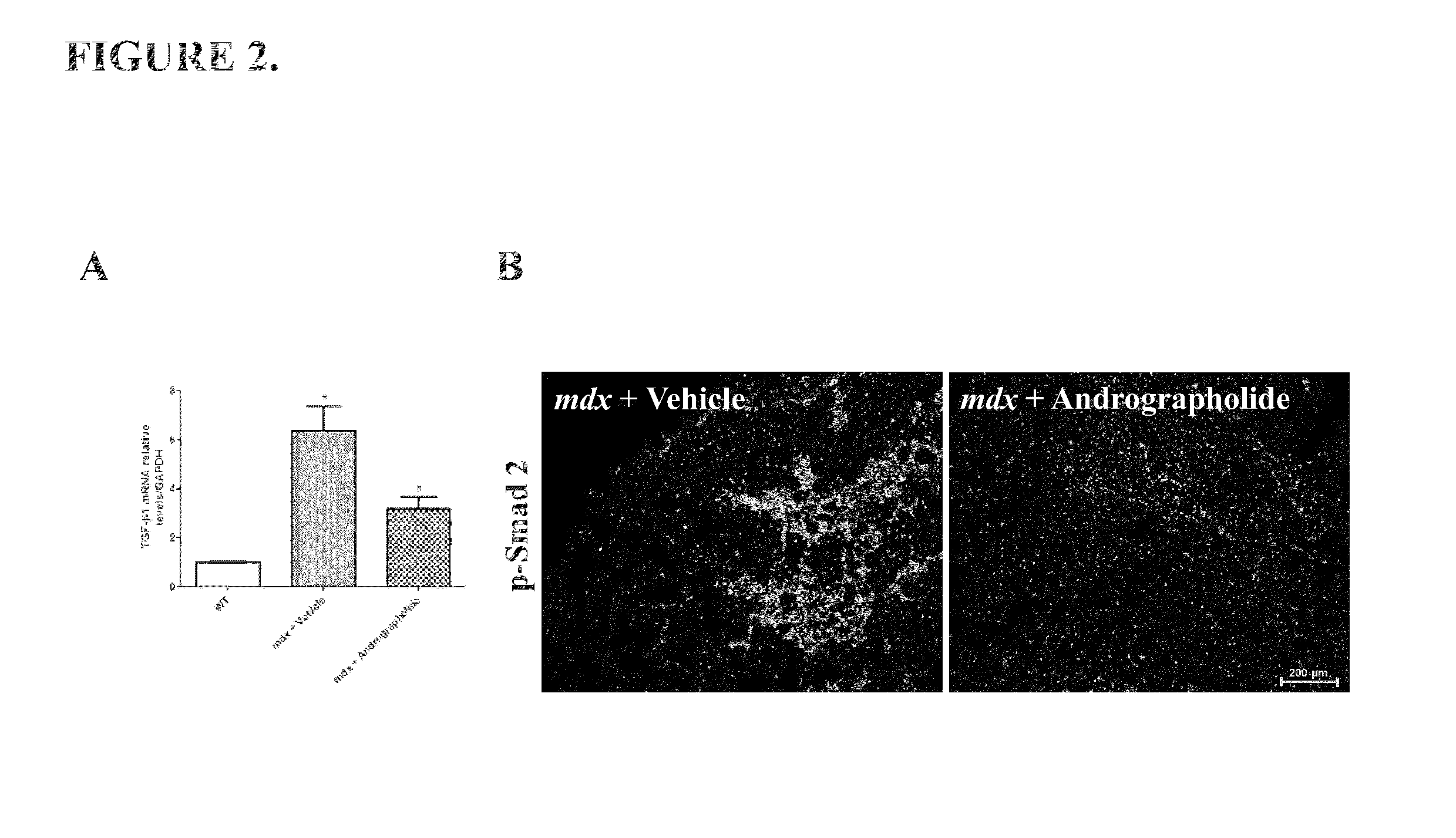
[0049]The pro-fibrotic cytokine TGF-β1 is augmented in mdx mice, which is related with the induction of skeletal muscle fibrosis (Andreetta et al., 2006). Therefore we evaluate if andrographolide could modulate the expression of TGF-β1 in vivo. FIG. 2A shows that andrographolide reduced the expression of TGF-β1 in mdx mice.

[0050]The canonical signaling pathway induced by TGF-β1 is through phosphorylation of smad proteins. Thus we evaluate the activity of TGF-β1 canonical signaling pathway by immunofluorescence of phosphorylated smad2 protein (p-Smad 2). FIG. 2B shows that andrographolide reduced the number of positive nuclei for phosphorylated smad2 p...

example 3

Effect of Andrographolide on CTGF Action In Vivo

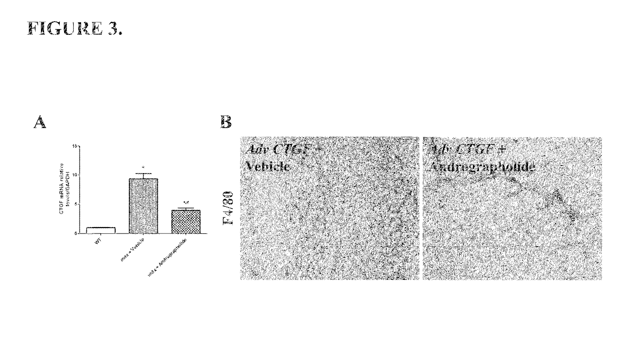
[0051]Another pro-fibrotic cytokine overexpressed in the skeletal muscle is CTGF (Morales et al., 2011). FIG. 3A shows that andrographolide reduced CTGF expression in mdx mice. Moreover, andrographolide inhibits the pro-inflammatory effects of CTGF in vivo. Overexpression of CTGF by an adenovirus induces inflammation and fibrosis in wild type muscles (WT), showing similar features of dystrophic muscles (Morales et al., 2011). However, andrographolide inhibited these effects. FIG. 3B shows that andrographolide reduced the number of F4 / 80 (a macrophages specific marker) positive cells (Tidball and Villalta, 2010).


PUM

PropertyMeasurementUnit
Timeaaaaaaaaaa
Timeaaaaaaaaaa
Strengthaaaaaaaaaa

Abstract

A method of treating a muscular dystrophy disease in a patient includes administering an effective amount of a botanical drug isolated from Andrographis paniculata in combination with cell therapy. The method improves skeletal muscle performance.

Description

FIELD OF THE INVENTION[0001]The current invention is related to a method for treating muscular dystrophies using a combination of therapies which found to be more effective than those therapies applied individually.[0002]Specifically, the current invention refers to the use of a botanical drug isolated from Andrographis paniculata together with a cell therapy for the treatment of Muscular Dystrophies, e.g., Duchenne muscular dystrophy (DMD).[0003]In this invention, we demonstrated the impact of andrographolide in the progression of dystrophic diseases, evaluating fibrosis induction, muscle strength and finally we demonstrated that andrographolide generates a propitious niche to increase stem cell therapy engraftment.BACKGROUND OF THE INVENTION[0004]Muscular Dystrophies are a group of muscular genetic diseases. The most severe is Duchenne muscular dystrophy (DMD). DMD is an X-linked recessive disorder that affects 1 of 3500 birth, for which there is no effective therapy (Kapsa et al....

Claims


Application Information

Patent Timeline
IPC IPC(8): A61K36/19A61K31/365A61K35/34
CPCA61K36/19A61K31/365A61K35/34A61P21/00
Inventor BRANDAN, ENRIQUECABRERA, DANIELGUTIERREZ, JAIMEMORALES, GABRIELACABELLO-VERRUGIO, CLAUDIOHANCKE, JUAN
Owner PONTIFISIA UNIVERSIDAD KATOLIKA DE CHILE
Learn More