
Methods for diagnosing vitreo-retinal disease

Active Publication Date: 2014-09-18
RIVERSIDE RES INST +1
View PDF4 Cites 1 Cited by
  • Summary
  • Abstract
  • Description
  • Claims
  • Application Information

AI Technical Summary

Benefits of technology

The patent describes a method for characterizing vitreous inhomogeneities, which are changes in the eye's vitreous body that can indicate the presence of disease. The method involves using ultrasound to collect data on the vitreous and analyzing it to determine parameters such as size, shape, and distribution. These parameters can be used to diagnose vitreo-retinal diseases at an early state before outcomes become unavoidable. The technique provides an objective means of diagnosing vitreo-retinal diseases and monitoring their progression. The global parameter derived from the ultrasound data can also be used to diagnose other health conditions of the eye.

Problems solved by technology

Macromolecular changes occurring during this process result in inhomogeneities within the vitreous.
Unfortunately, because no current diagnostic technology is able to detect the precursors of diabetic retinopathy, the disease is typically only detected in the aforementioned advanced states at which point treatments are costly, difficult to implement, carry some risk to patient or may be too late to prevent blindness.
The formation of liquid vitreous in myopia markedly destabilizes vitreous and threatens the retina because this process occurs relatively early in life and is not concurrent with dehiscence at the vitreo-retinal interface.
Myopic patients have a much greater risk of retinal detachment and, without treatment, blindness.
However, nothing is known about the size, number, or location of floaters as they relate to disease progression and normal aging.
Optical coherence tomography (OCT) permits visualization of abnormalities in the vicinity of the vitreo-retinal interface, but does not permit assessment of peripheral retinal pathology or imaging of the sclera and orbital tissues, nor does it allow assessment of motility of vitreous membranes in response to saccades, which is readily accomplished with real-time ultrasound.

Method used


Image

  • Methods for diagnosing vitreo-retinal disease
  • Methods for diagnosing vitreo-retinal disease
  • Methods for diagnosing vitreo-retinal disease

Examples


example 1

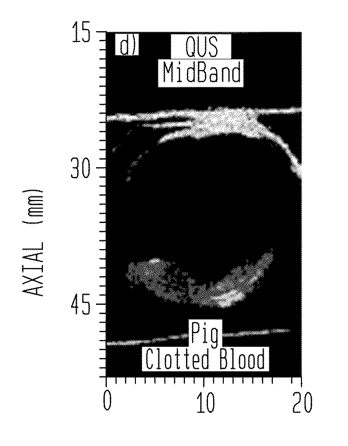
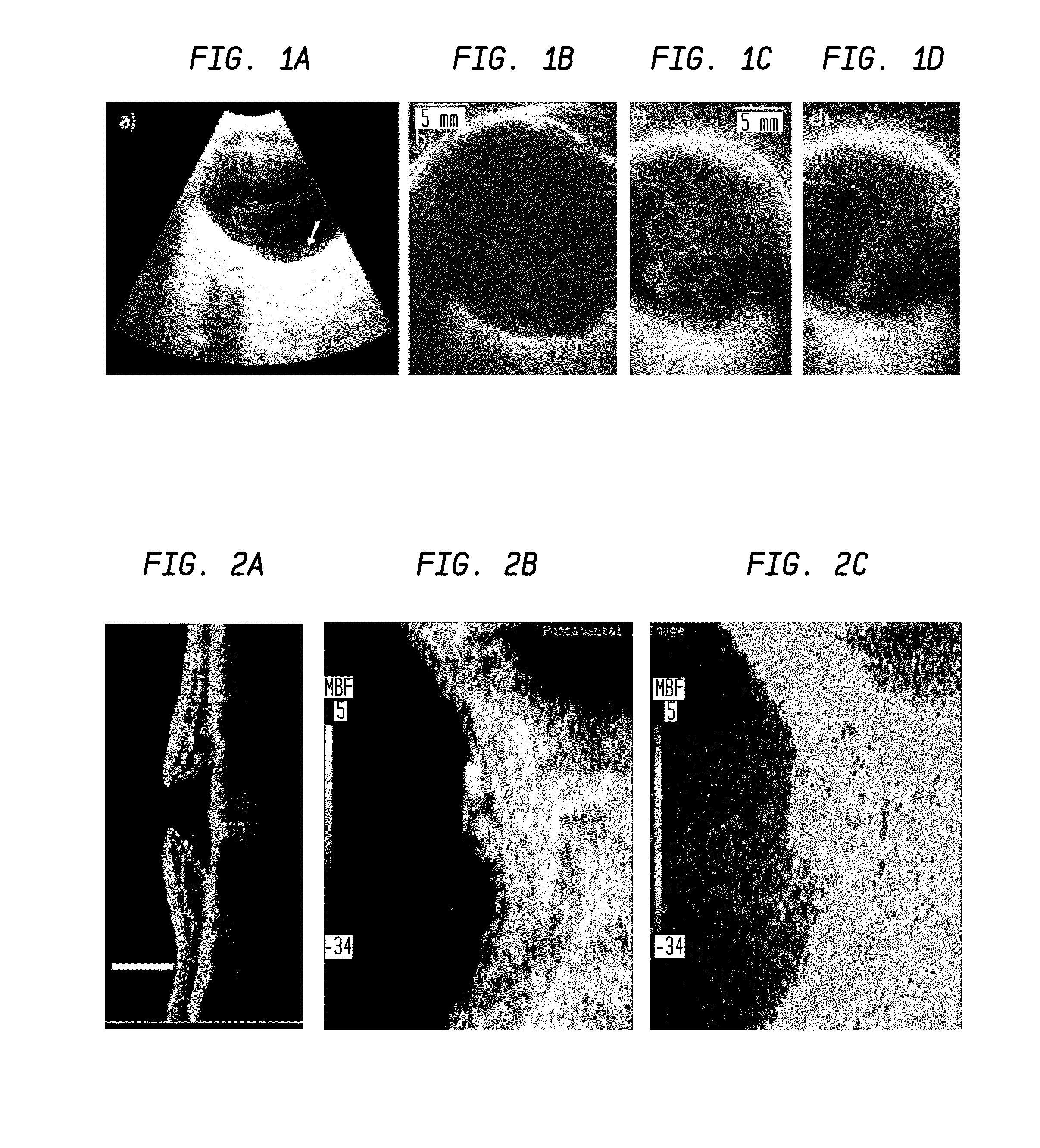
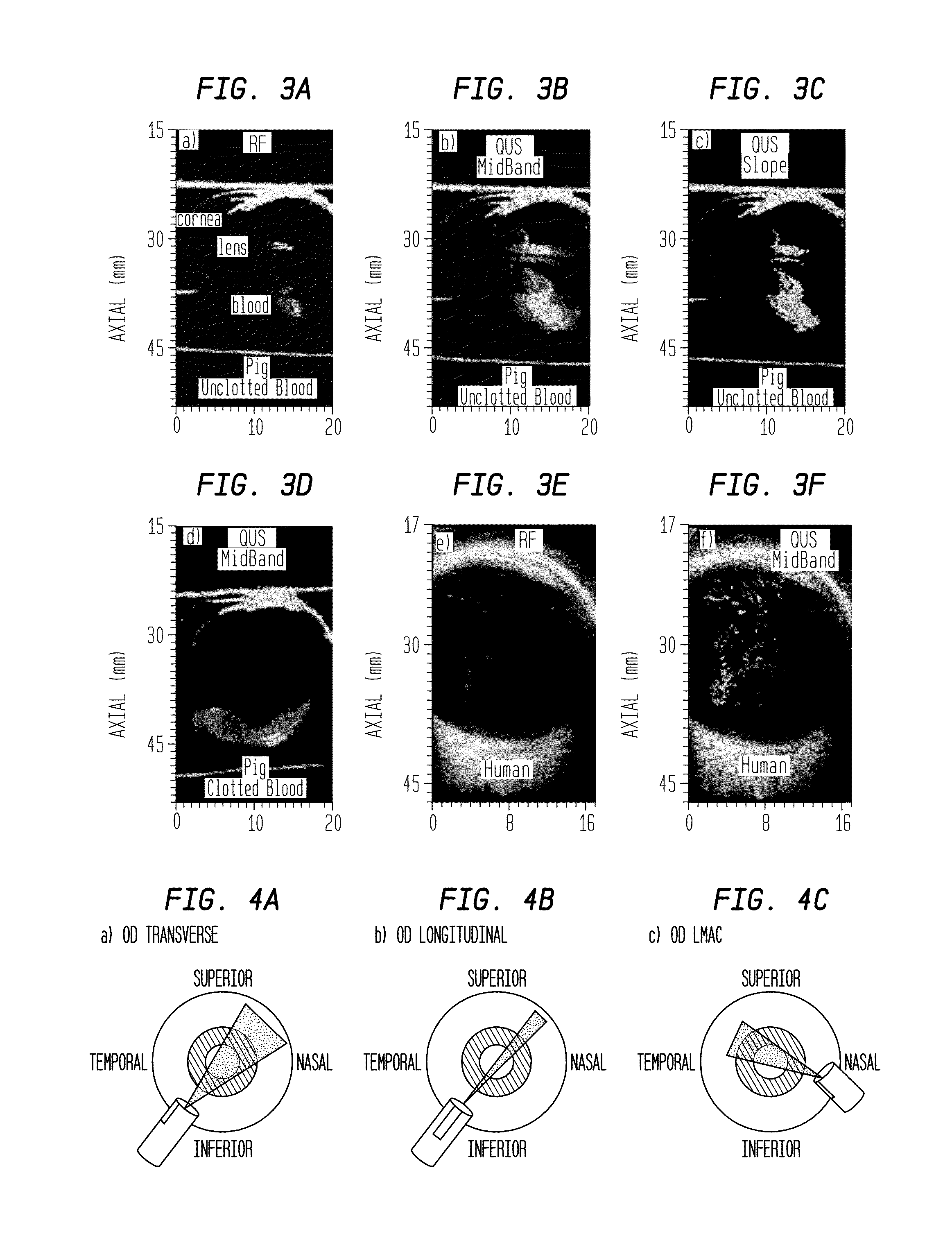
[0036]One example of a method to characterize inhomogeneities of the vitreous is provided. First, phase-resolved echo data are acquired from planes within the vitreous using a 20 MHz, high-frequency-ultrasound annular array. Planar sections are acquired in a series of parallel planes such that a 3D volume is sampled. Next, the data are normalized and processed to obtain local-ROI QUS estimates. The large collection of local-ROI estimates are reduced to a set of global parameters that have clinical relevance related to the state of the vitreous. The global parameters along with relevant non-ultrasound clinical global parameters obtained from numerous patients are entered into a database. The database contains cases of normal vitreous at various ages and disease cases. Using the global parameters in the database, a classifier is trained to distinguish between healthy vitreous and vitreo-retinal disease. Once the classifier is trained, a look-up table can be created such that new globa...

example 2

[0039]In another embodiment of the invention, image-processing methods are employed to derive global index or end value of inhomogeneities (floaters) in patients who are candidates for vitrectomy. Patients could have developed floater etiologies related to posterior vitreous detachment, myopic vitreopathy or other common conditions of the eye.

[0040]An ophthalmic, clinical ultrasound unit with a single-element, 15-MHz transducer with a 20-mm focal length and 7-mm aperture is used to acquire 100 image frames of log-compressed envelope data before scan conversion and video display. These data are digitally sampled at 40-MHz with 8-bit accuracy before envelope detection. All the settings on the ultrasound machine are kept the same in order to allow for direct comparison between patients. An exam was then performed by placing the ultrasound probe directly on the globe at the limbus, to avoid attenuation by the eyelid and lens and to minimize trauma to the cornea.

[0041]As illustrated in F...


PUM


Abstract

A quantitative method to analyze phase-resolved, raw backscatter echo data or the envelope of phase-resolved, raw backscatter echo data to characterize vitreous inhomogeneities as they relate to normal aging and vitreo-retinal disease. The technique can be applied to 2D or 3D data acquired from the vitreous. The approach provides an objective end value to characterize the vitreous and provide a tool for early diagnosis, monitoring and planning treatment of vitreo-retinal diseases.

Description

BACKGROUND OF THE INVENTION[0001]The young, healthy vitreous is a homogeneous, optically transparent gel filling the posterior segment of the eye. Gel vitreous volume increases during the first decade while the eye is growing in size and then remains stable until about the age of 40 years, when it begins to decrease in parallel with an increase in liquid vitreous. Macromolecular changes occurring during this process result in inhomogeneities within the vitreous. Concurrently, changes occurring at the vitreo-retinal interface allow the posterior vitreous cortex to detach from the internal ILL of the retina. When liquefaction and dehiscence occur simultaneously and in concert, the result is an innocuous posterior vitreous detachment (PVD). When the natural processes do not occur in concert, an anomalous PVD (APVD) may develop. The ability to diagnose precursors of APVD and to differentiate normal changes in vitreous related to aging from abnormal changes would permit early interventio...

Claims


Application Information

Patent Timeline
IPC IPC(8): A61B8/10
CPCA61B8/10A61B8/5223G16H50/30
Inventor KETTERLING, JEFFREY A.MAMOU, JONATHANSILVERMAN, RONALD H.SEBAG, JERRY
Owner RIVERSIDE RES INST
Learn More