
Image processing device and method for operating endoscope system

Active Publication Date: 2014-09-11
FUJIFILM CORP
View PDF4 Cites 7 Cited by
  • Summary
  • Abstract
  • Description
  • Claims
  • Application Information

AI Technical Summary

Benefits of technology

This patent is about an image processing device and method for operating an endoscope system that can better display the ductal structure without making blood vessels noticeable. The technical effect is an enhanced display of the ductal structure without making the blood vessels a significant component of the image.

Problems solved by technology

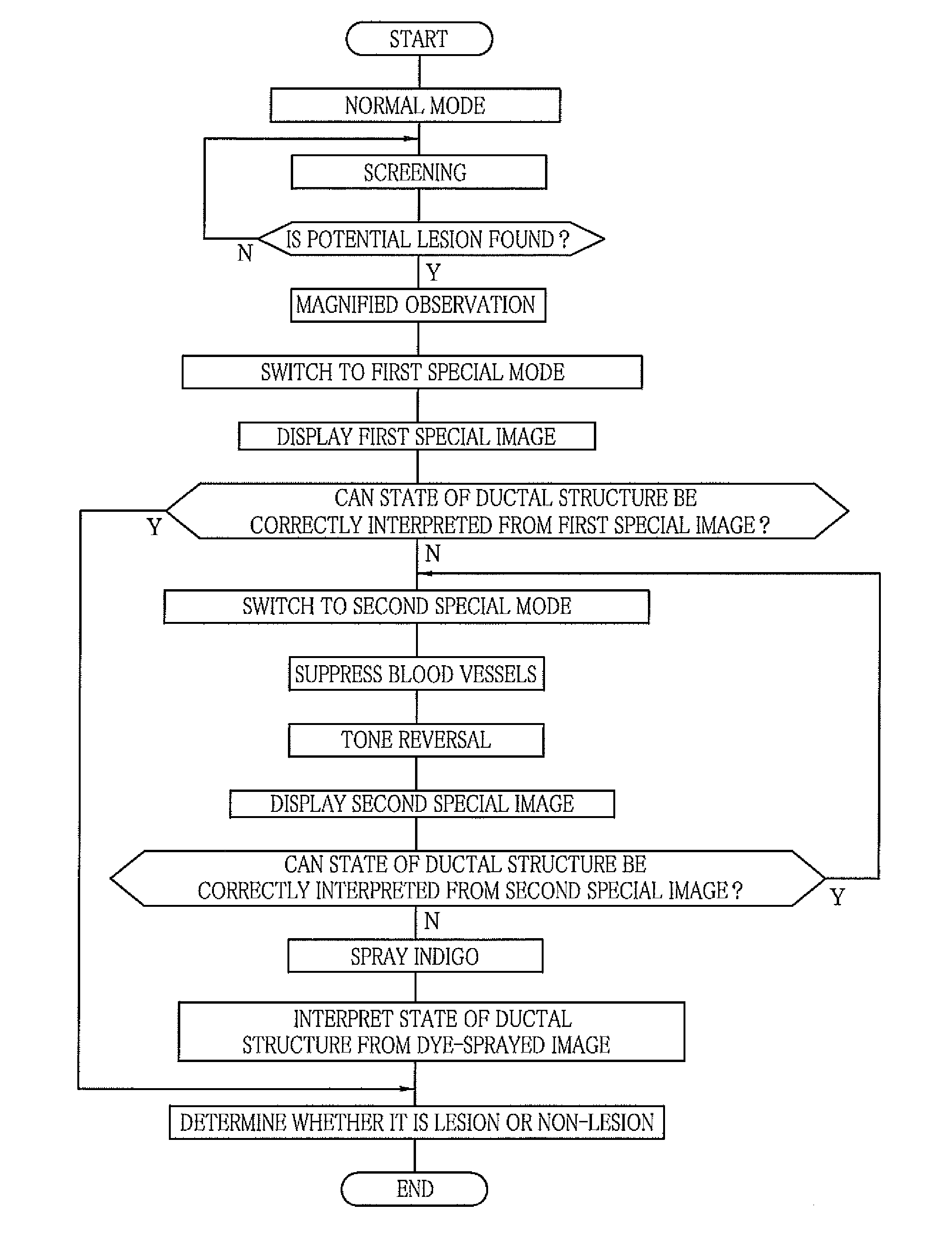
It is difficult to interpret the state of the ductal structure S. In this case, a dye such as an indigo is sprayed.
The doctor who is accustomed to the dye observation has difficulties in observing the ductal structure S displayed in a color different from that of the dye such as the indigo.
However, neither of the above-mentioned documents including the Japanese Patent No. 3572304 touches upon displaying the ductal structure with high clarity in the narrowband light observation and enhancing the ductal structure in the display without making the blood vessels conspicuous.

Method used


Image

  • Image processing device and method for operating endoscope system
  • Image processing device and method for operating endoscope system
  • Image processing device and method for operating endoscope system

Examples


first embodiment

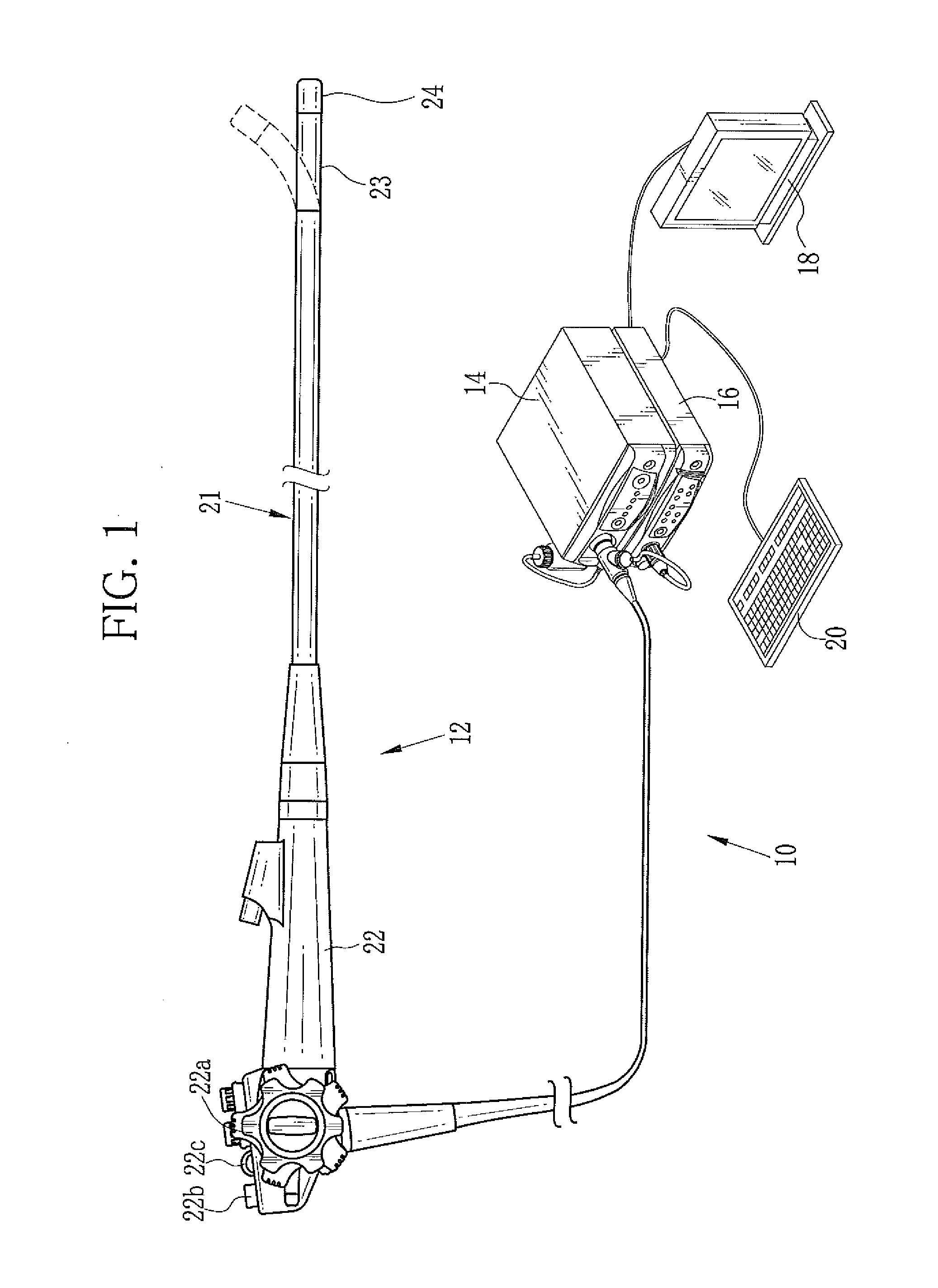
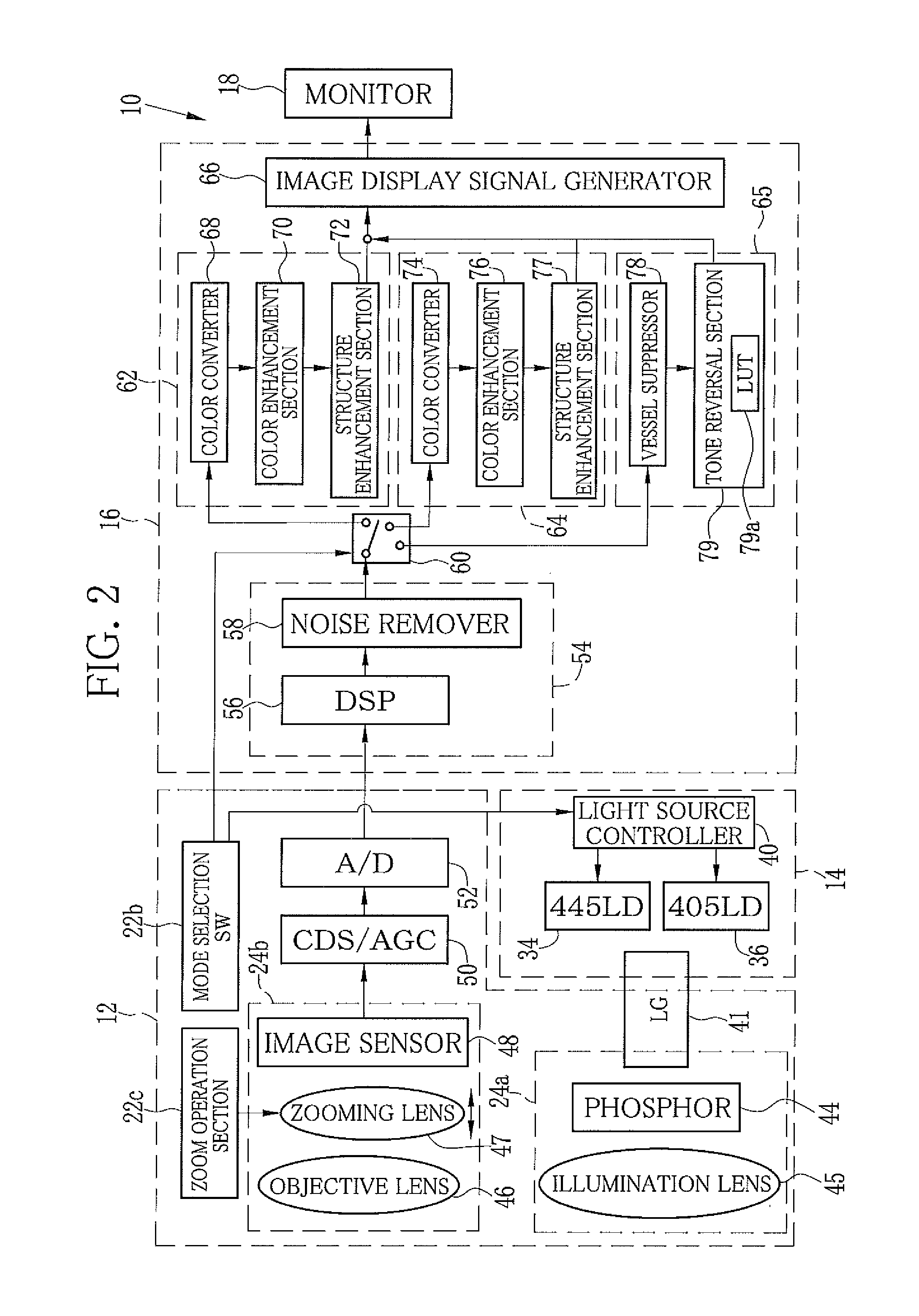
[0053]As shown in FIG. 1, an endoscope system 10 according to a first embodiment has an endoscope 12, a light source device 14, a processor device 16, a monitor 18, and a console 20. The endoscope 12 is optically connected to the light source device 14. The endoscope 12 is electrically connected to the processor device 16. The endoscope 12 has an insert section 21, a handle section 22, a bending portion 23, and a distal portion 24. The insert section 21 is inserted in a body cavity. The handle section 22 is provided in a proximal portion of the insert section 21. The bending portion 23 and the distal portion 24 are provided on a distal side of the insert section 21. The bending portion 23 is bent by operating an angle knob 22a of the handle section 22. The bending portion 23 is bent to direct the distal portion 24 to a desired direction.

[0054]In addition to the angle knob 22a, the handle section 22 is provided with a mode selection SW (switch) 22b and a zoom operation section 22c. T...

second embodiment

[0093]In the first embodiment, the tone reversal process made the ductal structure conspicuous, as if the indigo has been sprayed thereon. Additionally, the tone reversal process of the second embodiment makes the color of the mucous membrane close to a color obtained by the image capture using white light and makes the color of the ductal structure closer to the color of the indigo than that in the first embodiment. In the second embodiment, the RGB image data of the vessel-suppressed image is inputted and the tone is reversed based on tone curves 180a to 180c for the RGB image data shown in FIG. 19. Thereby the RGB image data of the suppressed-and-reversed image is outputted.

[0094]The tone curve 180a for the R image data is convex-shaped, so that the outputted R image data is slightly greater than the inputted R image data, in intermediate values. The tone curve 180c for the B image data is concave-shaped, so that the outputted B image data is slightly smaller than the inputted B ...


PUM


Abstract

A vessel suppression process is performed on RGB image data. A display of capillary vessels is suppressed by the vessel suppression process. After the vessel suppression process, tone of the RGB image data is reversed. Thereby, a suppressed-and-reversed image is produced. Even after the tone reversal, the capillary vessels do not interfere with observation of a ductal structure in the suppressed-and-reversed image, because the display of the capillary vessels is suppressed. In the suppressed-and-reversed image, the ductal structure is darker than a mucous membrane due to the tone reversal, so that the color of the ductal structure is close to that of an indigo.

Description

BACKGROUND OF THE INVENTION[0001]1. Field of the Invention[0002]The present invention relates to an image processing device and a method for operating an endoscope system, for performing diagnoses based on observation of a ductal structure and the like in a human body.[0003]2. Description Related to the Prior Art[0004]Diagnoses using endoscope systems are widely performed in current medical care. The endoscope system comprises a light source device, an electronic endoscope, and a processor device. Observation of a human body cavity is performed using the endoscope system. Such observation includes screening observation and magnified observation. In the screening observation, a potential lesion (site with a high possibility of being a lesion) such as a brownish area or redness is detected from a far view. The magnified observation is performed in a case where the potential lesion is detected. In the magnified observation, the endoscope zooms in on the potential lesion. Thereby the po...

Claims


Application Information

Patent Timeline
IPC IPC(8): G06T11/00
CPCG06T11/001G06T2207/10024G06T2207/10068G06T2207/30101A61B2576/00G06T7/0012A61B1/043G02B23/2461A61B1/0638G16H30/40A61B1/000094H04N23/56G06T5/90G06T2207/30004
Inventor KUBO, MASAHIRO
Owner FUJIFILM CORP
Learn More