Methods for Generating Cardiomyocytes
a technology of cardiomyocytes and fibroblasts, applied in the direction of genetically modified cells, skeletal/connective tissue cells, peptide/protein ingredients, etc., can solve the problem of limited current therapeutic approaches
- Summary
- Abstract
- Description
- Claims
- Application Information
AI Technical Summary
Benefits of technology
Problems solved by technology
Method used
Image
Examples
example 1
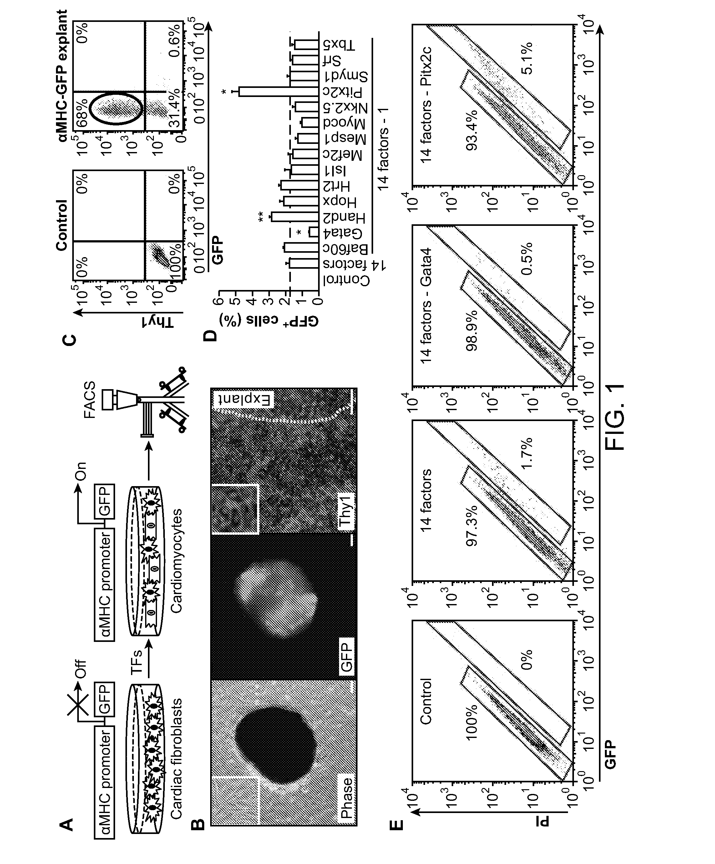
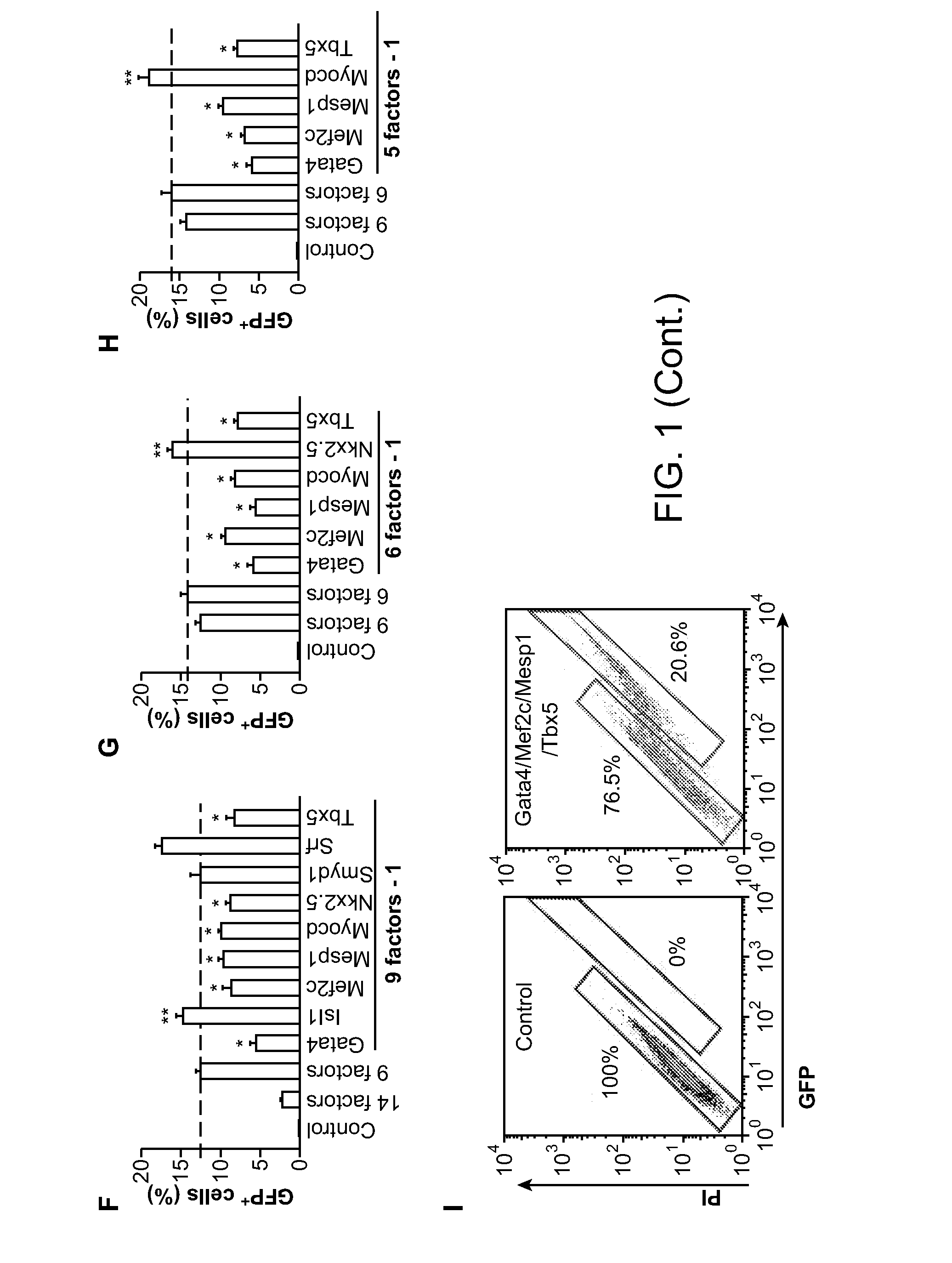
Direct Reprogramming of Cardiac Fibroblasts into Functional Cardiomyocytes by Defined Factors
Materials and Methods
Generation of αMHC-GFP and Isl1-YFP Mice
[0259]To generate α-myosin heavy chain-green fluorescent protein (αMHC-GFP) mice, enhanced green fluorescent protein-internal ribosome binding site-puromycinR (EGFP-IRES-Puromycin) cDNA were subcloned into the expression vector containing α-myosin heavy chain (α-MHC) promoter (Gulick et al. (1991) J Biol Chem 266, 9180-9185). Pronuclear microinjection and other procedures were performed according to the standard protocols (Ieda et al. (2007) Nat Med 13, 604-612. The transgene was identified by polymerase chain reaction (PCR) analysis (the forward primer, 5′-ATGACAGACAGATCCCTCCT-3′ (SEQ ID NO:11); the reverse primer, 5′-AAGTCGTGCTGCTTCATGTG-3′ (SEQ ID NO:12)). Isl1-yellow fluorescent protein (Isl1-YFP) mice were obtained by crossing Isl1-Cre mice and R26R-enhanced yellow fluorescent protein (R26R-EYFP) mice (Srinivas et al. (2001) B...
example 2
Direct Reprogramming of Cardiac Fibroblasts into Functional Cardiomyocytes
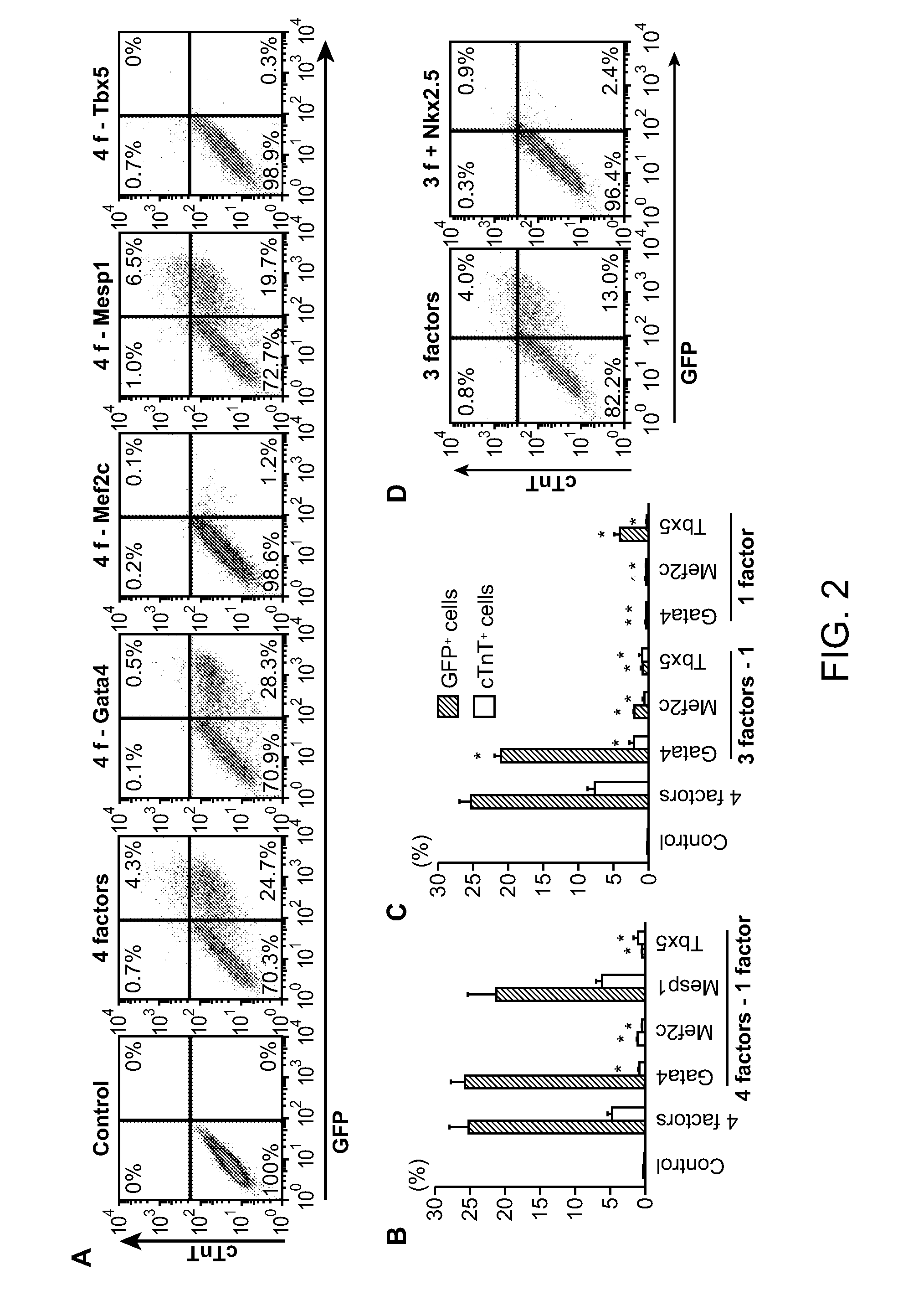
[0304]Using methods essentially as described in Example 1, exogenous Gata4, Tbx5, and Mef2c were introduced into mouse post-natal tail tip fibroblasts. About 20% to 30% of the post-natal tail tip fibroblasts were reprogrammed to myosin heavy chain-GFP+ cells (cardiomyocytes) following introduction of Gata4, Tbx5, and Mef2c (where Gata4, Tbx5, and Mef2c are collectively referred to as GMT).
example 3
In Vivo Reprogramming of Murine Cardiac Fibroblasts into Cardiomyocytes
Materials and Methods
[0305]Retrovirus Generation, Concentration and Titration.
[0306]Retroviruses were generated as described in Example 1. The pMXs retroviral vectors containing coding regions of Gata4, Mef2c, Tbx5, and dsRed were transfected into Plat-E cells with Fugene 6 (Roche) to generate viruses. Ultra-high titer virus (>1×101° plaque-forming units (p.f.u) per ml) was obtained by standard ultracentrifugation. Retroviral titration was performed using Retro-X qRT-PCR Titration Kit (Clontech).
[0307]Animals, Surgery, Echocardiography and Electrocardiography.
[0308]Postn-Cre; R26R-lacZ mice were obtained by crossing Periostin-Cre mice (Snider et al. (2009) Circulation Res. 105:934) and Rosa26-lacZ mice (Soriano (1999) Nat. Genet. 21:70). Postn-Cre; R26R-EYFP mice were obtained by crossing Periostin-Cre mice and Rosa26-EYFP mice (Srinivas et al. (2001) supra). All surgeries and subsequent analyses were performed i...
PUM
| Property | Measurement | Unit |
|---|---|---|
| time | aaaaa | aaaaa |
| time | aaaaa | aaaaa |
| time | aaaaa | aaaaa |
Abstract
Description
Claims
Application Information
 - R&D
- Intellectual Property
- Life Sciences
- Materials
- Tech Scout
- Unparalleled Data Quality
- Higher Quality Content
- 60% Fewer Hallucinations
Browse by: Latest US Patents, China's latest patents, Technical Efficacy Thesaurus, Application Domain, Technology Topic, Popular Technical Reports.
© 2025 PatSnap. All rights reserved.Legal|Privacy policy|Modern Slavery Act Transparency Statement|Sitemap|About US| Contact US: help@patsnap.com


