


Quick Research
Generate reliable direction feasibility study reports for your R&D in just a few steps.

Technical Q&A
Discover and master advanced knowledge NOW. Basics, ideas, possibilities, all at once.

Find Solutions
As an expert in R&D theories, this can generate solutions to your technical problems instantly.

Evaluate Feasibility
Analyze your overall solution with one click, know your potential R&D risks in advance.

Monitor Landscape
Get weekly tech updates, stay abreast of the latest tech innovations and key insights.
Colorimetric gelatinase assay
a gelatinase and colorimetric technology, applied in the field of colorimetric gelatinase assay, can solve the problems of inability to evaluate gelatinase activity, laborious, skillful and time-consuming (about a week) lab work, and minimal work in adapting the lateral flow assay format for detecting enzyme activity
- Summary
- Abstract
- Description
- Claims
- Application Information
AI Technical Summary
Benefits of technology
Problems solved by technology
Method used
Image


Examples
first embodiment
Lateral Flow
[0028]The first embodiment of the present invention relates to a lateral flow-based, colorimetric, gelatinase (MMP-2 and MMP-9) assay test. The assay contains at least the following four elements: a sample pad, a reagent pad, a membrane, and an absorbent pad, in that order.
[0029]The first element, i.e., the sample pad, has the capacity to transport fluid spontaneously and passively by capillary action. The sample pad draws up a volume of sample fluid obtained from, for example, wound exudate, urine, tears, saliva and equine synovial fluid.
[0030]The sample pad can be made of, for example, woven meshes and cellulose filters such as porous paper. Woven meshes, sometimes called screens, normally work very well to distribute the sample volume evenly to the reagent pad. They also typically have good tensile strength and handle well, even when wet. Meshes have very low bed volumes, meaning that they retain very little sample volume, normally 1-2 μL / cm2. On the other hand, it is...
second embodiment
Microwell Strip
[0053]The second embodiment of the present invention is a microwell strip-based embodiment. While metallic colloids of varying kinds can be used in this embodiment (e.g., silver colloid, gold colloid, etc.), colloidal gold will be discussed in the description of this embodiment as an exemplary metallic colloid. The scope of this embodiment is not limited to colloidal gold.
[0054]Colloidal gold is manufactured reproducibly and inexpensively at room temperature without use of dispersals which facilitates coating with gelatin. The metal hydrosol is prepared at room temperature in a size range of 50-55+ / −2 nm and has an intense ruby red coloration. Any larger particle size and the color violet making any blue color from flocculation less distinguished. Recombinant human gelatin (from FibroGen, 100 kD) is bloomed and melted in buffer at 50° C. then chemititrated at different pH levels onto the particles. This produces the lowest detection limit. Finally, additives such as e...
third embodiment
Whole Tube
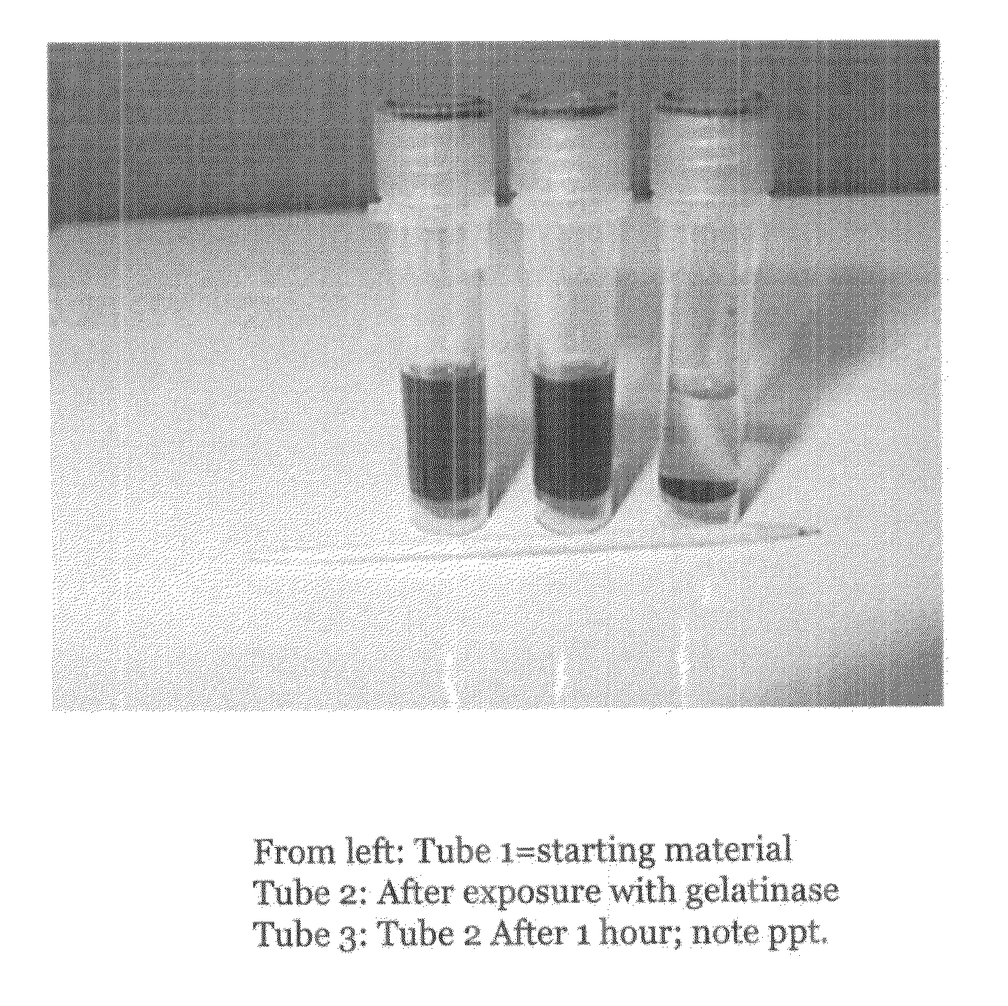
[0057]The third embodiment of the present invention is a whole tube-based embodiment. While metallic colloids of varying kinds can be used in this embodiment (e.g., silver colloid, gold colloid, etc.), colloidal silver will be discussed in the description of this embodiment as an exemplary metallic colloid. The scope of this embodiment is not limited to colloidal silver.
[0058]Beef gelatin, colloidal silver (300 ppm from Purest Colloids, NJ) and electrolyte buffers are chemititrated in the same manner as described above in the second embodiment. The mixture is contained in individual microvials of 1-2 ml capacity which have been previously treated with a siliconizing agent (e.g., Rain-X). The sample fluid (e.g., a wound, urine, tears, saliva, equine synovial fluid) collector in this embodiment is a dot of paper made from punching out a circle from absorbent paper. In this case, the circle of paper has only so much capacity to hold liquid (e.g., about 0.1 ml). It is removed ...
PUM
 Abstract
Description
Claims
Application Information
 - R&D Engineer
- R&D Manager
- IP Professional
- Industry Leading Data Capabilities
- Powerful AI technology
- Patent DNA Extraction
Browse by: Latest US Patents, China's latest patents, Technical Efficacy Thesaurus, Application Domain, Technology Topic, Popular Technical Reports.
© 2024 PatSnap. All rights reserved.Legal|Privacy policy|Modern Slavery Act Transparency Statement|Sitemap|About US| Contact US: help@patsnap.com