
Linear magnetic drive transducer for ultrasound imaging

a magnetic drive and ultrasound imaging technology, applied in the field of ultrasound imaging devices, can solve the problems of difficult to perform certain procedures that require the use of a second hand, small devices, and high cost of array transducer devices for current probe-based ultrasound systems, and achieve the effect of low manufacturing cost and high performance of ultrasound imagers

Active Publication Date: 2013-12-26
RGT UNIV OF MICHIGAN
View PDF9 Cites 35 Cited by
  • Summary
  • Abstract
  • Description
  • Claims
  • Application Information

AI Technical Summary

Benefits of technology

The present patent describes a low-cost ultrasound imaging system that is compact and easy to use in various medical applications, such as placement of central vein catheters or assessment of carotid artery stenosis. The system uses innovative engineering design, accurate track and hold, low-power and low-bandwidth analog to digital converters, a motion feedback electro-magnetic transducer drive system, and distributed intelligent signal processing architecture. It can also measure certain parameters, such as elasticity and shear adjustment, using a closed-loop control configuration. The server stores the raw data streams, and higher-level modules are provided to perform user-customizable analysis of the data. The system is designed to provide high-quality ultrasound images for diagnostic use.

Problems solved by technology

Generally speaking, current probe-based ultrasound systems are expensive array transducer devices that employ a large array of transducer elements used to scan an ultrasound beam over an area of interest.
The devices are small, but still cumbersome in that the operator (e.g., physician, pharmacist, or other health care provider) hold and scan the probe with one hand and hold the display / processing unit in the other hand.
This can make it difficult to perform certain procedures that require the use of another hand.
The result is that the ultrasound image is not always easily kept within the field of view of the operator; and, either way, the operator is limited in where they can position the display because it must be within their visual range to discern important details in the image, should ideally be within the working area of the procedure, and must be tethered to the ultrasound probe.
Some have proposed modifications to such devices, such as, a tethered interface that uses a removable connection, including a docking station; but the need to control the operation of the probe still requires tethering during operation.
Furthermore the devices are expensive, in large part because all image processing is performed on the portable device, which requires expensive signal processing circuitry and the associated programming.

Method used


Image

  • Linear magnetic drive transducer for ultrasound imaging
  • Linear magnetic drive transducer for ultrasound imaging
  • Linear magnetic drive transducer for ultrasound imaging

Examples


Embodiment Construction

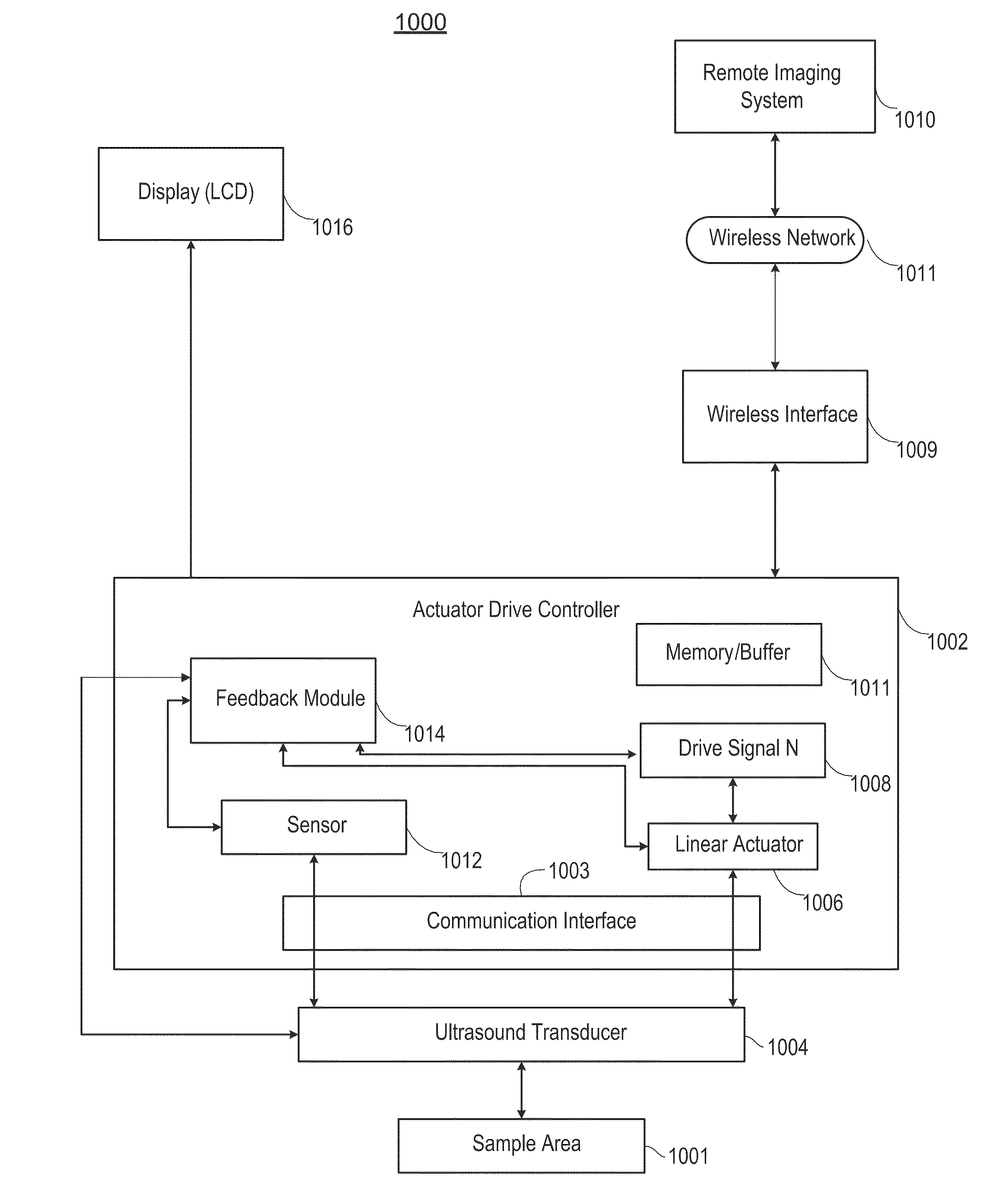
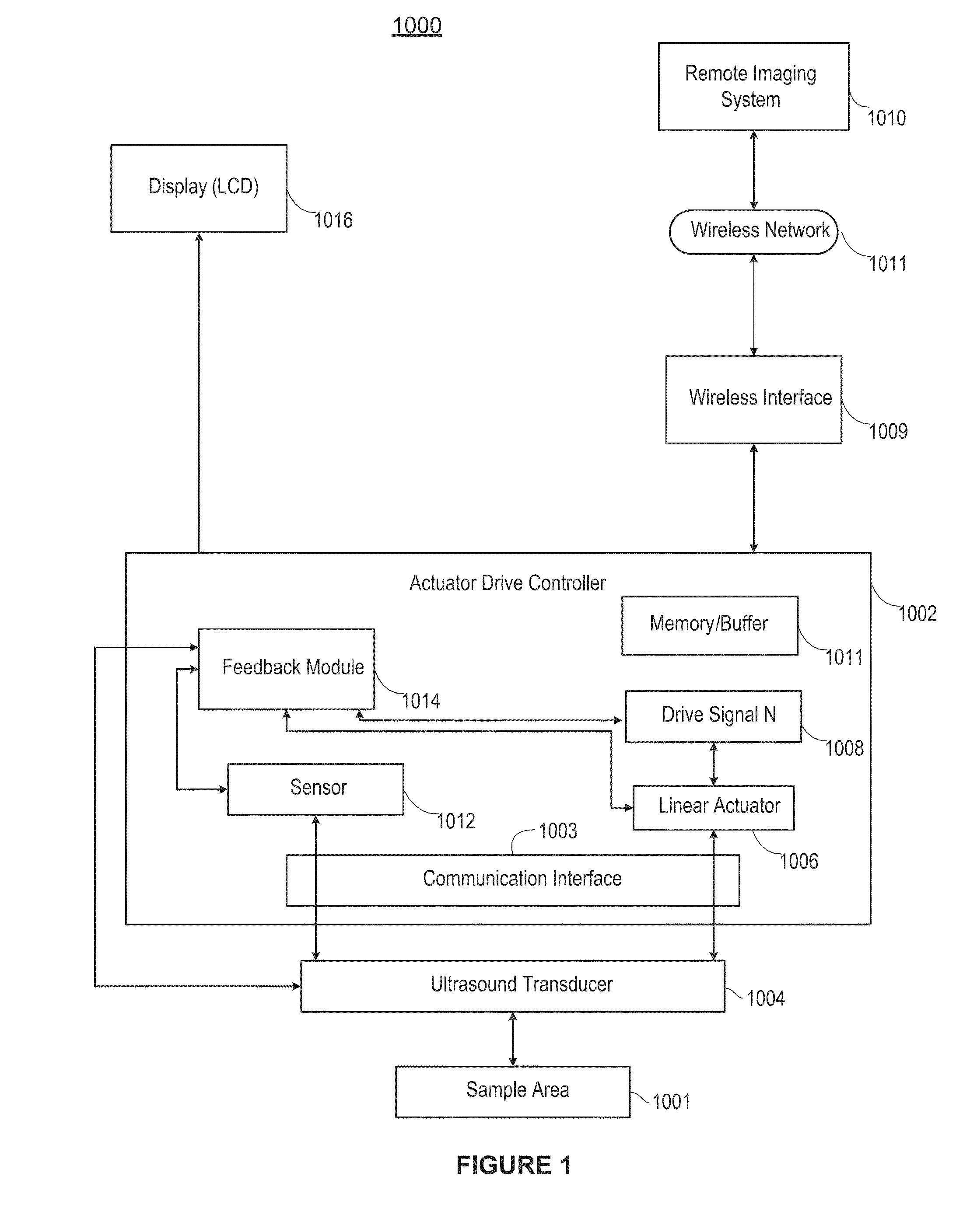
[0057]FIG. 1 illustrates an ultrasound Doppler imaging system 1000 in accordance with an example implementation of the present techniques. Generally, the techniques provide a first device that has a single (or perhaps few) transducer elements (i) to generate an ultrasound signal and (ii) to transmit the signal data to a second device for processing the signal, a receiver to receive the processed information, and a screen display to display processed data, where the receiver and display screen may be an integrated device including the first device, or a separate device connected thereto through a wired or wireless connection. The techniques may include, for example, a simplified mechanical drive system to collect ultrasound data from a plurality of locations or orientations, and a second device for processing the data received from the first device (i.e. for distributed signal processing) and transmitting the processed data to the first device for display (or to the receiver-display ...


PUM


Abstract

An ultrasound imaging system uses a magnetic linear motor driven ultrasound scanner, with accurate track and hold operation and / or other motion feedback, to scan a two dimensional or three dimensional area of a sample. The scanner is implemented in a low-power and low-bandwidth handheld device and is connected with a remote image processing system that receives raw data and performs full ultrasound image analysis and creation, allowing the handheld to be used for scanning, pre-processing, and display.

Description

CROSS-REFERENCE TO RELATED APPLICATIONS[0001]This application claims the benefit of U.S. Application Ser. No. 61 / 645,025, filed May 9, 2012, entitled “Linear Magnetic Drive Transducer For Ultrasound Imaging,” which is hereby incorporated by reference in its entirety.STATEMENT REGARDING FEDERALLY SPONSORED RESEARCH OR DEVELOPMENT[0002]This invention was made with government support under HL101881 awarded by the National Institutes of Health. The government has certain rights in the invention.FIELD OF THE INVENTION[0003]The invention relates generally to ultrasound imaging devices and, more particularly, to linear actuator-based ultrasound devices.BACKGROUND[0004]Ultrasound imaging has been a core technology used in the detection and treatment of many diseases and injuries. Ultrasound imagers use high-frequency sound waves to view soft tissues such as blood vessels, muscles, and internal organs, in real-time. The transducer sends out high-frequency sound waves that reflect off of body...

Claims


Application Information

Patent Timeline
IPC IPC(8): A61B8/00A61B8/08
CPCA61B8/4483A61B8/461A61B8/4427A61B8/488A61B8/0891A61B8/4461A61B8/4472G01S15/8945G01S15/8995G01S7/003G01S7/52065G01S15/8915G01S7/52042A61B8/56G01N29/222A61B8/483
Inventor WEITZEL, WILLIAMKRUGER, GRANTTHELEN, BRIANRUBIN, JONATHANKOZIOL, LEOBROOK, ROBERTMITRA, MAINAK
Owner RGT UNIV OF MICHIGAN
Learn More