Detection of a polypeptide dimer by a bivalent binding agent
a polypeptide dimer and bivalent binding technology, applied in the field of bivalent binding agents, can solve the problems homo- or heterodimer detection of such receptor molecules, and achieve the effect of solving the problem of a single, non-dimerized background of single molecules
- Summary
- Abstract
- Description
- Claims
- Application Information
AI Technical Summary
Benefits of technology
Problems solved by technology
Method used
Image
Examples
example 1
Bivalent Binding Agent to Troponin T
[0259]1.1 Monoclonal Antibodies and Fab′-Fragments
[0260]Two monoclonal antibodies binding to human cardiac Troponin T at different, non-overlapping epitopes, epitope A′ and epitope B′, respectively, were used. Both these antibodies are used in the current Roche Elecsys™ Troponin T assay, wherein Troponin T is detected in a sandwich immuno assay format.
[0261]Purification of the monoclonal antibodies from culture supernatant was carried out using state of the art methods of protein chemistry.
[0262]The purified monoclonal antibodies are protease digested with either pre-activated papain (anti-epitope A′ MAb) or pepsin (anti-epitope B′ MAb) yielding F(ab′)2 fragments that are subsequently reduced to Fab′-fragments, i.e. A and B, respectively, in Formula I (A-a′:a-S-b:b′-B), with a low concentration of cysteamin at 37° C. The reaction is stopped by separating the cysteamin on a Sephadex G-25 column (GE Healthcare) from the polypeptide-containing part o...
example 2
Bivalent Binding Agent to Phosphorylated IGF-1R
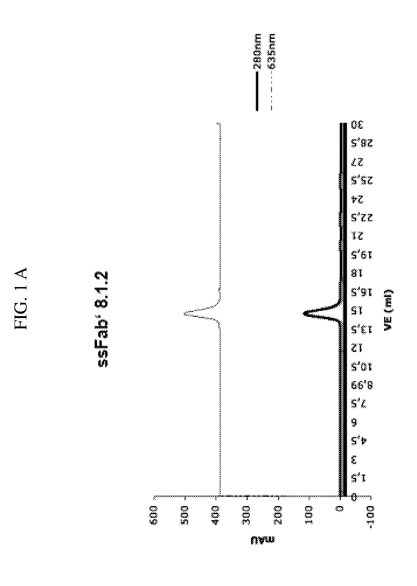
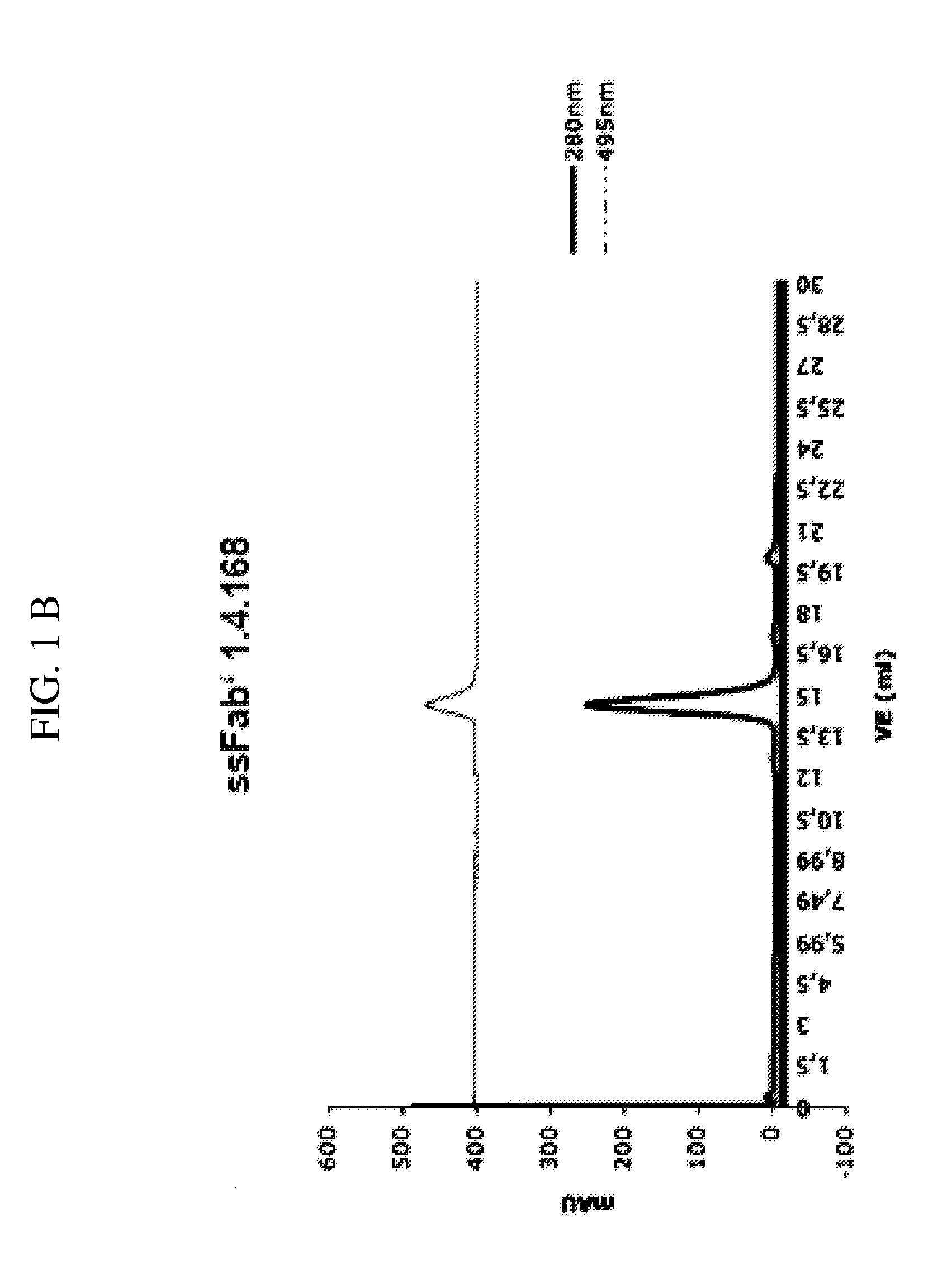
[0296]2.1 Monoclonal Antibody Development (mAb 8.1.2 and mAb 1.4.168)
[0297]a) Immunization of Mice
[0298]BALB / C mice are immunized at week 0, 3, 6 and 9, respectively. Per immunization 100 μg of the conjugate comprising the phosphorylated peptide pIGF-1R (1340-1366) (SEQ ID NO:11) is used. This peptide had been phosphorylated at tyrosine 1346 (=1346-pTyr) and coupled to KLH via the C-terminal cysteine (=Aoc-Cys-MP-KLH-1340) to yield the conjugate used for immunization. At weeks 0 and 6, respectively, the immunization is carried out intraperitoneally and at weeks 3 and 9, respectively, subcutanuosly at various parts of the mouse body.
[0299]b) Fusion and Cloning
[0300]Spleen cells of immunized mice are fused with myeloma cells according to Galfre G., and Milstein C., Methods in Enzymology 73 (1981) 3-46. In this process ca 1×10⁸ spleen cells of an immunized mouse are mixed with 2×10⁷ myeloma cells a(P3×63-Ag8653, ATCC CRL1580) and centrifug...
example 3
Bivalent Binding Agent—Binding to the Homodimeric Form of Insulin-Like Growth Factor 1
[0366]The insulin-like growth factor 1 receptor (IGF1-R) belongs to the family of tyrosin kinase receptors and it is activated by IGF1. The receptor is homodimeric.
[0367]An IGF1-R monomer consists of one extracellular a subunit which is covalently linked to a transmembrane β subunit via a disulfide bridge. Two monomers are covalently linked by a disulfite bridge between the a subunits of two monomers. The IGF1 receptor plays an important role in cancer, aging and insulin signalling.
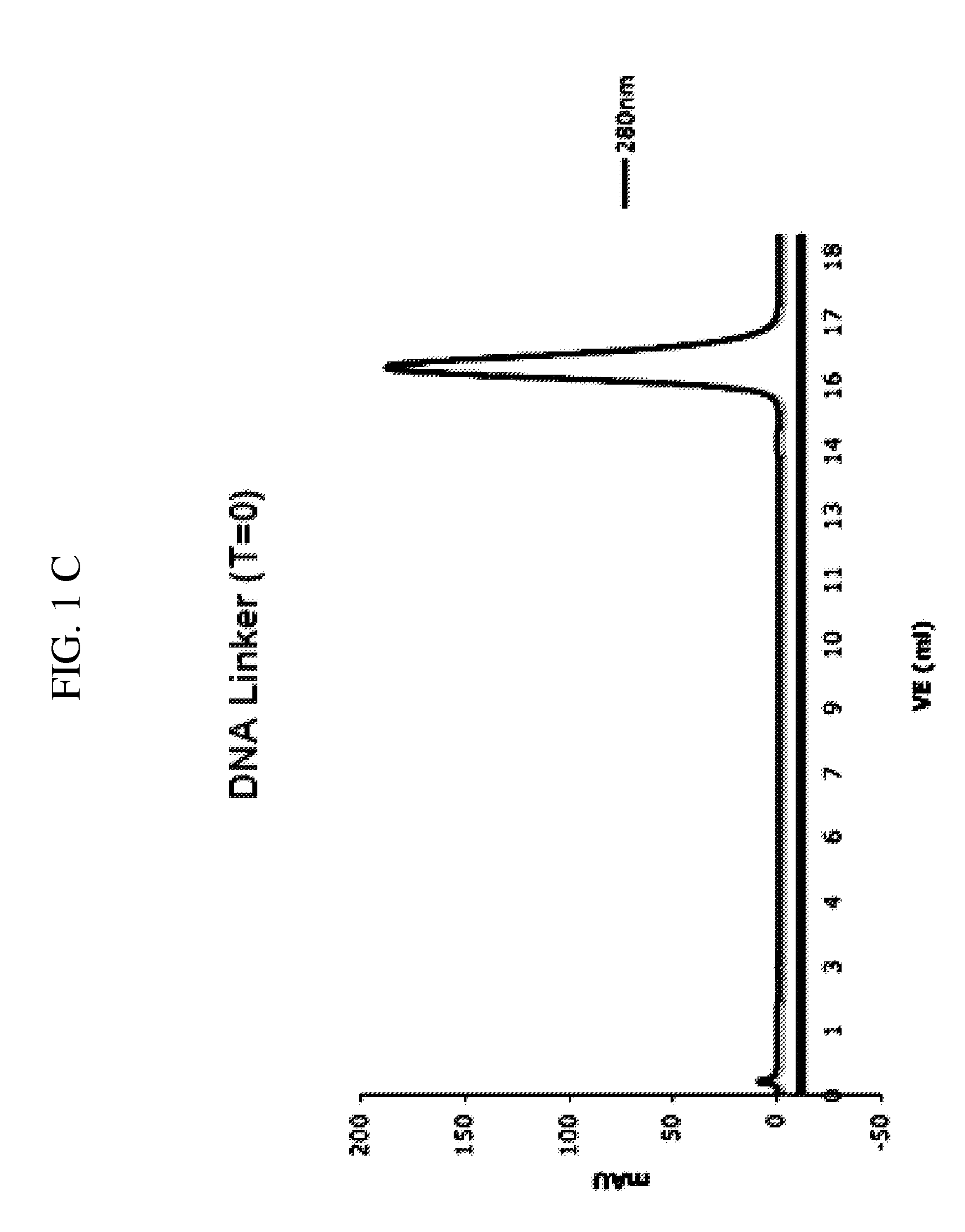
[0368]The dimer-binder, we developed, comprises two identical Fab′-fragments. Both these Fab′-fragments—when used individually—bind to their target peptide (IGF1-R) only with low complex stability. In order to produce a bivalent binding agent the Fab′-fragment is conjugated to a hybridizable oligonucleotide. An adaptor DNA capable of hybridizing with both the linker DNA as well as with this oligonucleotide is used as a b...
PUM
| Property | Measurement | Unit |
|---|---|---|
| Length | aaaaa | aaaaa |
Abstract
Description
Claims
Application Information
 - R&D
- Intellectual Property
- Life Sciences
- Materials
- Tech Scout
- Unparalleled Data Quality
- Higher Quality Content
- 60% Fewer Hallucinations
Browse by: Latest US Patents, China's latest patents, Technical Efficacy Thesaurus, Application Domain, Technology Topic, Popular Technical Reports.
© 2025 PatSnap. All rights reserved.Legal|Privacy policy|Modern Slavery Act Transparency Statement|Sitemap|About US| Contact US: help@patsnap.com


