
Detection of adenylate cyclase

a technology of adenylate cyclase and detection method, which is applied in the field of disease diagnosis, can solve the problems of cytokine dysregulation and immune dysfunction, shock, respiratory failure, death, etc., and achieve the effect of gentle and rapid separation

Inactive Publication Date: 2013-08-15
UNITED STATES OF AMERICA
View PDF1 Cites 3 Cited by
  • Summary
  • Abstract
  • Description
  • Claims
  • Application Information

AI Technical Summary

Benefits of technology

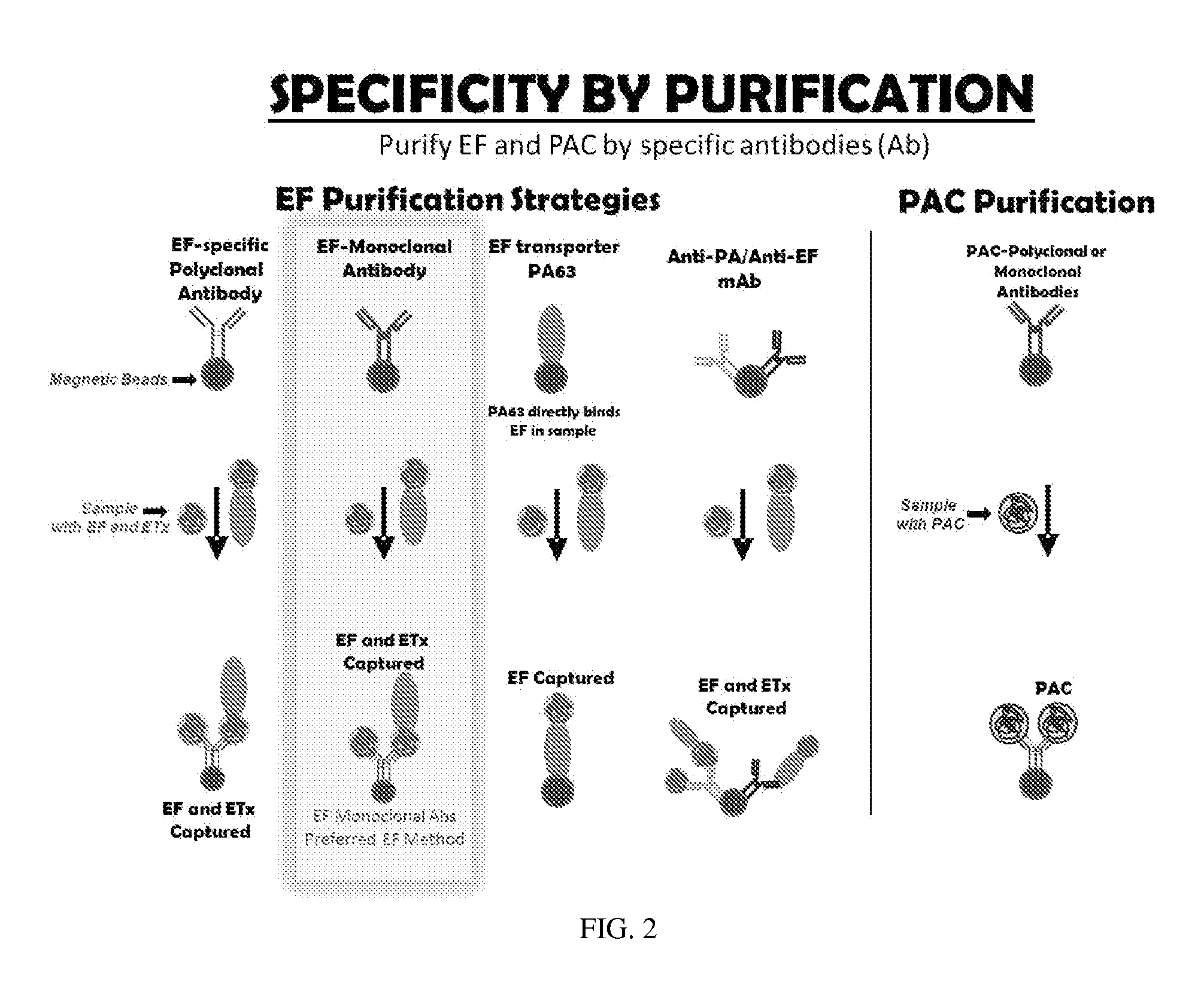
The patent describes a method for isolating and concentrating a specific form of an envelope protein called EF from a biological sample. This is done by using special beads that are attached to an antibody specific to either EF or a related protein called PA-EF. The beads can be magnetic, which makes it easy to separate them from other components of the sample. The purification can be carried out on a solid substrate like a microtiter plate. The antibodies used can be derived from various organisms like humans, mice, rabbits, etc. The patent also mentions that some embodiments can use both monoclonal and polyclonal antibodies.

Problems solved by technology

All three forms can progress to a systemic infection leading to shock, respiratory failure, and death.
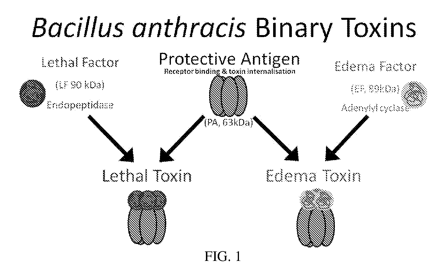
The cleavage of these proteins disrupts a signaling pathway and leads to cytokine dysregulation and immune dysfunction.
It is also known that there are fatal anthrax cases where administration of antibiotics and clearance of bacteria have failed to rescue the patient.
Assays for EF activity such as competitive enzyme assays (Duriez, E, et al., Anal. Chem., 2009; 81:5935-5941) or radiometric assays (Gottle, M, et al., Biochemistry, 2010; 49:5494-503), are impractical for high-throughput screening of compound collections and rapid diagnosis of host infection.

Method used


Image

  • Detection of adenylate cyclase
  • Detection of adenylate cyclase
  • Detection of adenylate cyclase

Examples


example 1

[0055]Preparation of Tosyl-Activated Magnetic Beads.

[0056]Tosyl-activated magnetic beads are obtained from Invitrogen. 20-100 μl of bead suspension are used to covalently link immunoglobulin (IgG) from a 100 μl sample containing IgG to the beads according to the manufacturer's protocol. To separate the beads, the reaction tube is placed on a magnet for 1 min and the resulting supernatant discarded by aspiration. The beads are resuspended in phosphate buffered saline with 0.05% Tween20, pH 7.3 (PBS-TW) and stored until ready for use. Thorough washing is achieved by repeating the magnetic pelleting and resuspension steps three times

example 2

[0057]Coating Tosyl-Activated Beads with Desired Anti-EF or Anti PAC Antibody.

[0058]Anti-EF, anti-PA, or anti-PAC (or other antibodies) are coated onto magnetic beads forming magnetic antibody beads (e.g. MABs). EF or PAC-specific MABs are prepared using mouse monoclonal anti-EF IgG or anti-PAC IgG according to the manufacturer's protocol (Invitrogen) using 40 μg IgG / 100 μl magnetic bead suspension.

example 3

[0059]Purification and Concentration of EF from Serum.

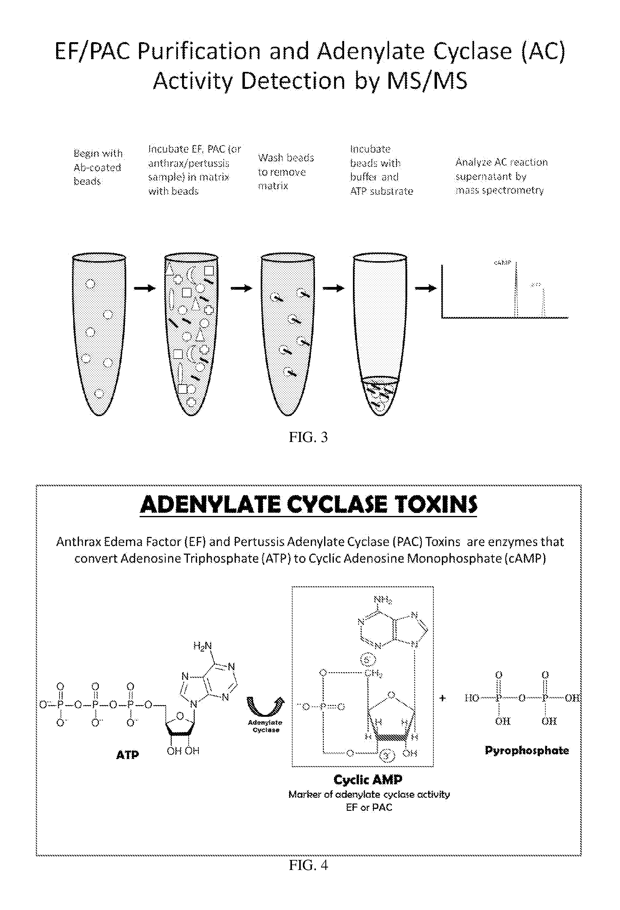
[0060]A serum, plasma, pleural fluid or other biological sample is obtained from a patient or infected animal. The sample is diluted 1:5 in 500 or 1000 μl PBS-TW and mixed gently with 20 μl EF MABs for 1 hour. The beads with EF and / or ETx bound antibody are retrieved, washed three times in PBS-TW and reconstituted in PBS-TW for further analyses by enzymatic reaction and mass spectrometry, as shown in FIGS. 2 and 3.

[0061]One approach for isolation of EF uses protective antigen (PA) antibody (anti-PA IgG) on MABs for total EF (EF+ETx) retrieval. The first step begins with addition of free activated PA63 that binds to free EF converting it into complexed form, ETx, rendering all EF as the complexed form ETx. Then a PA-MAB that is specific for the distal cell receptor binding portion of PA63 as depicted in FIG. 2 where the antibody binds to PA63 remote from the PA-EF (ETx) interface is used to capture the total EF as converted to ETx...


PUM

PropertyMeasurementUnit
volumesaaaaaaaaaa
pHaaaaaaaaaa
volumeaaaaaaaaaa

Abstract

One major problem in diagnosis methods presently available for anthrax is that these methods require several days to produce a result, are rendered unusable after antibiotic use, or are not quantifiable. The only existing treatment for anthrax requires administration soon after infection at a time when patients are exhibiting only mild flu-like symptoms. Thus, by the time a diagnosis is made a patient may be days beyond the time when treatment would be effective. The present invention reduces diagnosis time to as little as four hours providing same day identification of anthrax radically increasing the odds of delivering proper treatment and patient recovery. The rapid identification of anthrax edema factor activity exhibited by the invention is also amenable to in vivo screening protocols for the discovery and development of anthrax vaccines, anti-toxins and edema factor inhibitors. The invention isolates and concentrates edema factor and edema toxin from nearly any sample. By capitalizing on the adenylate cyclase activity of edema factor the invention amplifies output signals producing reliable detection of low concentrations of edema factor previously unachievable. The invention involves novel purification and detection techniques and substrates for rapid, reproducible, and quantitative measurements of anthrax edema factor, and other adenylate cyclases in biological samples.

Description

CROSS-REFERENCE TO RELATED APPLICATIONS[0001]This application is a U.S. National Phase of PCT / US2011 / 059,739, filed Nov. 8, 2011, which claims priority to U.S. provisional patent application 61 / 411,056, filed Nov. 8, 2010, both of which are incorporated herein in their entirety.GOVERNMENT INTEREST[0002]The invention described herein may be manufactured, used, and licensed by or for the United States Government.FIELD OF THE INVENTION[0003]The invention relates generally to disease diagnostics, and in particular to methods for detecting infection of anthrax in a patient and screening anthrax therapeutics.BACKGROUND OF THE INVENTION[0004]Anthrax is caused by infection with Bacillus anthracis, a spore-forming, rod-shaped bacterium. The dormant spore-form is highly resistant to extreme conditions, high temperatures, and a variety of chemical treatments. The spores gain entry either through an open wound, causing cutaneous disease, or by ingestion, causing gastrointestinal disease or are ...

Claims


Application Information

Patent Timeline
Patent Type & Authority Applications(United States)
IPC IPC(8): G01N33/569
CPCC12Q1/527G01N2333/988G01N2333/32G01N33/56911
Inventor BOYER, ANNE E.LINS, RENATO C.KUKLENYIK, ZSUZSANNAGALLEGOS-CANDELA, MARIBELQUINN, CONRAD P.BARR, JOHN R.
Owner UNITED STATES OF AMERICA
Learn More