
System and method for ultrasound analysis of biological structures

a biological structure and ultrasound technology, applied in the field of ultrasound apparatus, systems and methods for quantitative analysis of biological structures, can solve the problems of fatigue fractures commonly occurring in the lower limb, unreliable current technology used for detecting fatigue fractures, difficult and costly, etc., to reduce the transducer count, reduce the cost, and reduce the effect of pulse coupling

Inactive Publication Date: 2013-08-08
NEW JERSEY INSTITUTE OF TECHNOLOGY
View PDF2 Cites 0 Cited by
  • Summary
  • Abstract
  • Description
  • Claims
  • Application Information

AI Technical Summary

Benefits of technology

The LTP technology enhances detection resolution, reduces radiation exposure, and provides accurate, cost-effective quantitative analysis of bone fractures and soft tissue, minimizing errors and increasing accuracy by analyzing signal parameters such as amplitude, flight time, and correlation, effectively detecting fractures and tumors with improved spatial resolution and signal-to-noise ratio.

Problems solved by technology

Fatigue fractures are also known to occur in a deconditioned person who just started a new exercise program, such as military recruits.
Fatigue fractures commonly occur in lower limbs as a result of the ground reaction forces that must be dissipated during running, walking, marching or jumping.
However, current technology used in detecting fatigue fractures is often unreliable, difficult and costly.
Misdiagnosis in fracture detection partly arises from fractures that are simply difficult to see with x-rays.
Many types of fractures are easily missed in children.
The presence of a fatigue fracture may not show up on plain radiograph film for up to 2 to 10 weeks after symptom onset, which increases the likelihood of a nonunion.
However, using multiple x-rays at different vantage points to detect fatigue fractures requires the patient to absorb increased amounts of harmful radiation.
The inability and uncertainty in detecting fatigue fractures using plain radiograph films leave physicians to resort to costlier methods, such as bone scan, MRI or CT.
Using bone scan, MRI or CT to carry out the diagnosis increases the cost of the treatment.
Although these tests can be performed repeatedly throughout the fracture life span, the cost of the treatment and the time spent by physicians and technicians interpreting the data makes such an approach prohibitive.
Along with the cost and time required for qualitative methods there exists the possibility of errors in the readings.
These errors can be induced due to physicians' technique, perception, knowledge, judgment or communication.
Along with the errors, there are also differences in diagnoses among physicians.
In these devices only one parameter of acoustic signal is investigated, thus the signal cannot be quantified properly to yield a proper diagnosis.
Similarly, soft tissue analysis, detection and screening methods, such as those used for tumor detection, including x-ray (mammography), ultrasound and CT are expensive, inconvenient to use, and relatively unsafe due to their ionizing energy.
All of the prior art methods are based on visual examination and therefore require extensive time / effort and are subject to interpretation errors.
Traditionally, in order to achieve high enough resolution, the operating frequency must be increased, into the tens of megahertz range, thereby increasing the cost of instrumentation while reducing the depth of sound penetration and the amount of tissue information.
Furthermore, successive signal packets tend to interfere with each other due to transducer transients which further limit the detection resolution.
Pulse aliasing is a major problem: different echoes combine to form a single echo signal, thereby obscuring true signal characteristics (flight time, amplitude, etc.).

Method used


Image

  • System and method for ultrasound analysis of biological structures
  • System and method for ultrasound analysis of biological structures
  • System and method for ultrasound analysis of biological structures

Examples


Embodiment Construction

[0059]The following is a detailed description of the invention provided to aid those skilled in the art in practicing the present invention. Those of ordinary skill in the art may make modifications and variations in the embodiments described herein without departing from the spirit or scope of the present invention. Unless otherwise defined, all technical and scientific terms used herein have the same meaning as commonly understood by one of ordinary skill in the art to which this invention belongs. The terminology used in the description of the invention herein is for describing particular embodiments only and is not intended to be limiting of the invention. All publications, patent applications, patents, figures and other references mentioned herein are expressly incorporated by reference in their entirety.

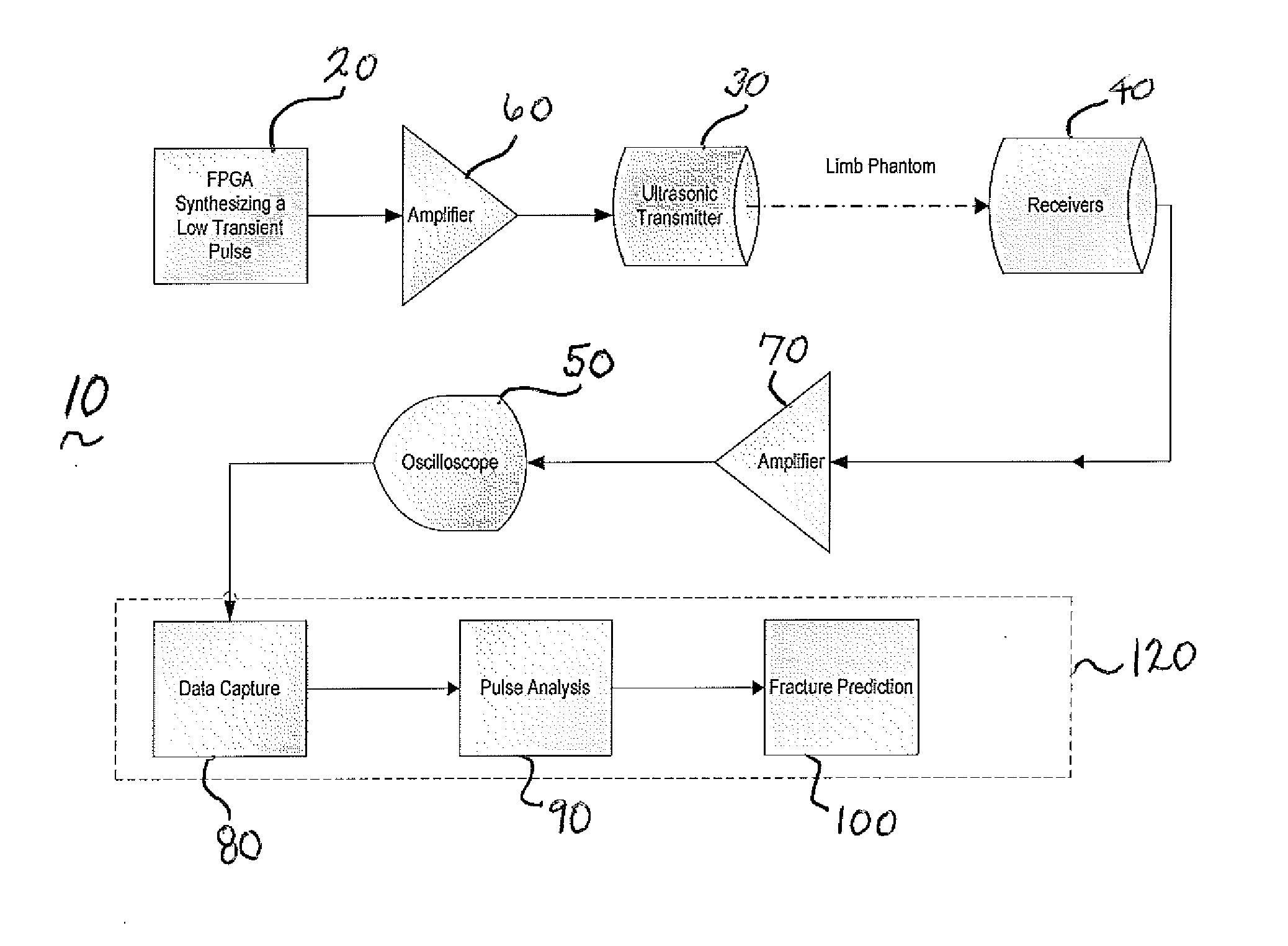
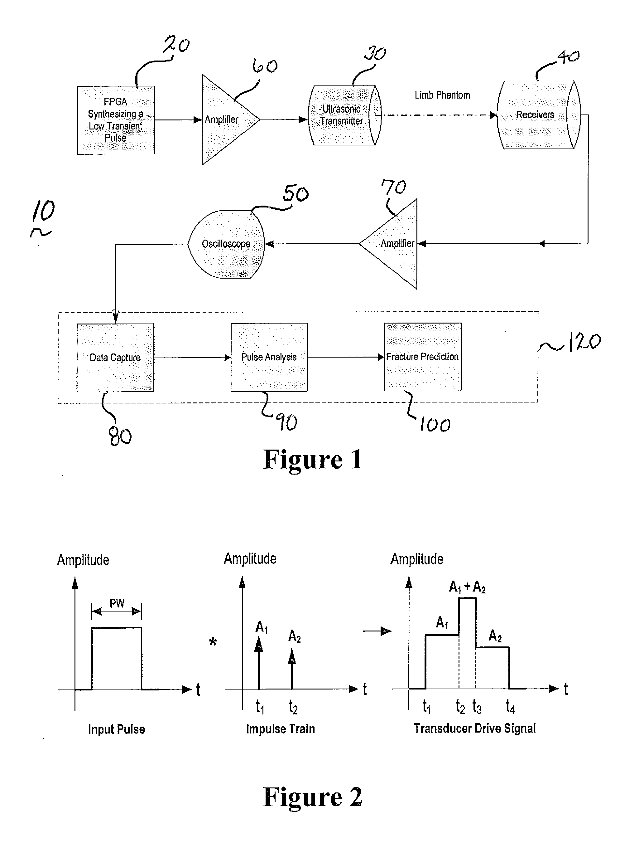
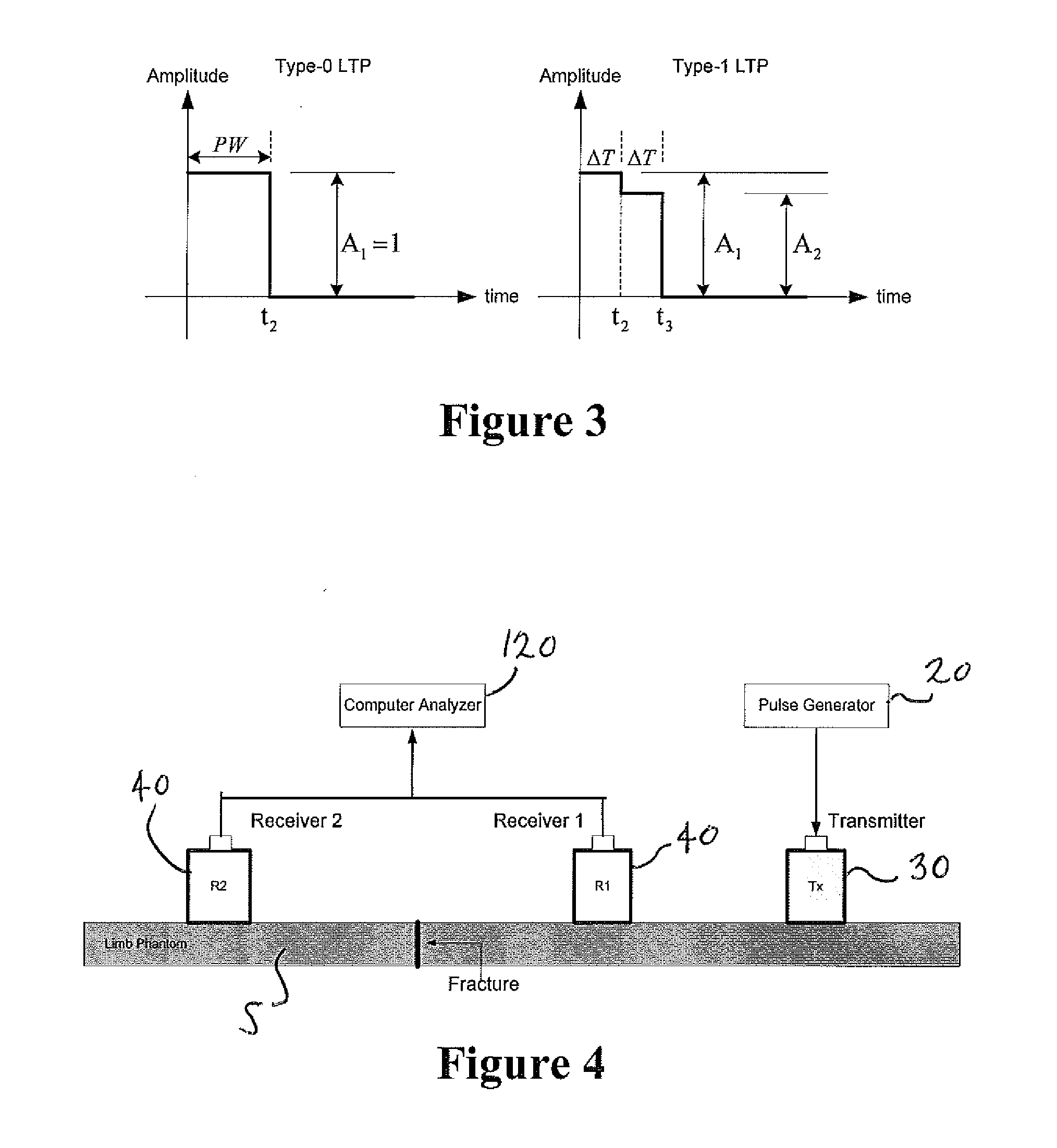
[0060]Now referring to FIG. 1 a system in accordance with at least one embodiment of the present invention is depicted. System 10 includes a signal generator 20, transmitter 30...


PUM


Abstract

Ultrasound systems and methods for detecting features of biological soft tissue are described. Systems and methods may employ low transient pulse technology. Methods employ detection and analysis of behavioral patterns of different signal parameters such as flight time, maximum amplitude, phase angle, and correlation for tumor detection and tissue analysis. Flight time and frequency components may be employed in tumor detection methods.

Description

CROSS REFERENCE TO RELATED APPLICATIONS[0001]This is a continuation of U.S. patent application Ser. No. 12 / 625,078 filed Nov. 24, 2009 and claims the benefit of U.S. Provisional Patent Application Ser. Nos. 61 / 118,261, filed Nov. 26, 2008 and 61 / 121,322, filed Dec. 10, 2008, the entireties of which are incorporated by reference herein.FIELD OF THE INVENTION[0002]This invention relates to ultrasound apparatus, systems and methods for detection and quantitative analysis of biological structures, particularly to analysis of skeletal and soft tissue structures using low transient pulse technology.BACKGROUND OF THE INVENTION[0003]Fatigue fractures, also known as stress fractures, are one type of incomplete fracture in bone. Fatigue fractures are often seen as fine disruptions of normal bone architecture. These micro fractures are caused due to an activity which exerts repetitive sub-threshold loading which over time exceeds bone's intrinsic ability to heal itself, for example running or ...

Claims


Application Information

Patent Timeline
Patent Type & Authority Applications(United States)
IPC IPC(8): A61B8/08
CPCA61B8/08A61B8/52G01S7/52036A61B8/0875
Inventor CHANG, TIMOTHY M.
Owner NEW JERSEY INSTITUTE OF TECHNOLOGY
Learn More