
Single-insertion, multiple sample biopsy device with integrated markers

a biopsy device and multiple sample technology, applied in the field of tissue biopsy sampling device, can solve the problems of inability to obtain more than, inaccurate post-biopsy diagnosis, pain and scarring of the body site, etc., and achieve the effect of preventing a vacuum

Active Publication Date: 2012-12-20
CR BARD INC
View PDF4 Cites 26 Cited by
  • Summary
  • Abstract
  • Description
  • Claims
  • Application Information

AI Technical Summary

Benefits of technology

Enables the collection of multiple tissue samples with reduced patient discomfort and accurate marker placement, improving diagnostic accuracy by allowing for multiple sample collection without additional needle insertions and ensuring precise marker deposition at the biopsied site.

Problems solved by technology

However, there are circumstances where there may be a need to obtain more than one samples.
While the known biopsy needle can be re-inserted multiple times, such technique can cause pain and scarring of the body site.
The additional step and device may not allow the marker to be deposited at the actual biopsied site, which can lead to inaccurate post-biopsy diagnosis.

Method used


Image

  • Single-insertion, multiple sample biopsy device with integrated markers
  • Single-insertion, multiple sample biopsy device with integrated markers
  • Single-insertion, multiple sample biopsy device with integrated markers

Examples


Embodiment Construction

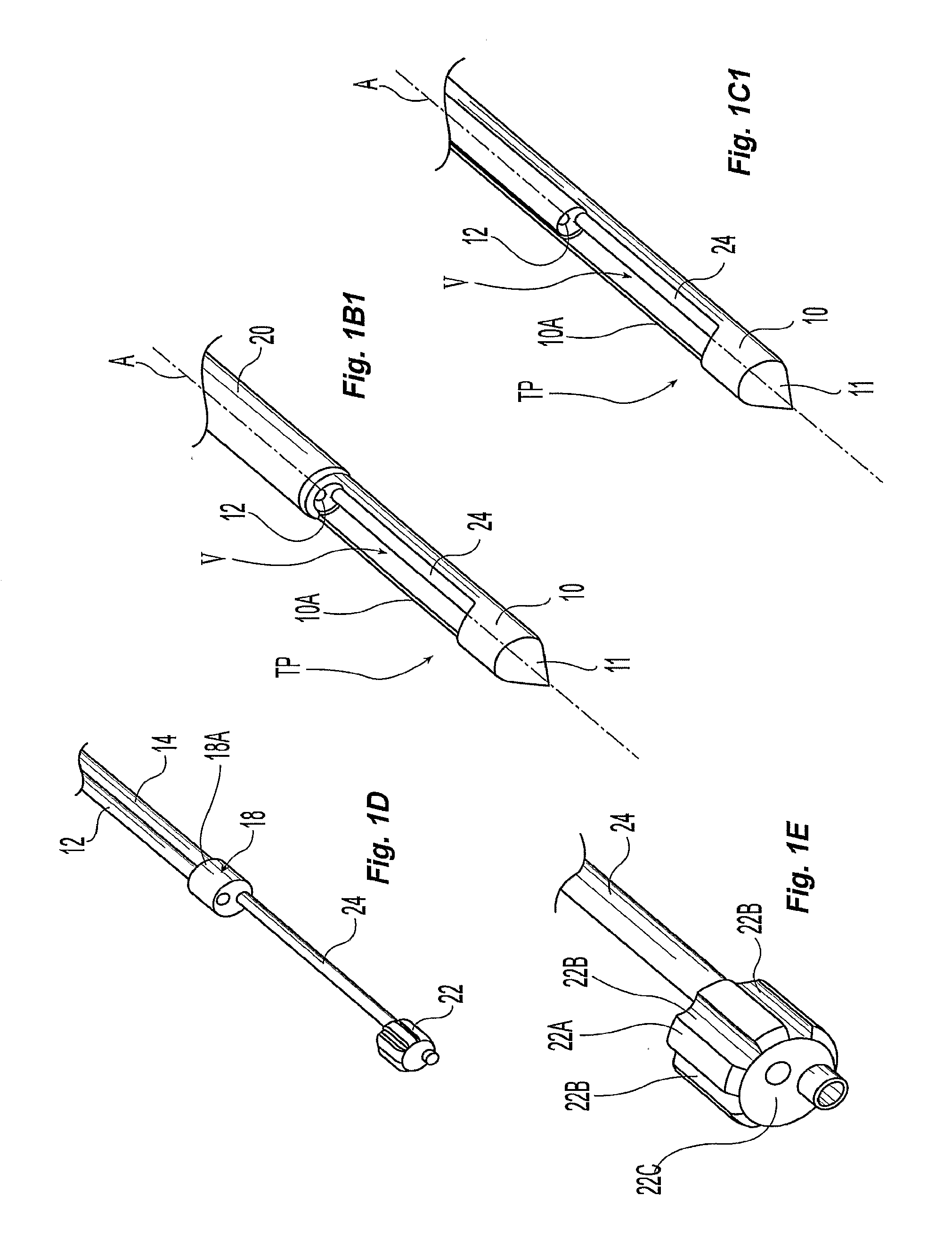
[0074]FIGS. 1-6 illustrate the preferred exemplary embodiments. In particular, FIG. 1 shows a perspective view of a stylet 10 coupled to the single-insertion, multiple samples biopsy device 100 provided with a transport subassembly 200A. The transport subassembly 200A includes the stylet, which has a tip 11 at the distal end and an outer cutting cannula 20 covering a substantial portion of the stylet 10 and a first port 10A. Extending through a hollow portion of the stylet 10 are two flexible lumens 12 and 14 coupled to a common pulley 16 proximate a second port 10B. The transport subassembly 200A can be coupled to ancillary components of the device 100 such as respective saline 37 reservoir and pump and vacuum and air pressure pump 39, a motor drive 200A, and switches and sensors as shown in FIG. 1A.

[0075]Referring to FIG. 1D, the flexible lumens 12 and 14 are coupled to a first bulkhead 18.

[0076]A second bulkhead 22 is coupled to the first bulkhead via a rigid lumen 24. One of the...


PUM


Abstract

The present invention provides for exemplary embodiments of a single-insertion, multiple sample biopsy device. Exemplary embodiments of a single-insertion, multiple sampling device with integrated marker release.

Description

PRIORITY DATA AND INCORPORATION BY REFERENCE[0001]This application claims benefit of priority to U.S. Provisional Patent Application Ser. No. 60 / 707,229 filed Aug. 10, 2005 which is incorporated by reference in its entirety.FIELD OF THE INVENTION[0002]This invention relates to a tissue biopsy sampling device.BACKGROUND OF THE INVENTION[0003]Often, it is either desirable or necessary to obtain specimens of tissue from humans and other animals, particularly in the diagnosis and treatment of patients with cancerous tumors, premalignant conditions, and other diseases or disorders. For example, when it is discovered that suspicious conditions exist, either by means of x-ray or ultrasound imaging in various tissues of the body, a physician typically performs a biopsy to determine if the cells at the suspected site are cancerous.[0004]A biopsy can be done either by an open or percutaneous technique. Open biopsy is an invasive procedure using a scalpel, whereby either a portion (incisional ...

Claims


Application Information

Patent Timeline
Patent Type & Authority Applications(United States)
IPC IPC(8): A61B10/02
CPCA61B10/0275A61B10/0283A61B2090/3987A61B2010/0225A61B2010/0208
Inventor TAYLOR, JONTHOMPSON, STANLEY O.COONAHAN, TIMOTHY J.GRAY, GREGORY A.
Owner CR BARD INC
Learn More