Computerized methods for tissue analysis using n-gene profiling

a tissue analysis and computerized technology, applied in the field of tissue analysis, can solve the problems of requiring excessive time, subject to human errors in analysis, and difficult for pathologists to attempt an analysis,

Active Publication Date: 2012-10-11
RGT UNIV OF MINNESOTA
View PDF6 Cites 23 Cited by
  • Summary
  • Abstract
  • Description
  • Claims
  • Application Information

AI Technical Summary

Benefits of technology

[0011]Methods are disclosed for use in computerized tissue analysis using a number of diagnostic quality di

Problems solved by technology

In practice, certain single gene assays utilize a single biomarker for application on a tissue section and manual quantification by a trained pathologist, requiring excessive time and subject to human errors in the analysis.
Furthermore, it is often problematic for a pathologist to attempt an analysis based on a single biomarker.
Present methods for pathology analysis are limited to physical notation and analysis by a trained practitioner.
Currently, there has yet to be developed a method for automation of tissue analysis using one or more biomarkers on a computerized platform.

Method used


Image

  • Computerized methods for tissue analysis using n-gene profiling
  • Computerized methods for tissue analysis using n-gene profiling
  • Computerized methods for tissue analysis using n-gene profiling

Examples


example 1

IHC Assays

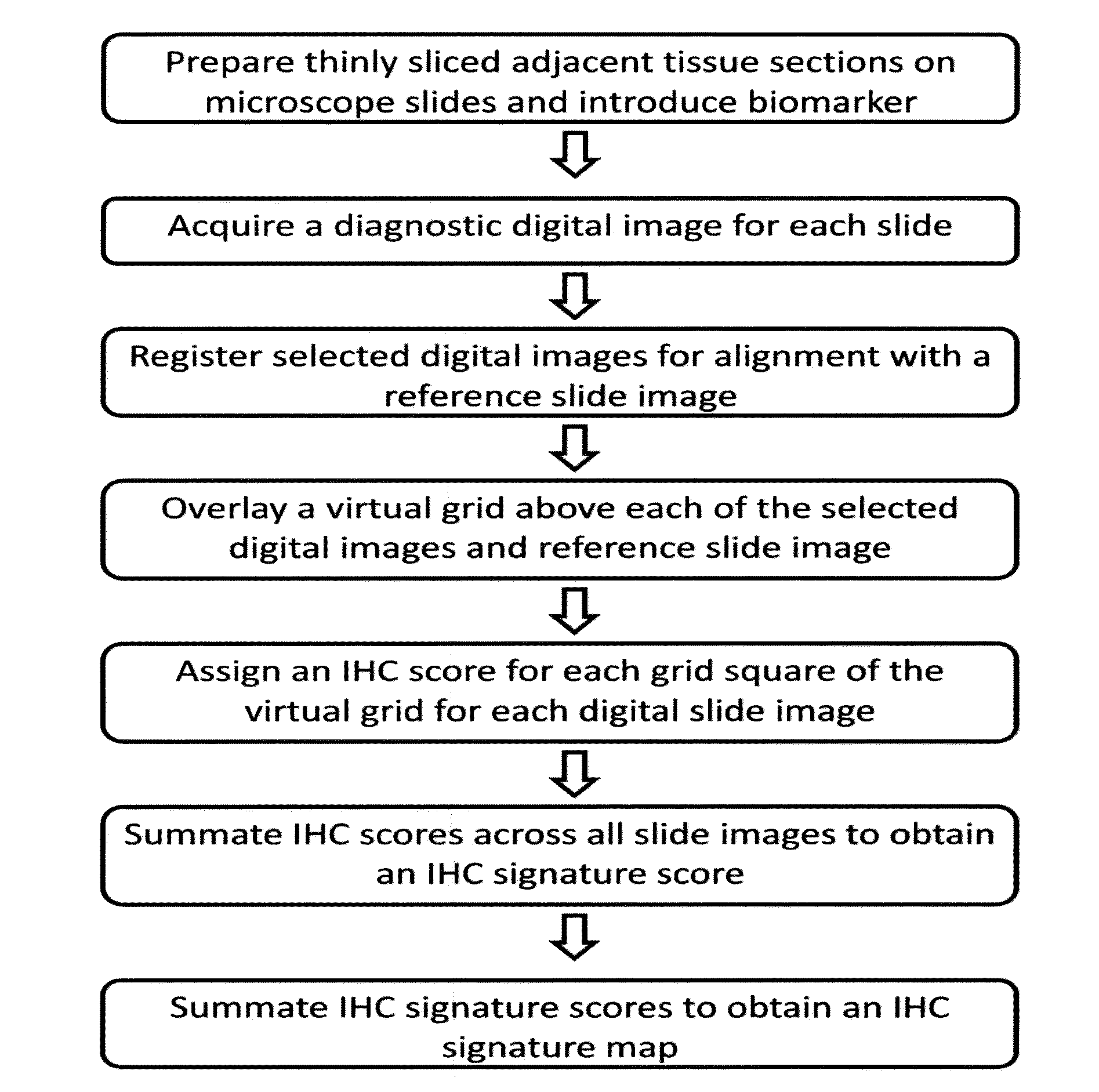
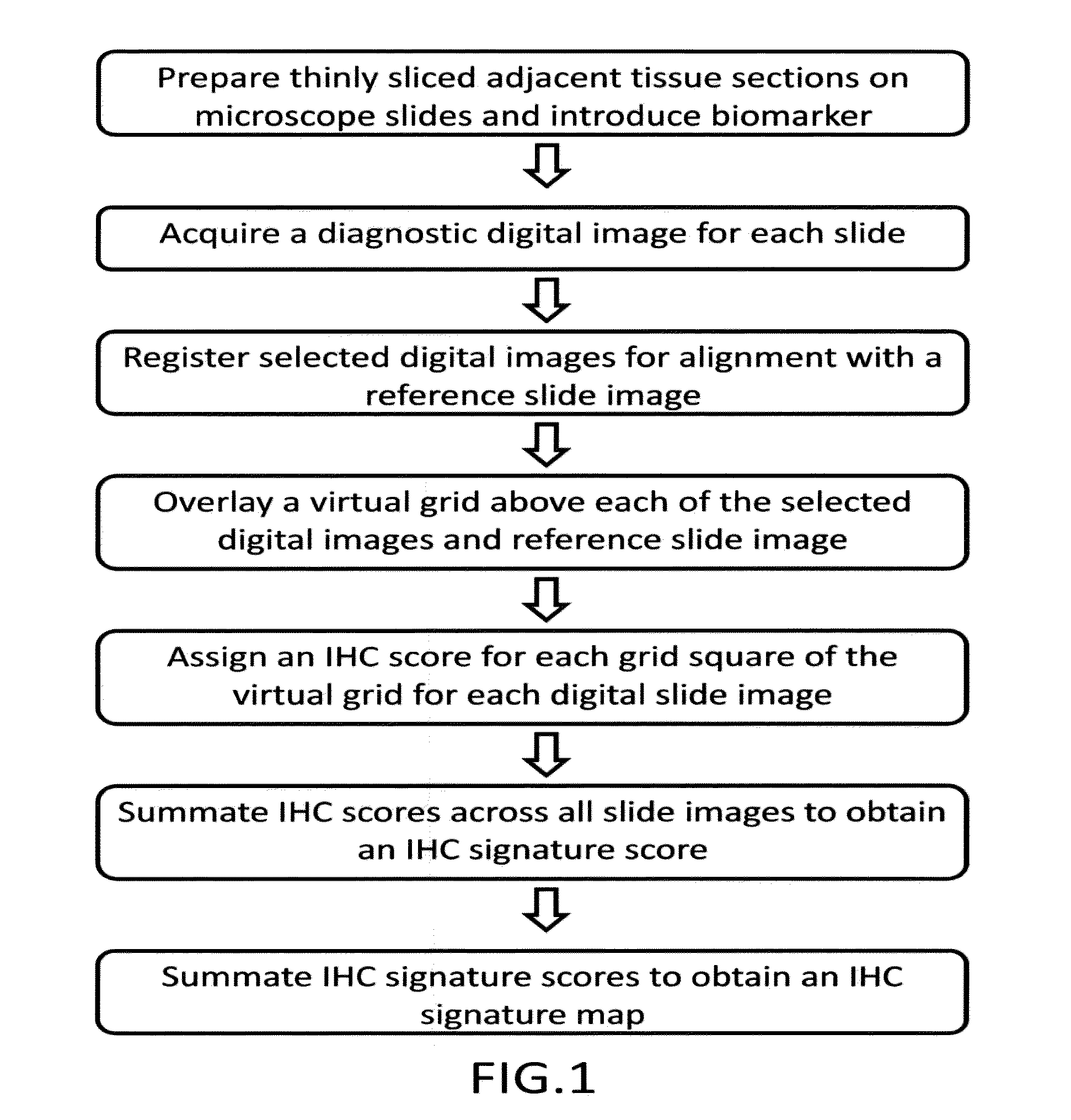
[0041]Unstained sections of formalin-fixed, paraffin-embedded prostate tissue containing discrete areas of prostatic adenocarcinoma of different histologic grades were obtained. 3 micrometer (n+1) adjacent sections were cut from the paraffin block. One section (the reference slide) was stained with hematoxylin and eosin (H&E). Adjacent sections were stained with primary antibody directed against either prostate specific acid phosphatase using monoclonal antibody clone PASE / 4LT and a heat-induced epitope retrieval method using citrate buffer, or neuron-specific enolase using monoclonal antibody clone BBS / NC / V1-h14 and a heat-induced epitope retrieval method using citrate buffer. [[CD34 clone QBend / 10 without epitope retrieval; Ki67 clone MM1 with citrate buffer; Mud 1 clone MaG96 with citrate buffer]] After washing, brown precipitate at sites of primary antibody binding was developed using a peroxidase-conjugated second step antibody and a 3,3-diaminobenzidine (DAB) reagent...


PUM


Abstract

A computerized method for immunohistochemistry analysis of tissue utilizes digital images of multiple adjacent tissue sections aligned within a computerized software and processed with an algorithm to quantify a two-dimensional IHC signature score for each respective slide image. In various embodiments, the IHC score is performed over several adjacent sections and further processed to produce a three-dimensional IHC quantification referred to as an IHC signature map.

Description

CROSS-REFERENCE TO RELATED APPLICATIONS[0001]This application claims priority to U.S. Provisional Ser. No. 61 / 473,706, filed Apr. 8, 2011, the contents of which are hereby incorporated by reference.STATEMENT REGARDING FEDERALLY SPONSORED RESEARCH OR DEVELOPMENT[0002]This invention was made with government support under National Cancer Institute (NCI) grants R01 CA131013 and R01 CA119092; Minnesota Medical Foundation grant 3824-9202-08; NCI grants P30 CA77598 and P50 CA101955. The government has certain rights in the invention.FIELD OF THE INVENTION[0003]This invention relates generally to tissue analysis; and more particularly, to computerized methods for analysis of tissue objects.BACKGROUND OF THE INVENTION[0004]Pathologists utilize various immunohistochemistry (IHC) assays for quantification of cellular material, often in terms of expression values within targeted tissue areas, such as for example the relative number of carcinoma cells versus background stroma and benign epitheli...

Claims


Application Information

Patent Timeline
IPC IPC(8): G06K9/00
CPCG06T7/0012G06K9/00127G06T2207/30024G06T2207/10056G06T2207/20021G06T2207/10024G06V20/69
Inventor METZGER, GREGORYSCHMECHEL, STEPHEN C.DANKBAR, STEPHENHENRIKSEN, JONATHAN
Owner RGT UNIV OF MINNESOTA
Why Eureka
Try Eureka