Method and magnetic resonance system for combining signals acquired from different acquisition coils

Active Publication Date: 2012-05-10
SIEMENS HEALTHCARE GMBH
View PDF8 Cites 16 Cited by
  • Summary
  • Abstract
  • Description
  • Claims
  • Application Information

AI Technical Summary

Benefits of technology

[0011]It is an object of the present invention to reduce at least some of the disadvantages cited in the preceding, and in particular to avoid the imaging of unwanted structures and / or of aliasing artifacts.
[0020]The at least two MR signals can also be provided and combined in the form of at least two MR image data sets (data files). One MR image data set can then be acquired with one of the acquisition coils and correspond to MR coil signals transformed into image space, or correspond to a combination of MR coil signals acquired with various individual coils that is transformed into image space. For example, magnitude images corresponding to the MR signals can thus be combined in a simple manner in image space.
[0024]In another embodiment, the method furthermore includes the provision of the MR coil signals acquired with the at least two acquisition coils and the implementation of a principal component analysis of the at least two coil signals to determine linearly independent principal components of the MR coil signals. Particularly in the case of larger coil arrays, correlations of the MR coil signals acquired with adjacent coils can occur, such that the determination of the linearly independent principal components for such a signal matrix is advantageous. The MR coil signals can then be combined such that at least two linearly independent MR mode signals are formed that correspond to the principal components in order to provide the at least two MR signals. The number of MR signals to be processed can therefore be reduced and, moreover, they are linearly independent, such that when combining the MR signals, an arbitrary weighting of the MR signal portions corresponding to the individual acquisition coils is enabled in principle. An effective suppression of the unwanted MR signal portions can thus be realized with reduced computing cost.
[0031]It is also possible to acquire only one k-space line (or a spoke in the case of a radial scan) so that the MR signals respectively correspond to a projection profile in image space. By using the combined MR signals in which the unwanted signal portions are suppressed, the imaging of unwanted structures can be suppressed in the reconstructed image data.
[0033]The method can be implemented repeatedly in order to determine a time series of combined image data. In the time series of image data, the movement of the imaged structure can be determined by the position of the image structure of the examination subject being determined (detected) in the image data of the time series. A “self-gating”—i.e. of the type known as a self-triggering—can thus be realized. The time series delivers information about the movement of the imaged structure with which a movement correction can subsequently take place. K-space can be scanned radially, with a k-space line (along the z-axis, for example) being evaluated to determine the movement of the structure. This is possible in principle for each spoke of the radial scan, so the movement can be determined in corresponding directions without interfering structures. A robust, automatic evaluation of the data can take place via suppression of the imaging of unwanted structures.

Problems solved by technology

In contrast to conventional methods, no optimally homogeneous irradiation / detection is thus sought; rather, specific MR coil signals are deliberately given less consideration than others.

Method used


Image

  • Method and magnetic resonance system for combining signals acquired from different acquisition coils
  • Method and magnetic resonance system for combining signals acquired from different acquisition coils
  • Method and magnetic resonance system for combining signals acquired from different acquisition coils

Examples


Embodiment Construction

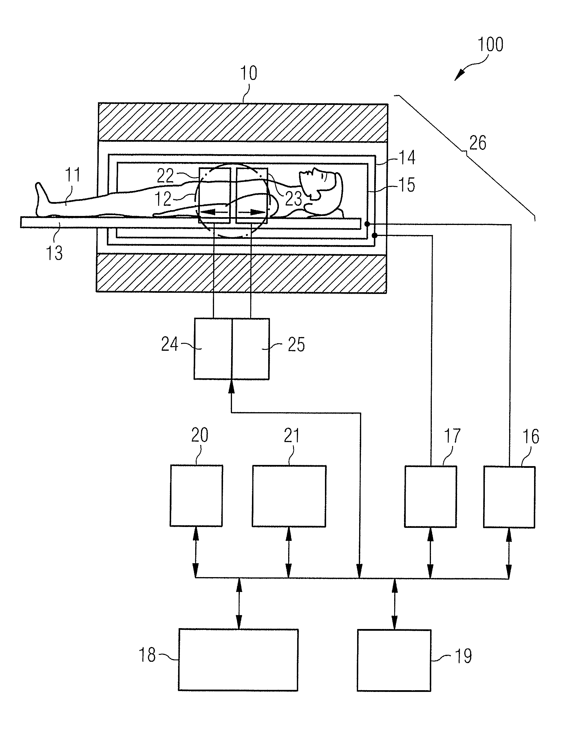
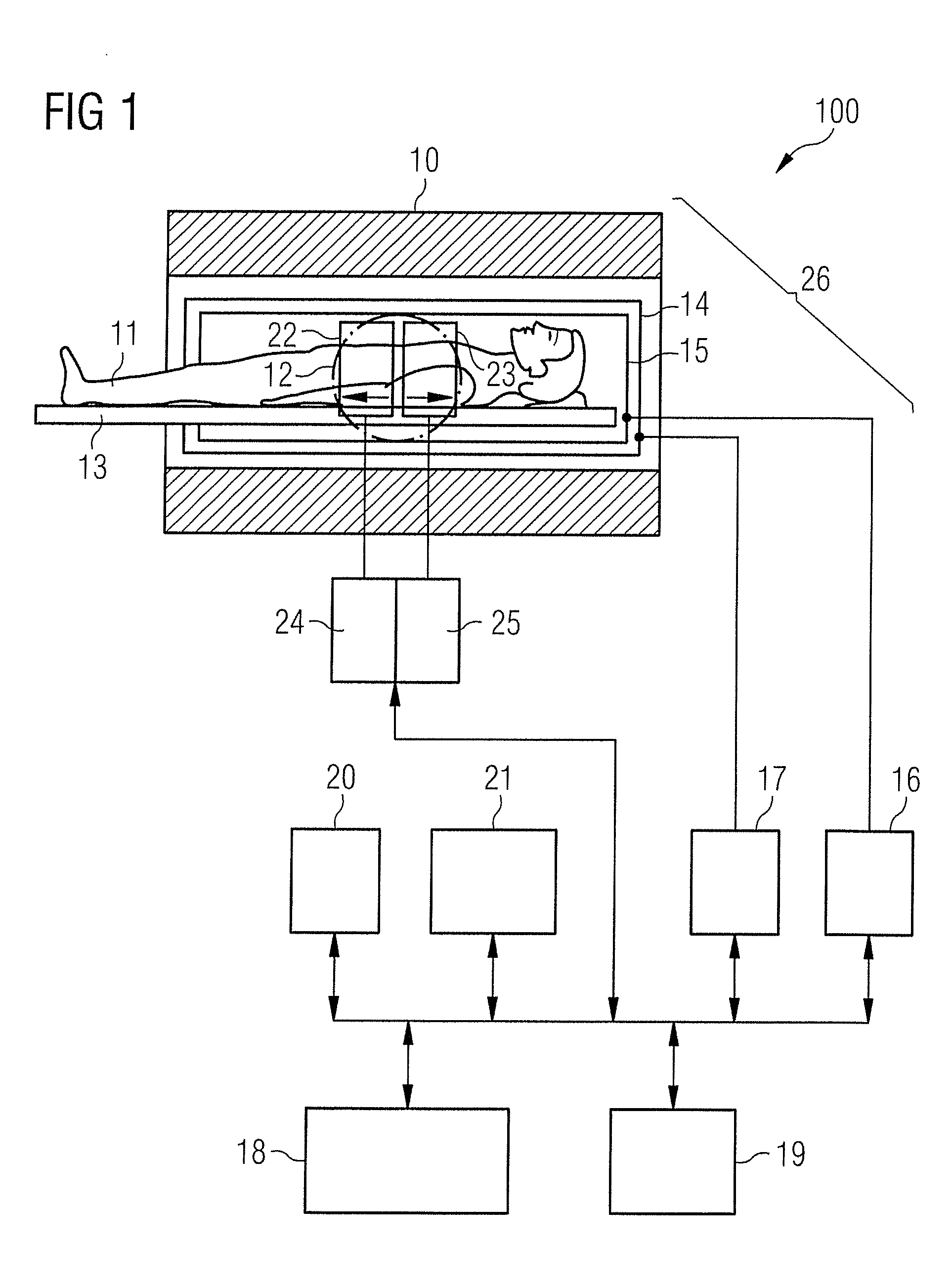
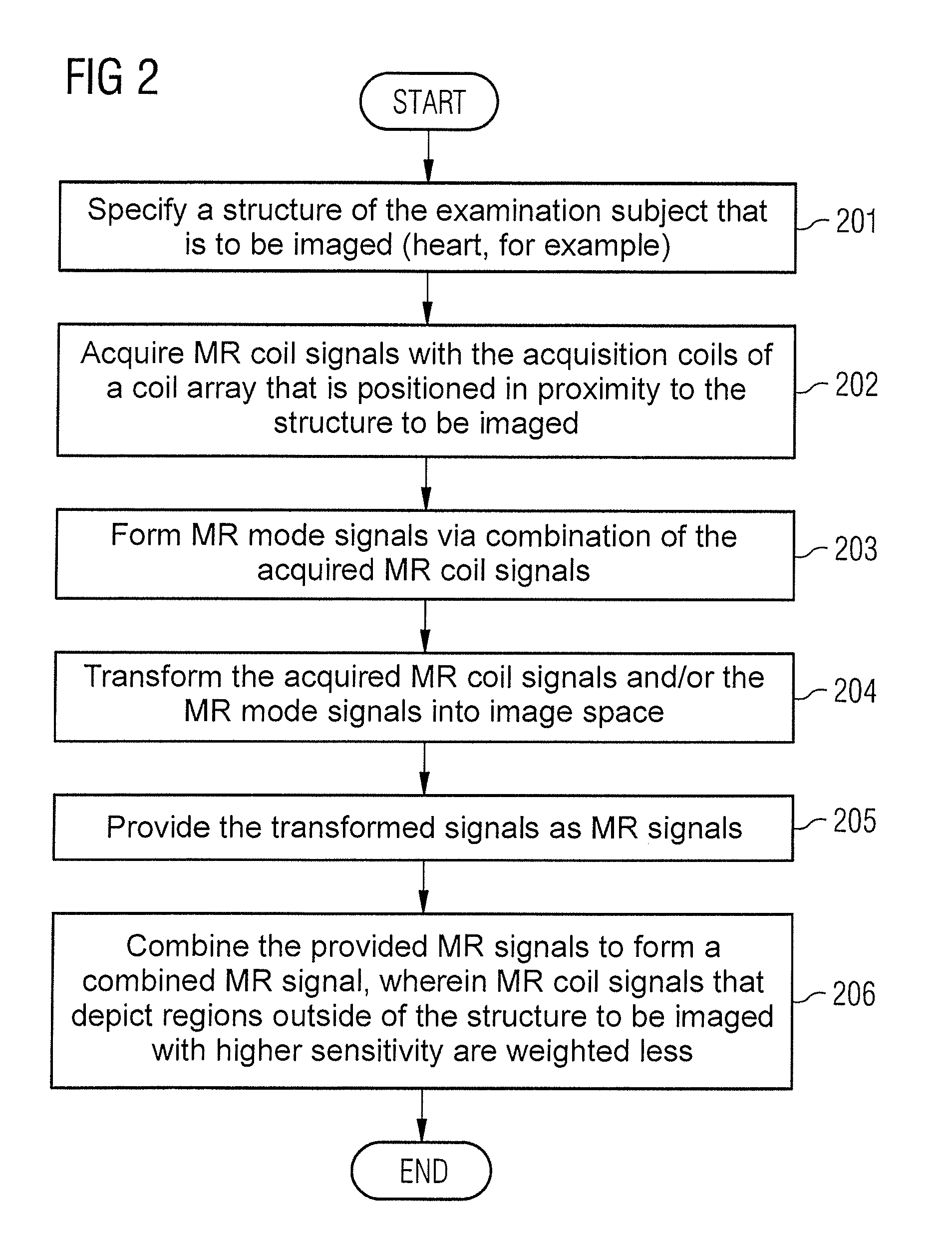
[0049]With the embodiments of the invention that are described in the following, it should be avoided that reconstructed MR image data depict unwanted structures or artifacts. In the prior art, the mapping of unwanted structures is achieved via the placement of regional saturation slices (REST) over these structures, for example, as is described in the publication by C. Stehning et. al., “Free-Breathing Whole-Heart Coronary MRA with 3D Radial SSFP and Self-Navigated Image Reconstruction”, Mag. Reson. Med. 54:476-480 (2005). As an example, FIG. 10 illustrates a detail of a radial three-dimensional acquisition of a phantom with sagittal slice selection. The image in FIG. 10 shows how the suppression of an unwanted lateral signal with such a method leads to the situation that the image data represent a compromise between the sharpness of the edges at which the suppression is applied and the signal oscillations across the imaged subject. The edges of the subject at the right and left im...


PUM


Abstract

In a method and magnetic resonance (MR) apparatus for combining MR signals that were acquired with different acquisition coils from a region of an examination subject at least two MR signals that are based on MR signals acquired with at least two different acquisition coils are provided to a processor. Due to the spatially differing arrangement of the respective acquisition coils, the at least two MR signals image the region of the examination subject with different sensitivity profiles. The provided MR signals are combined, such that unwanted MR signal portions are suppressed, to form a combined MR signal with the suppression of unwanted MR signal portions being implemented by MR signal portions that were acquired with an acquisition coil that detects the unwanted MR signal portions with increased sensitivity in comparison to other acquisition coils being weighted less in the combined MR signal than other MR signal portions.

Description

BACKGROUND OF THE INVENTION[0001]1. Field of the Invention[0002]The present invention concerns a method for combination of magnetic resonance (MR) signals that were acquired with different acquisition coils from a region of an examination subject, as well as a magnetic resonance system for implementation of such a method.[0003]2. Description of the Prior Art[0004]Magnetic resonance tomography (MRT) is an imaging modality that enables the imaging of structures inside an examination subject (in particular even of soft tissues) with high resolution. In MRT, protons in the examination subject are aligned in a basic magnetic field (B0) so that a macroscopic magnetization arises that is subsequently excited by means of one or more induction coils, and a spatial coding of the acquired signal is achieved by the switching (activation) of slice selection, phase coding and / or frequency coding gradients before or during the signal / data acquisition. The induction signal acquired with an acquisit...

Claims


Application Information

Patent Timeline
IPC IPC(8): G01R33/44
CPCA61B5/055G01R33/4838G01R33/56545G01R33/5611G01R33/5608
Inventor LITTMANN, ARNEPICCINI, DAVIDEZENGE, MICHAEL
Owner SIEMENS HEALTHCARE GMBH
Why Eureka
Try Eureka