Eureka AIR delivers breakthrough ideas for toughest innovation challenges, trusted by R&D personnel around the world.

Method For Image Reconstruction Using Low-Dimensional-Structure Self-Learning and Thresholding

Inactive Publication Date: 2012-04-26
BETH ISRAEL DEACONESS MEDICAL CENT INC
View PDF2 Cites 52 Cited by
  • Summary
  • Abstract
  • Description
  • Claims
  • Application Information

AI Technical Summary

Benefits of technology

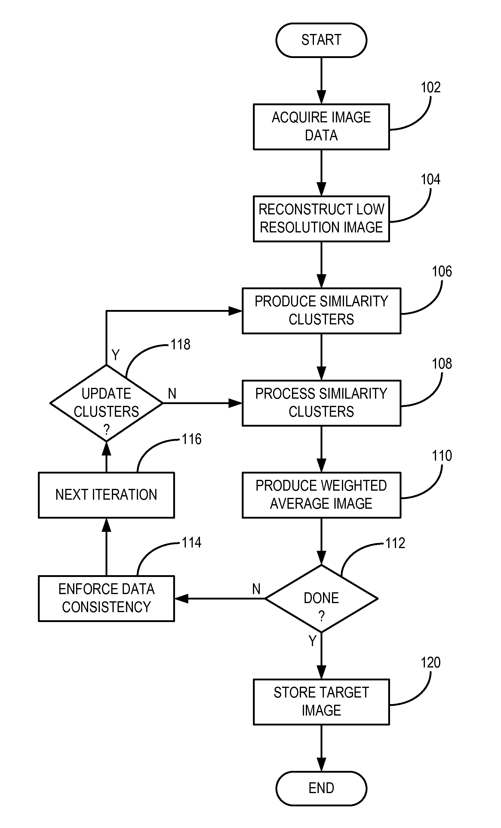
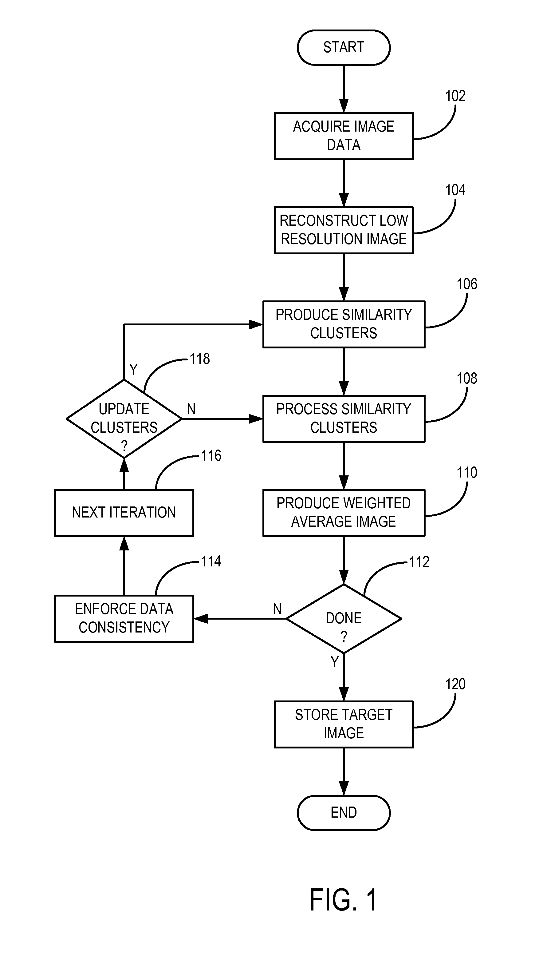
[0008]The present invention overcomes the aforementioned drawbacks by providing a method for image reconstruction that utilizes a priori information available from, for example, a low resolution image reconstructed from a portion of an acquired image data set. An improved compressed sensing reconstruction method is provided for the reconstruction of target images from undersampled image data. The method uses the structure and anatomical features in the target image being reconstructed to improve the reconstruction. By using the information from the target image itself, various features of the image can be sparsely represented in an adaptive fashion, without the need for training data. Once such an adaptive representation is achieved, aliasing artifacts and noise can be removed from the target image. Furthermore, learning the structure in this self-contained manner provides robustness to variations among patients, exams, and contrast levels.

Problems solved by technology

Despite all these efforts, long scan time is still a main challenge for MRI, especially in cardiac imaging where alternative modalities such as multi-detector CT benefit from rapid acquisition and high patient throughput.
Although sparsity is a necessary condition for ll-norm reconstruction, it is extremely difficult to know whether a transform can efficiently represent the underlying target image characteristics.
For instance, wavelets cannot capture smooth transitions sparsely, whereas finite differences have problems with sharp edges.
Thus, minimization of sparsity with a fixed transform domain may result in blurring and other image artifacts.
Even if the transform dictionary is generated using the computationally expensive dictionary learning algorithms, the effectiveness of such transforms may degrade due to inter and intra-patient variability, especially for cardiac MRI where contrast and signal levels vary significantly between different acquisitions.
Furthermore, such techniques fail to provide a reconstruction strategy that can be applied to various different anatomical features without the need for training data or prior assumption about underlying features.
Moreover, since no local information is used, these methods are generally not robust to estimation errors in coil sensitivity maps.

Method used


Image

  • Method For Image Reconstruction Using Low-Dimensional-Structure Self-Learning and Thresholding
  • Method For Image Reconstruction Using Low-Dimensional-Structure Self-Learning and Thresholding
  • Method For Image Reconstruction Using Low-Dimensional-Structure Self-Learning and Thresholding

Examples


Embodiment Construction

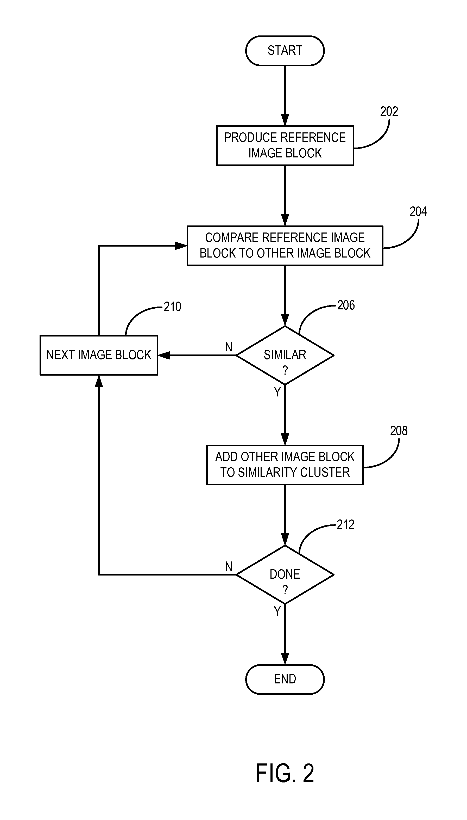
[0018]An improved method for reconstructing images from undersampled image data, such as k-space data acquired with a magnetic resonance imaging (“MRI”) system; x-ray projection data acquired with an x-ray imaging system, such as a computed tomography (“CT”) system; or the like, is provided. The method is generally referred to as “low-dimensional-structure self-learning and thresholding” (“LOST”), and utilizes structure from an underlying image to improve the reconstruction process. Generally, a low resolution image from a substantially fully-sampled portion of the image data, such as the central portion of k-space, is reconstructed, from which image blocks containing similar image characteristics, such as anatomical characteristics, are identified. These image blocks are arranged into “similarity clusters,” which are subsequently processed for de-aliasing and artifact removal using underlying low-dimensional properties. Alternatively, other methods can be implemented to reconstruct...


PUM


Abstract

A method for reconstructing an image of a subject from undersampled image data that is acquired with an imaging system, such as a magnetic resonance imaging system or computed tomography system, is provided. From the acquired undersampled image data, an image of the subject is reconstructed and used to guide further image reconstruction. For example, a low resolution image is reconstructed from a portion of the undersampled image data, such as from a portion corresponding to the center of k-space when MRI is used. From this image, a number of similarity clusters are produced and processed. The processing may be by hard thresholding, Wiener filtering, principal component pursuit, or other similar techniques. These processed similarity clusters are then used to reconstruct a final, target image of the subject using, for example, a weighted average combination of the similarity clusters.

Description

STATEMENT REGARDING FEDERALLY SPONSORED RESEARCH[0001]This invention was made with United States government support awarded by the following agency: National Institutes of Health, NIH EB008743. The United States government has certain rights in this invention.FIELD OF THE INVENTION[0002]The field of the invention is methods and systems for imaging, including medical imaging. More particularly, the field of the invention relates to methods and systems for image reconstruction, including reconstruction of medical images such as magnetic resonance or x-ray computed tomography images.BACKGROUND OF THE INVENTION[0003]Accelerated magnetic resonance imaging (“MRI”) acquisition techniques such as partial Fourier, non-Cartesian, parallel imaging, and time-frequency acquisitions have been widely studied over the past few decades. Partial Fourier methods use phase information to reduce scan time. Non-Cartesian sampling techniques rely on more efficient k-space transversals and have incoherent ...

Claims


Application Information

Patent Timeline
IPC IPC(8): G06K9/00
CPCG06T11/008G06T5/002G06T5/10G06T2207/30004G06T2207/10081G06T2207/10088G06T2207/20056G06T2200/12G06T5/70
Inventor AKCAKAYA, MEHMETNEZAFAT, REZA
Owner BETH ISRAEL DEACONESS MEDICAL CENT INC
Why Eureka
Eureka Blog
Learn More