Methods for feature analysis on consecutive tissue sections

a tissue section and feature analysis technology, applied in image analysis, image enhancement, instruments, etc., can solve the problems of increasing the probability of errors, tedious steps, handicapping precision advantages, etc., and achieve the effect of reducing the time required by pathologists

Active Publication Date: 2012-03-29
FLAGSHIP BIOSCI
View PDF14 Cites 84 Cited by
  • Summary
  • Abstract
  • Description
  • Claims
  • Application Information

AI Technical Summary

Benefits of technology

[0010]The following specification discloses improved methods for quantitative pathology analysis, including aligning consecutive tissue cross-sections and transferring regions of interest (ROIs) from one tissue section of a reference slide, to a target slide containing an adjacent tissue cross-section using computer software tied to an imaging system. The regions of interest need only to be outlined on the reference slide, either manually or with digital tissue analysis. Additionally, whole slide digital images of tissue sections being cut closely, and ideally sequentially, will be aligned above and below the reference slides, allowing the regions of interest to be transferred to these target slides.
[0011]This invention allows multiple biomarkers to be measured, or more accurate features to be identified, with only the reference slide requiring annotation of regions of interest, thereby reducing time required by a pathologist for tedious annotation of multiple slides. It is an important improvement in quantitative histopathology assessments, especially in both cancer research and toxicologic pathology, although not limited to these areas.

Problems solved by technology

This step is tedious and error-prone, whether it is done manually or with the use of a computer.
And with each slide this process has to be repeated, further adding to increased error probability.
This precision advantage unfortunately is handicapped by the extreme difficulty that the computer has in intelligently deciding on which region of a slide to make this colorimetric measurement.
In practice, especially in oncology clinical trials, samples are collected under non-uniform conditions and with many uncontrolled variables of sample collection, fixation, and even immunohistochemistry processing.
This makes the ability to run tissue analysis across non-uniform samples extremely difficult.
Neither of these approaches are cost-effective, as pathologist time is one of the most costly in tissue diagnostics.

Method used


Image

  • Methods for feature analysis on consecutive tissue sections
  • Methods for feature analysis on consecutive tissue sections
  • Methods for feature analysis on consecutive tissue sections

Examples


Embodiment Construction

[0017]In the following description, for purposes of explanation and not limitation, details and descriptions are set forth in order to provide a thorough understanding of the embodiments of the invention. However, it will be apparent to those skilled in the art that the present invention may be practiced in other embodiments that depart from these details and descriptions.

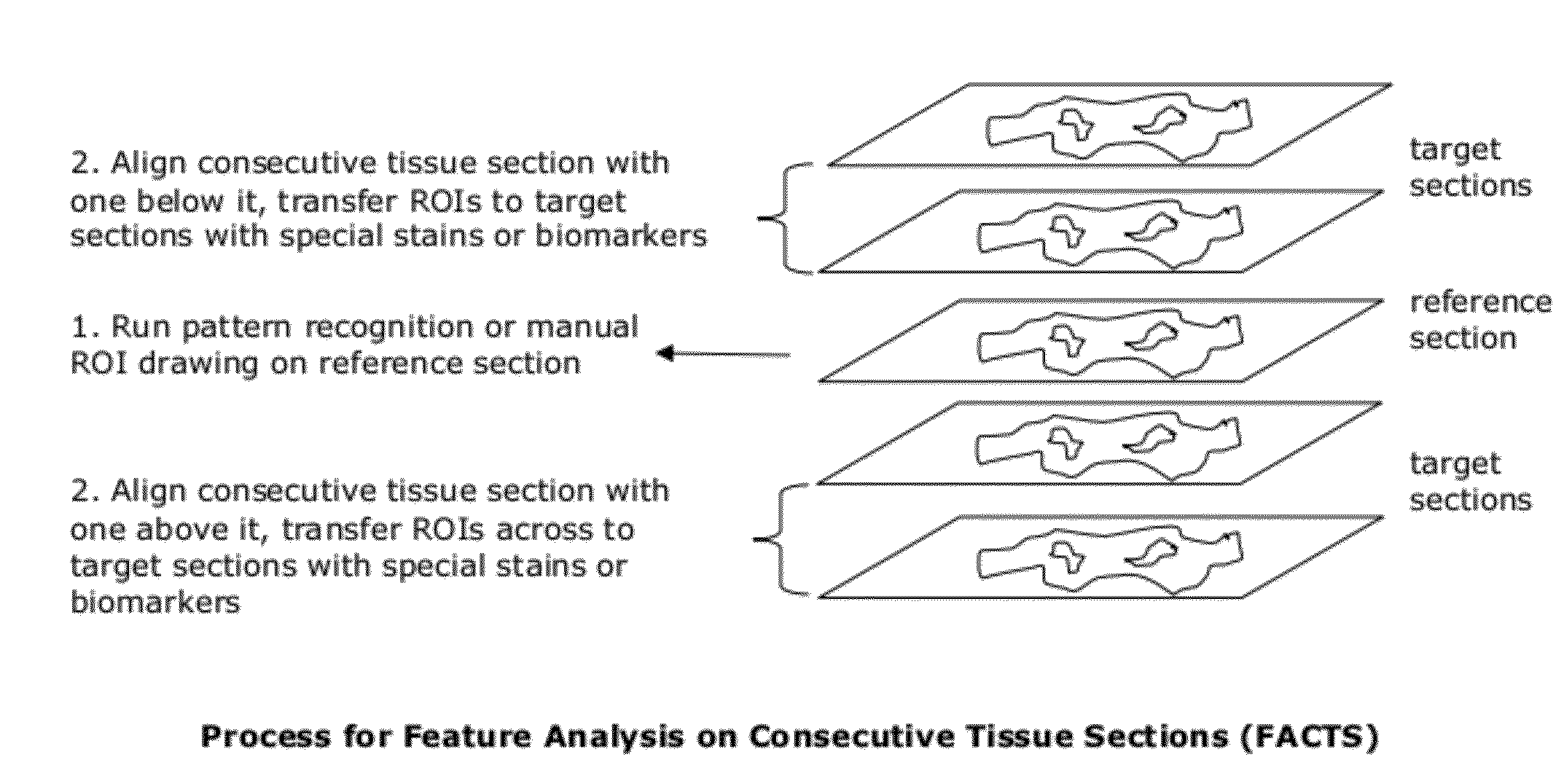
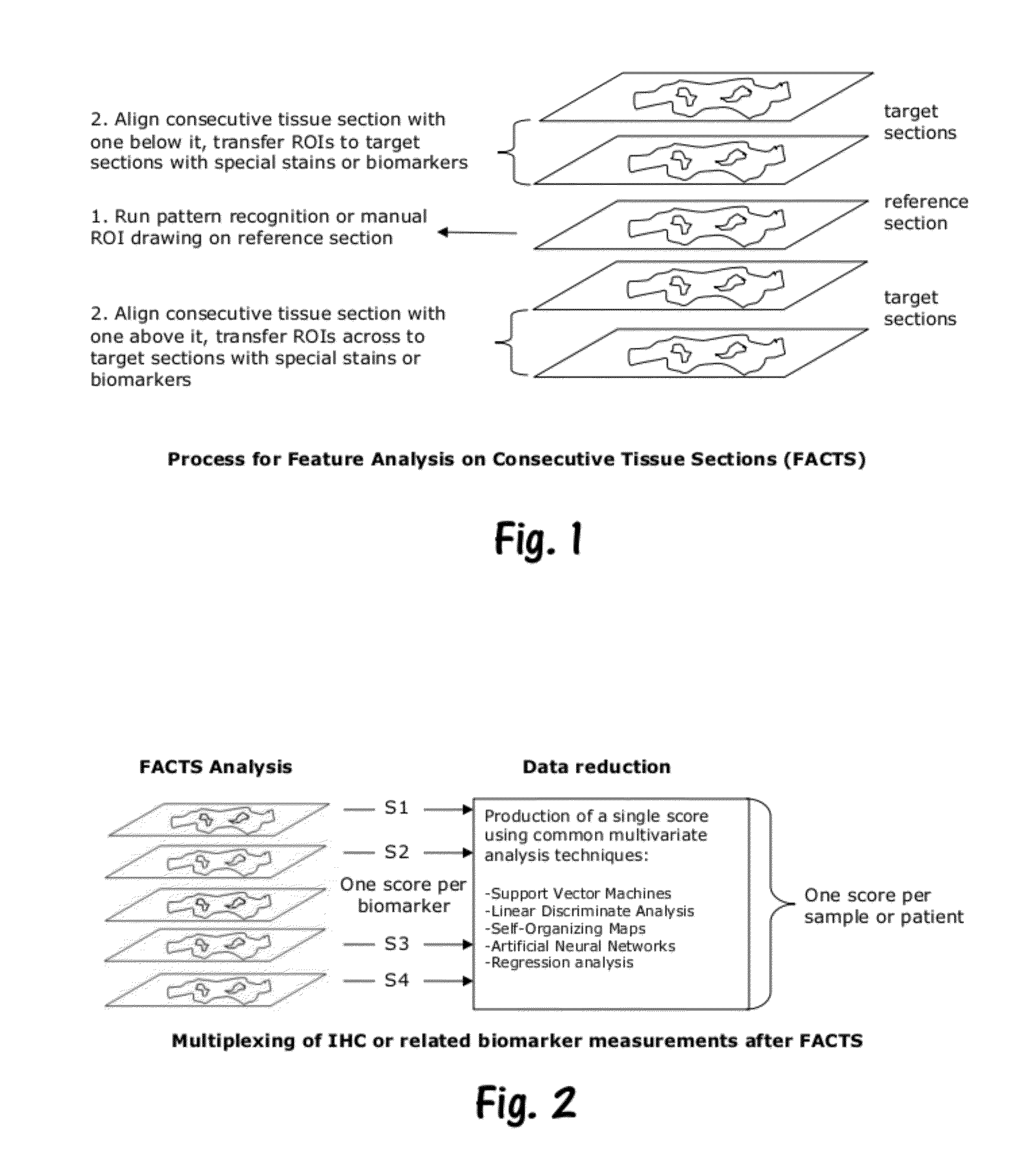
[0018]In various embodiments of the invention, the above problems and limitations have been overcome with the use of a technique herein referred to as “Feature Analysis on Consecutive Tissue Sections”, or FACTS.

[0019]In a general embodiment of the invention, a method for feature analysis on consecutive tissue sections is provided, wherein a tissue block is sectioned into thin adjacent, cross-sections. Ideally, the adjacent cross sections are maintained in consecutive order, however it is possible to non-consecutive but relatively nearby “local” sections. Each of the cross sections is prepared on a microscope slide ...


PUM


Abstract

Feature analysis on consecutive tissue sections (FACTS) includes obtaining a plurality of consecutive tissue sections from a tissue sample, staining the sections with at least one biomarker, obtaining a digital image thereof, and identifying one or more regions of interest within a middle of the consecutive tissue sections. The digital images of the consecutive tissue sections are registered for alignment and the one or more regions of interest are transferred from the image of the middle section to the images of the adjacent sections. Each image of the consecutive tissue sections is then analyzed and scored as appropriate. Using FACTS methods, pathologist time for annotation is reduced to a single slide. Optionally, multiple middle sections may be annotated for regions of interest and transferred accordingly.

Description

CROSS-REFERENCE TO RELATED APPLICATIONS[0001]This application claims priority to U.S. Provisional Ser. No. 61 / 387,298, filed Sep. 28, 2010; U.S. Provisional Ser. No. 61 / 434,428, filed Jan. 19, 2011; and U.S. Provisional Ser. No. 61 / 446,942, filed Feb. 25, 2011; the entire contents of which are hereby incorporated by reference.BACKGROUND OF THE INVENTION[0002]1. Field of the Invention[0003]This invention relates to methods for analysis of biological tissue sections; and more particularly to improved methods for quantitative pathology analysis across multiple consecutive tissue sections for a variety of applications.[0004]2. Related Art[0005]Quantitative pathology analysis includes a variety of known techniques for analysis of biological samples for the purposes of diagnosis and quantification of the presence of one or more diseases within a tissue specimen. For example, a pathologist might analyze a tissue sample to diagnose the presence of a cancer or other disease, or the pathologi...

Claims


Application Information

Patent Timeline
IPC IPC(8): G06K9/00
CPCG06T7/0014G06T7/0038G06T2207/30096G06T2207/20104G06T2207/10056G06T7/38
Inventor POTTS, STEVENJOHNSON, TREVORVOELKER, FRANKYOUNG D, GEORGE
Owner FLAGSHIP BIOSCI
Why Eureka
Try Eureka