
System for image scanning and acquisition with low-dose radiation

a low-dose radiation and imaging system technology, applied in the field of medical imaging system and shape adaptive collimator, can solve the problems of unfavorable patient imaging, unfavorable patient imaging, and unnecessary radiation exposure to an area outside the roi, and achieve the effect of reducing patient x-ray exposur

Active Publication Date: 2012-03-08
SIEMENS HEALTHCARE GMBH
View PDF12 Cites 36 Cited by
  • Summary
  • Abstract
  • Description
  • Claims
  • Application Information

AI Technical Summary

Benefits of technology

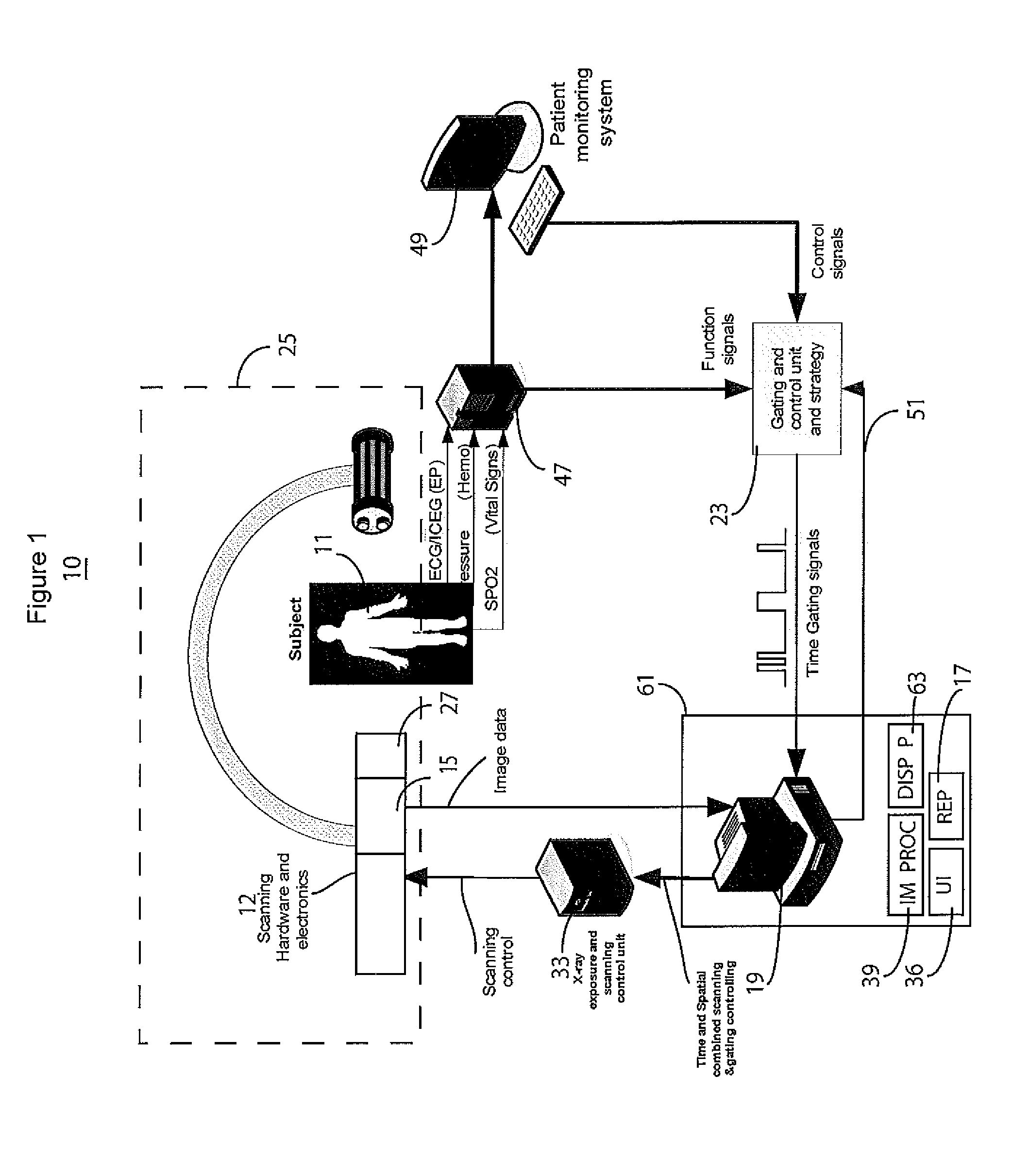
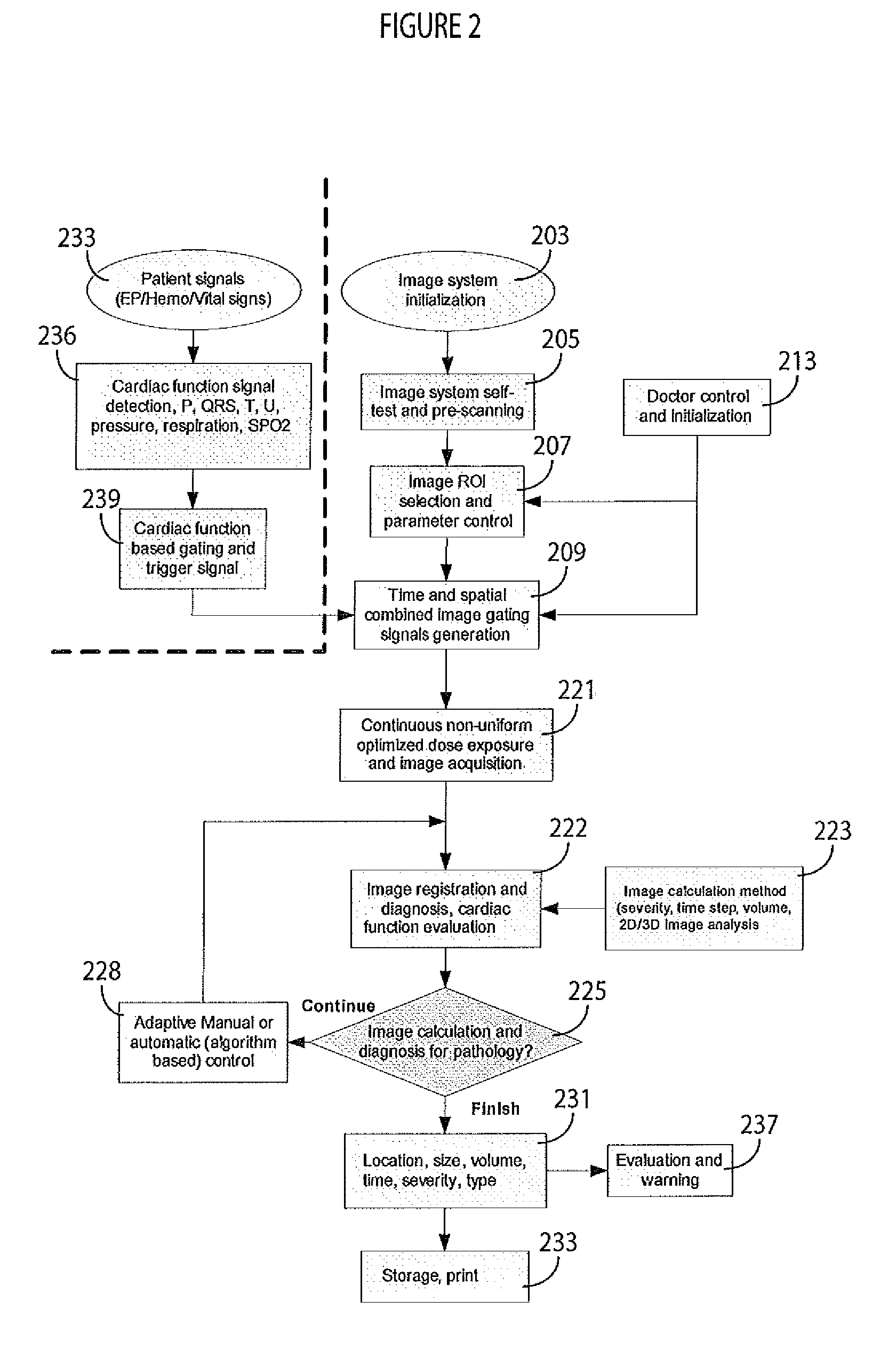
[0005]A system uses an adaptive adjustable image radiation filter or collimator to control non-uniform image radiation exposure and beam shape / focus and intensity, low and high dose exposure of areas in a single image and adaptive radiation dose optimization using X-ray beam spatial control and beam exposure time gating and triggering in response to hemodynamic, electrophysiological and vital sign signals. A medical imaging system adaptively acquires anatomical images using a shape adaptive collimator, a synchronization processor, an X-ray image acquisition device and a display processor. The shape adaptive collimator includes multiple different portions of X-ray absorbent material automatically adjustable to alter the dimensions of a spatial cross section of an X-ray beam of radiation into a non-rectangular shape, in response to a control signal indicating shape of a region of interest of patient anatomy. The synchronization processor provides a heart rate related synchronization signal derived from a patient cardiac function associated parameter. The synchronization signal enables adaptive variation in timing of image acquisition within an individual heart cycle and between successive heart cycles of each individual image frame of multiple sequential image frames. The X-ray image acquisition device uses the shape adaptive collimator for acquiring anatomical images of the region of interest with reduced patient X-ray exposure in response to the synchronization signal. The display processor presents images, acquired by the acquisition device to a user on a reproduction device.

Problems solved by technology

Known X-ray imaging and fluoroscopic imaging typically capture images randomly or based on time and may lead to unnecessary X-ray exposure.
However known image scanning and acquisition systems usually employ uniformly timed image acquisition and may fail to capture anatomical features during an ECG Q wave or S wave, for example, since the image scanning is triggered in response to an R wave.
However, especially in heart monitoring cases, an anatomical region of interest (ROI) area is usually not a rectangle or square which results in unnecessary radiation exposure to an area outside the ROI.

Method used


Image

  • System for image scanning and acquisition with low-dose radiation
  • System for image scanning and acquisition with low-dose radiation
  • System for image scanning and acquisition with low-dose radiation

Examples


Embodiment Construction

[0014]An imaging system improves medical imaging by capturing patient tissue images with low-dose X-ray radiation. The imaging system achieves non-uniform image scanning and acquisition with adaptive and automated control of radiation exposure timing and spatial ROT (region of interest) area. The image radiation timing is controlled and adjusted in response to patient signals (such as ECG, ICEG, Hemodynamic, and vital sign signals, for example) and radiation spatial information is determined by a user or image analysis in response to ROI selection. A programmable image radiation filter or collimator is used to accurately control non-uniform image radiation exposure by adjusting beam shape and focus and intensity as well as to determine low and high radiation dose areas in a single image and adaptive radiation dose optimization. The system reduces redundant beam scanning and determines an accurate image scanning time, provides stable image capture and cardiac tissue and function char...


PUM


Abstract

A medical imaging system adaptively acquires anatomical images using a shape adaptive collimator including multiple different portions of X-ray absorbent material automatically adjustable to alter the dimensions of a spatial cross section of an X-ray beam of radiation into a non-rectangular shape, in response to a control signal. The synchronization processor provides a heart rate related synchronization signal derived from a patient cardiac function related parameter. The synchronization signal enables adaptive variation in timing of image acquisition within an individual heart cycle and between successive heart cycles of each individual image frame of multiple sequential image frames. The X-ray image acquisition device uses the shape adaptive collimator for acquiring anatomical images of the region of interest with reduced patient X-ray exposure in response to the synchronization signal. A display processor presents resultant images.

Description

[0001]This is a non-provisional application of provisional application Ser. No. 61 / 380,751 filed Sep. 8, 2010, by H. Zhang.FIELD OF THE INVENTION[0002]This invention concerns a medical imaging system and shape adaptive collimator for adaptively acquiring anatomical images with reduced patient X-ray exposure in response to adaptive variation in timing of image acquisition within an individual heart cycle and between successive heart cycles of each individual image frame of a sequence of image frames.BACKGROUND OF THE INVENTION[0003]Angiography (or arteriography) imaging is used to monitor and characterize cardiac tissue and functions, including morphology, blood flow, anatomy and arterial luminal size by displaying static and dynamic image silhouettes. This provides the ability to assess cardiac and coronary arterial function and to visualize a heart system (Ventricle, Atrium, Vessel), which facilitates diagnosis and determination of cardiac disease. It is desirable to minimize radia...

Claims


Application Information

Patent Timeline
IPC IPC(8): A61B6/00
CPCA61B5/024A61B5/7285A61B6/06A61B6/4035Y10S378/901A61B6/503A61B6/507A61B6/541A61B6/542A61B6/469
Inventor ZHANG, HONGXUAN
Owner SIEMENS HEALTHCARE GMBH
Learn More