Systems and methods for detection of disease including oral scopes and ambient light management systems (ALMS)

Inactive Publication Date: 2011-11-10
LED MEDICAL DIAGNOSTICS
View PDF0 Cites 19 Cited by
  • Summary
  • Abstract
  • Description
  • Claims
  • Application Information

AI Technical Summary

Benefits of technology

Exemplary procedures include the following:1. The patient sits upright in a chair during the procedure. He / she will typically not be in a supine position, although such can be used if desired. The supine position causes the patient's tongue to fall back into the throat restricting tissue exposure.2. The light source, scope, etc., preferably moves freely without the vestibular device either substantially constraining the movement or occluding the view of the dentist or other practitioner.3. The practitioner may, if desired, be able to grab and move the tongue without introducing stray light and without disrupting the procedure.
Exemplary device constructions include the following:4. The vestibule device can be composed of a draping material or other suitable shroud material, which can rigid or floppy or in-between as desired and depending on the given embodiment, non-rigid devices such as internal stiffeners can be included for shape. Systems and methods of attachment to the viewing scope, to a separate light source (if any), and to a tool (if any). Systems and methods of attachment to the patient can also be included.a. Attachment of the 
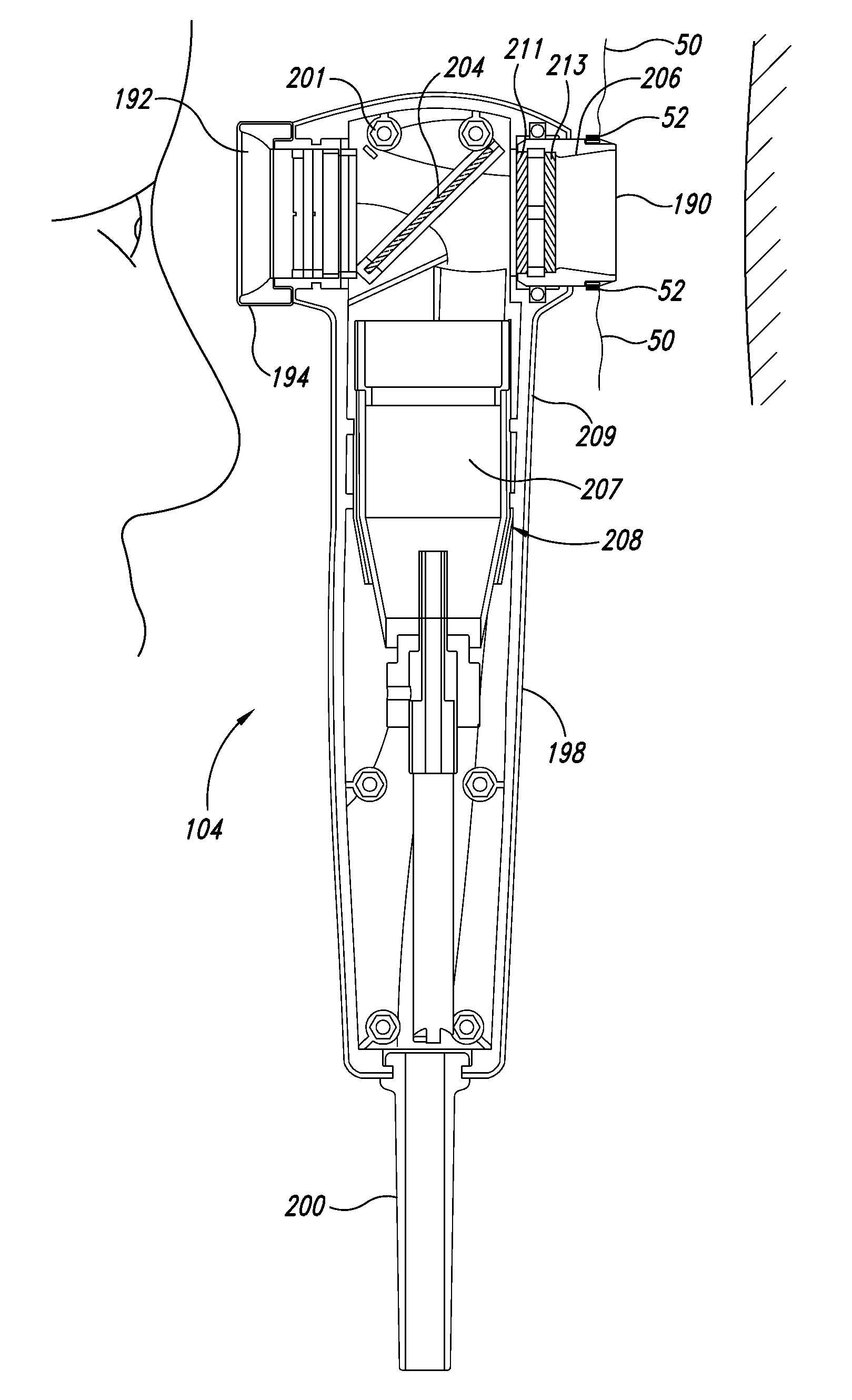
vestibule device to the scope: The scope or other investigative device enters the vestibular device to become, in some embodiments, a part of the ALMS, through a port or hole in the drape, typically in-line with the mouth or other target tissue. An attachment and / or seal enhances darkness and confident manipulation of the vestibular device and other elements of the systems without introduction of unwanted light.b. Attachment of the 
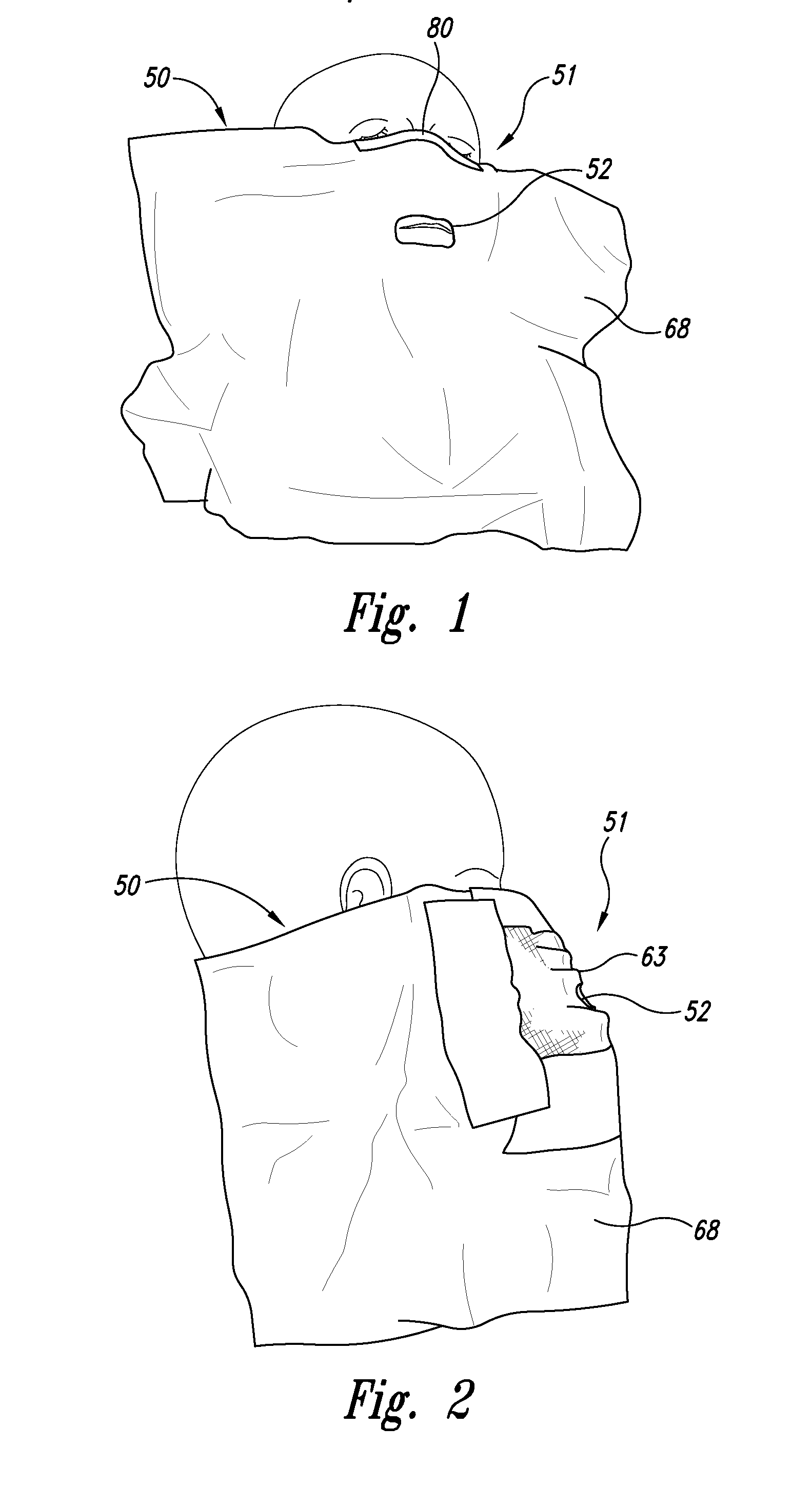
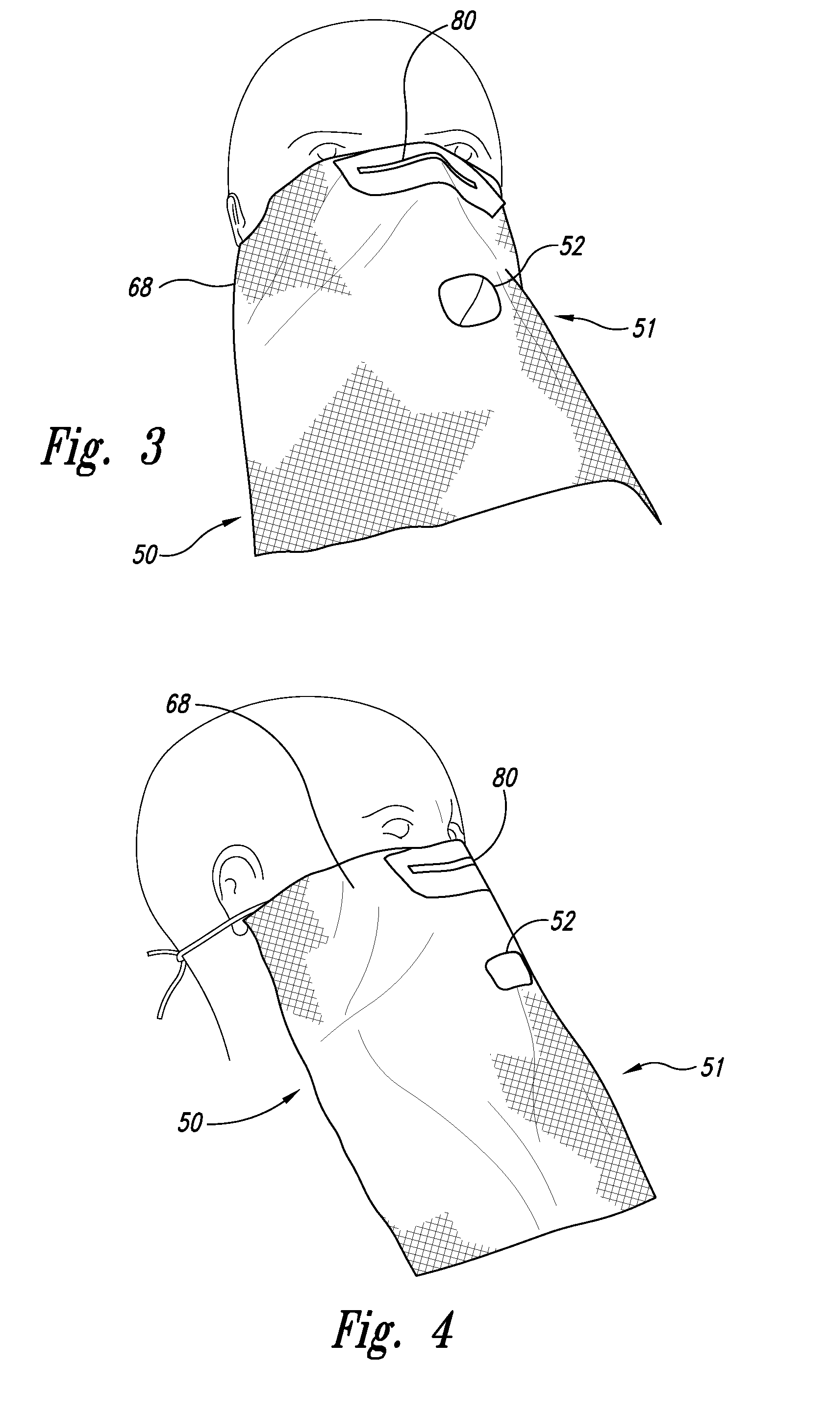
vestibule device to the patient. A malleable / shapeable piece of material that conforms to the curves of the nose and cheeks can be provided, such as an elastic that goes around the patients head or ear clips similar to frames of glasses. Attachment to the patient's head can allow the dentist to have more freedom to move the device to different angles. Molding around the nose and cheeks acts as a seal that improves the reduction of ambient light.5. In some embodiments the vestibular device is disposable to inhibit the spread of bacteria, viruses or material between patients.6. Particularly in embodiments where the vestibular device may be disposable, it should be inexpensive.
Exemplary dimensions for the vestibular device include the following:16. The vestibular devices are preferably not an encumbrance to the patient and the practitioner; e.g., does not interfere with movement during the procedure.17. The vestibular device is typically large enough that it will drape over the patient's mouth, and possibly body, and block out light that could enter at various points such as at the shoulders (in embodiments where the shoulders are of concern). The vestibular device, which can be disposable in whole or in part, is large enough to prevent light from entering when the device is moved during the procedure.18. The vestibular device will, in some embodiments, be attached to the patient and thereby in certain embodiments should not have significant perceptible or uncomfortable weight, for example less than 100 g.
Exemplary interfacing for the vestibular device include the following:19. The vestibular device can couple / integrate with the investigative device such that it does not fall away from the investigative device unintentionally.20. The vestibular device can comprise a port or other access point of allowing the investigating device to access the patient's mouth or other body part under investigation.21. The vestibular device can interface to the patient by being formed to make a seal with his / her face or other body part. In one embodiment this is accomplished via a malleable / shapeable piece of material in the mask.22. The draping material may be large enough and flexible enough that the health practitioner can grab and manipulate the tongue or other body from beneath the draping material without introducing ambient light.23. Space is typically created between the patient and the light source. To do this, for example, the draping material can be relatively stiff, reinforced with another material, more of the same material and / or stitching or will have a brim or other space-making structure.

Problems solved by technology

Detection of cancer, including precancerous and early cancerous cells, has always been a difficult and uncertain process.
However, the detection of autofluorescence in traditional dental and medical environments has been problematic because the autofluorescence signature itself is very very small compared to the ambient light typically present in an examination room, operating room, etc.

Method used


Image

  • Systems and methods for detection of disease including oral scopes and ambient light management systems (ALMS)
  • Systems and methods for detection of disease including oral scopes and ambient light management systems (ALMS)
  • Systems and methods for detection of disease including oral scopes and ambient light management systems (ALMS)

Examples


example

Example 1

Use of a Viewing Scope to Identify Primary Dysplasia in the Mouth

Use of a viewing scope to identify primary dysplasia in the mouth (N=49) based on autofluorescence. The light path of the scope had in order a collimator, 430+ / −30 nm filter (filter 1), a dichroic filter (filter 2), light to avoid absorber, glass window, mucosal tissue or other target tissue, dichroic filter (filter 2 (the light passes back past the same dichroic filter), 475 long pass filter (filter 3), 590 nm notch filter (filter 4), eyepiece ocular. The scope was used with a vestibular device with a port that operably linked to the scope and that blocked substantially all ambient light from reaching the target.

For primary lesions in patients with no history of oral cancer, the scope correctly identified the majority of dysplasia (70%), particularly high-grade preinvasive lesions (86%). About 18% of non-dysplastic lesions were also positive using the scope. The scope also improved the ability of the clinicia...


PUM


Abstract

Methods and systems related to detecting disease, such as oral cancer, in a patient, using a viewing scope to investigate a patient's tissues. The systems and methods excite and detect fluorescence from the tissue. The fluorescence can then be evaluated, and the possibility of certain diseases such as cancer can be determined. The devices include an ambient light management system (ALMS, often referred to as a “vestibular device”) that manages background light in the health practitioner's office. This device can be used with scope systems for fluorescence based detection of abnormal tissue.

Description

BACKGROUNDDetection of cancer, including precancerous and early cancerous cells, has always been a difficult and uncertain process. One of the approaches to identifying cancerous cells has been to measure the cells' autofluorescence signature, because cancerous cells have a distinct autofluorescence signature relative to healthy cells. Other diseases also have changes in their autofluorescent signature. However, the detection of autofluorescence in traditional dental and medical environments has been problematic because the autofluorescence signature itself is very very small compared to the ambient light typically present in an examination room, operating room, etc.The following are some references relating to the detection of cancer in the oral cavity. As with all other references cited herein, including in the Cross Reference to Related Applications, these references are incorporated herein in their entirety for all their teachings and for all purposes. The inclusion of such refe...

Claims


Application Information

Patent Timeline
IPC IPC(8): A61B1/06A61B1/00A61B1/24A61B1/32A61B5/00A61B5/107A61H21/00G01N21/64
CPCA61B1/00142A61B5/0077A61B1/24A61B1/32A61B5/0071A61B5/0088A61B5/107A61B5/411A61B5/444A61B2560/0456A61B1/00128A61B1/043A61B1/0615A61B1/0646A61B1/00193
Inventor GILHULY, TERENCE J.WHITEHEAD, PETER
Owner LED MEDICAL DIAGNOSTICS
Why Eureka
Try Eureka