Novel diagnostic method
a diagnostic method and diagnostic method technology, applied in the field of new diagnostic methods, can solve the problems of significant restnosis, post-ptca closure of the vessel, and 500,000-600,000 deaths annually
- Summary
- Abstract
- Description
- Claims
- Application Information
AI Technical Summary
Benefits of technology
Problems solved by technology
Method used
Image

Examples
example 1
MALDI-TOF Mass Spectrometry
[0234]Matrix-assisted laser desorption / ionization mass spectrometry was carried out using the Voyager De-Pro (Applied Biosystems, Darmstadt) with a linear time of flight analyzer. The instrument was equipped with a 337 nm nitrogen laser, a potential acceleration source and a 1.4 m flight tube. Detector operation was in the positive-ion mode. Samples (5 μl) were mixed with equal volumes of the matrix solution. For matrix solution sinapinic acid was used, prepared by solving 20 mg sinapinic acid (Sigma-Aldrich) in 1 ml acetonitrile / 0.1% TFA in water (1 / 1, v / v). A small volume (≈1 μl) of the matrix-analyte-mixture was transferred to a probe tip.
[0235]For long-term testing of Glu1-cyclization, MCP-1 peptides were incubated in 100 μl 0.1 M sodium acetate buffer, pH 5.2 or 0.1 M Bis-Tris buffer, pH 6.5 at 30° C. Peptides were applied in 0.15 mM to 0.5 mM concentrations, and 0.2 U QC was added. At different times, samples were removed from the assay tube, peptide...
example 2
Proteolytic Degradation of Human MCP-1(1-76) by Dipeptidyl-Peptidase 4 (DP4), Aminopeptidase P, and by Proteases Present in Human Serum
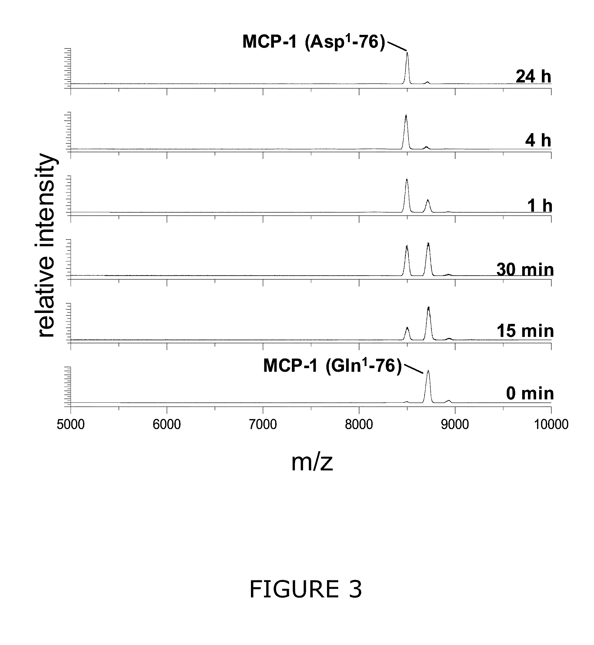
N-Terminal Degradation of MCP-1 by Recombinant Human DP4 in Absence and Presence of a QC-Specific Inhibitor
[0236]Recombinant human MCP-1(1-76) (SEQ ID NO: 1) starting with an N-terminal glutamine (Peprotech) was dissolved in 25 mM Tris / HCl pH 7.6 in a concentration of 10 μg / ml. The MCP-1 solution was either pre-incubated with recombinant human QC (0.0006 mg / ml) for 3 h at 30° C. and subsequently incubated with recombinant human DP4 (0.0012 mg / ml) at 30° C. or incubated with DP4 without prior QC application. In addition, the incubation of Gln1-MCP-1 with recombinant human QC was carried out in the presence of 10 μM of 1-(3-(1H-imidazol-1-yl)propyl)-3-(3,4-dimethoxyphenyl)thiourea hydrochloride. Resulting DP4 cleavage products were analyzed using Maldi-TOF mass spectrometry after 0 min, 15 min, 30 min, 1 h, 2 h and 4 h.
N-Terminal Degradation by Recombi...
example 3
Degradation of Human MCP-2, MCP-3 and MCP-4 N-Terminal Degradation of Human MCP-2 by DP4
[0241]Human recombinant MCP-2 (SEQ ID NO: 11) carrying an N-terminal glutaminyl instead of a pyroglutamyl residue (Peprotech) was dissolved in 25 mM Tris / HCl, pH 7.6, in a concentration of 10 μg / ml. MCP-2 was either pre-incubated with recombinant human QC (0.0006 mg / ml) for 3 h at 30° C. and subsequently incubated with recombinant human DP4 (0.0012 mg / ml) at 30° C. or incubated with recombinant human DP4 (0.0012 mg / ml) without pre-incubation with QC. Resulting DP4 cleavage products were analyzed using Maldi-TOF mass spectrometry after 0 min, 15 min, 30 min, 1 h, 2 h, 4 h and 24 h.
N-Terminal Degradation of Human MCP-3 by DP4
[0242]Human recombinant MCP-3 (SEQ ID NO: 12) carrying an N-terminal glutaminyl instead of a pyroglutamyl residue (Peprotech) was dissolved in 25 mM Tris / HCl, pH 7.6, in a concentration of 10 μg / ml. MCP-3 was either pre-incubated with recombinant human QC (0.0006 mg / ml) for 3 h...
PUM
| Property | Measurement | Unit |
|---|---|---|
| pH | aaaaa | aaaaa |
| concentrations | aaaaa | aaaaa |
| Concentrations | aaaaa | aaaaa |
Abstract
Description
Claims
Application Information
 - R&D
- Intellectual Property
- Life Sciences
- Materials
- Tech Scout
- Unparalleled Data Quality
- Higher Quality Content
- 60% Fewer Hallucinations
Browse by: Latest US Patents, China's latest patents, Technical Efficacy Thesaurus, Application Domain, Technology Topic, Popular Technical Reports.
© 2025 PatSnap. All rights reserved.Legal|Privacy policy|Modern Slavery Act Transparency Statement|Sitemap|About US| Contact US: help@patsnap.com