
Material for treatment of cerebral infarction and brain tissue regeneration method

Inactive Publication Date: 2011-07-21
NAT CENT FOR GERIATRICS & GERONTOLOGY
View PDF0 Cites 20 Cited by
  • Summary
  • Abstract
  • Description
  • Claims
  • Application Information

AI Technical Summary

Benefits of technology

[0034]A material for treatment of cerebral infarction and a brain tissue regeneration method according to the present invention are an unprecedented, original, and novel material for treatment of cerebral infarction and brain tissue regeneration method. The material for treatment of cerebral infarction and the brain tissue regeneration method according to the present invention effectively ameliorate angiopathy at a cerebral infarction region and improve brain function. As a result, the symptoms of cerebral disorder such as stroke can be improved effectively. Thus, they make a great contribution to saving lives in an aging society and offer immeasurable benefits.

Problems solved by technology

However, the rtPa intravenous drip treatment has the disadvantage that it is used in only approximately 2% of cases due to adverse reactions of intracerebral hemorrhage.
However, under the present circumstances, no drug used in this therapy is covered by national health insurance.
However, these techniques have a difficulty in offering efficient functional recovery in cerebral infarction regions.

Method used


Image

  • Material for treatment of cerebral infarction and brain tissue regeneration method
  • Material for treatment of cerebral infarction and brain tissue regeneration method
  • Material for treatment of cerebral infarction and brain tissue regeneration method

Examples


example 1

[0110]This Example shows nerve regeneration by porcine dental pulp tissue-derived CD31− / CD146− SP transplantation into rats after cerebral infarction.

[0111]Middle cerebral artery occlusion was performed using SD (Sprague-Dawley) rats to prepare rat models of cerebral infarction.

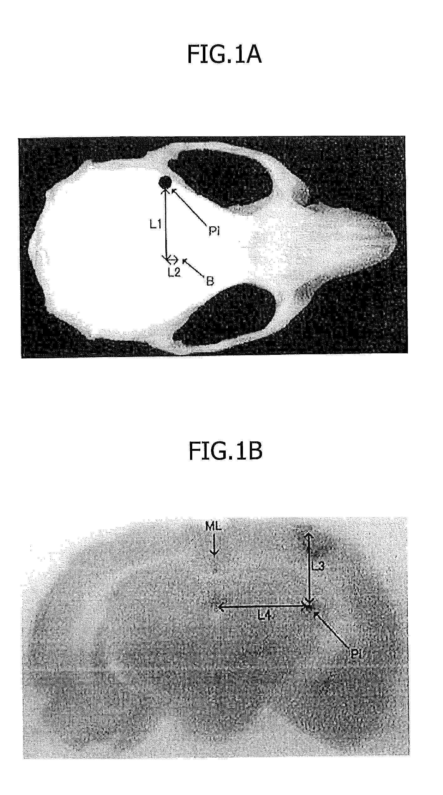
[0112]CD31-negative SP cells were fluorescently labeled with DiI and then transplanted 24 hours after cerebral infarction into an injection site Pi of the brain striatum in the brain tissues, as shown in FIGS. 1A and 1B, A PBS-injected control was used.

[0113]In FIG. 1A, B represents bregma, which is a point of intersection of the sagittal suture with the coronal suture of the cranium. L1 represents 6.0 mm, and L2 represents 1.0 mm.

[0114]FIG. 1B is a macro image of a coronal section. L3 represents 5.0 mm, and L4 represents 6.0 mm. ML represents a midline.

[0115]On day 21 after cell transplantation, perfusion fixation was performed, and frozen sections were prepared according to a routine method and immunostaine...

example 2

[0144]Unlike Example 1, human dental pulp tissue-derived dental pulp cells are used in this Example. Table 1 shows the properties of human dental pulp tissue-derived CD31− SP cells and porcine dental pulp tissue-derived CD31− SP cells.

TABLE 1HumanHuman totalPorcineCD31−dental pulpCD31−SP cellcellSP cell(%)(%)(%)CD310.000.060.00CD1460.0040.580.00CD240.1023.87—CD340.010.0169.00 CD4492.5091.60—CD9098.6772.690.20CD10521.234.35—CD1170.020.060.00CD1330.010.470.00CD1500.210.100.00CD2710.030.0194.00 SSEA10.290.06—

[0145]As shown in Table 1, the human dental pulp tissue-derived CD31−SP cells highly expressed CD90 and CD150 compared with porcine dental pulp tissue-derived CD3 V SP cells, in flow cytometry. Moreover, the human dental pulp tissue-derived CD31−SP cells highly expressed CD105 compared with human total dental pulp cells.

[0146]This Example shows nerve regeneration by the cell transplantation of human dental pulp tissue-derived CD31− / CD146− SP cells and human dental pulp tissue-deriv...


PUM


Abstract

Disclosed is a therapeutic material for cerebral infarction, which can recover a vascular disorder in an area affected by cerebral infarction to improve the brain function. The therapeutic material for cerebral infarction includes a dental pulp stem cell comprising at least one member selected from a CD105-positive cell, an SP cell, a CD24-positive cell, a CD133-positive cell, a CD271-positive cell and a CD150-positive cell. The therapeutic material may additionally include a protein secreted from a dental pulp cell. A transplanted dental pulp stem cell cannot be differentiated directly into a neural progenitor cell or a neurocyte, but is involved indirectly in the promotion of differentiation to eliminate an area affected by cerebral infarction, thereby recovering the affected area into a normal area.

Description

TECHNICAL FIELD[0001]The present invention relates to a material for treatment of cerebral infarction and to a brain tissue regeneration method using the material for treatment of cerebral infarction.BACKGROUND ART[0002]Cerebral angiopathy is the second leading cause of death in Japan and is responsible for the greatest cause of being bed-ridden, while not causing death. Therefore, the aging society is in urgent need of development of therapy appropriate for cerebral angiopathy.[0003]Among cerebral angiopathies, ischemic cerebrovascular disease such as cerebral infarction or cerebral thrombosis occurs with the highest frequency. Mortality due to cerebral infarction is greater than the total mortality due to myocardial infarction and ischemic heart disease.[0004]If the cause of paralysis is cerebral infarction, current treatment is thought to be effective within 3 hours after development, at present.[0005]In addition, rtPa (recombinant tissue plasminogen activator) intravenous drip t...

Claims


Application Information

Patent Timeline
IPC IPC(8): A61K35/12A61P25/00A61K35/32
CPCA61K35/32A61L27/3878A61L27/3834A61P9/00A61P9/10A61P25/00A61P25/28A61P43/00
Inventor NAKASHIMA, MISAKOSUGIYAMA, MASAHIKO
Owner NAT CENT FOR GERIATRICS & GERONTOLOGY
Learn More