
Surgery assistance system

a technology of surgical assistance and auxiliary equipment, applied in the field of surgical assistance system, can solve the problems of even a few millimeter errors and adverse effects, and achieve the effect of facilitating navigation and being more precis

Inactive Publication Date: 2011-05-26
HAMAMATSU UNIV SCHOOL OF MEDICINE
View PDF4 Cites 10 Cited by
  • Summary
  • Abstract
  • Description
  • Claims
  • Application Information

AI Technical Summary

Benefits of technology

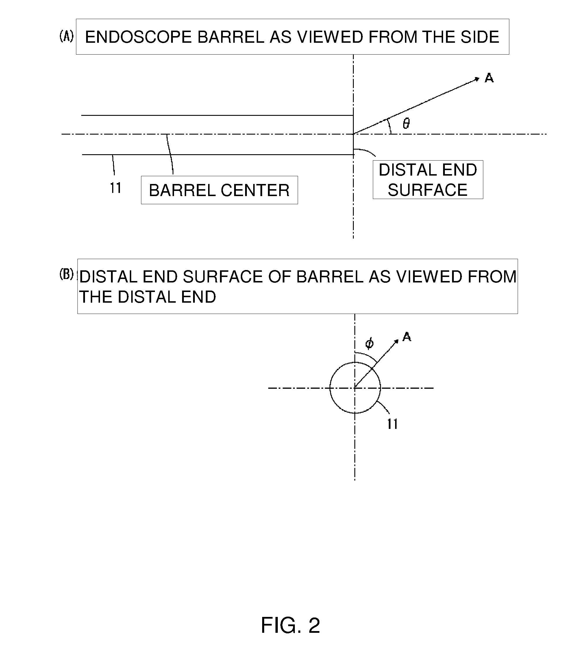
[0064]The present invention is configured as described above, and is therefore capable of measuring an actual optical axis position of a rigid endoscope in advance and performing surgery navigation which takes into account the actual optical axis position even when the actual optical axis is offset from the nominal value (hereinafter, referred to as offset of the optical axis). Specifically, since data obtained by measuring the actual optical axis position are used rather than the conventional method of moving and adjusting the display image of the endoscope to adjust the offset of the optical axis, the optical axis of the rigid endoscope and the intersection of the optical axis with the body cavity can be displayed on a surgery navigation screen. The system is also simplified by using the 3-dimensional shape measurement device for measuring the 3-dimensional surface shape of the patient without modification to measure the actual optical axis position. Since the actual optical axis position can be measured immediately prior to surgery, even when the amount of misalignment of the optical axis changes with each surgical operation as repeated disinfections take place for use in surgery, using the data of the actual optical axis position obtained immediately prior to surgery enables surgery navigation to be made more precise.
[0065]Moreover, by measuring the exact position of the actual optical axis (real optical axis), it is possible to assist surgery by displaying the point at which the optical axis is centered on the endoscope screen (the true center of the observation screen), as well as displaying which region of the image of the navigation device (preoperative CT or another image) in which the true center of the observation screen is positioned.

Problems solved by technology

Since surgery is a precision operation, an error of even a few millimeters can have adverse effects.

Method used


Image

  • Surgery assistance system
  • Surgery assistance system
  • Surgery assistance system

Examples


Embodiment Construction

[0077]Preferred embodiments of the surgery assistance system of the present invention will be described in detail with reference to the drawings. Reference symbols are used consistently to refer to the same elements in the drawings, and no redundant description thereof will be given. The embodiments described are also not necessarily shown to scale in the drawings.

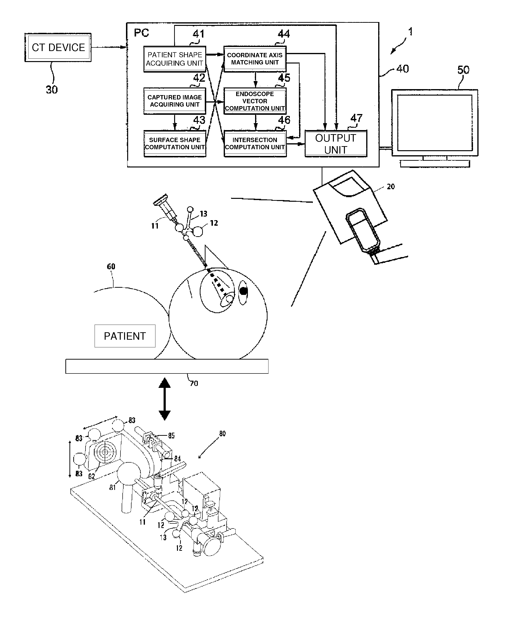
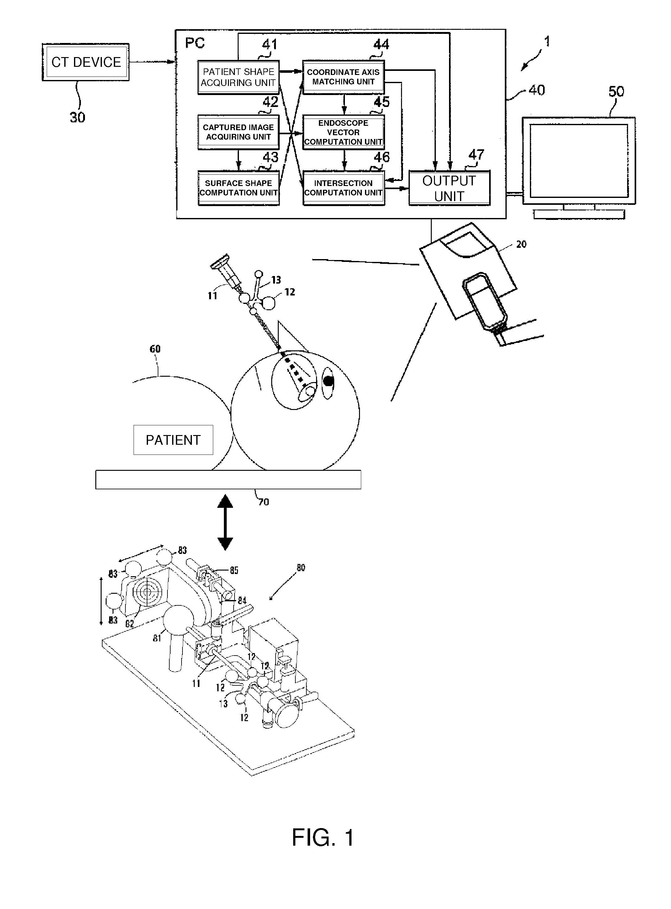
[0078]FIG. 1 is a view showing the overall configuration of an embodiment of the surgery assistance system 1 of the present invention. The surgery assistance system 1 is a device for providing a surgeon or another user with information relating to an image captured by an endoscope during surgery on a patient 60. The surgery assistance system 1 according to the present embodiment is used for surgery that involves imaging by a rigid endoscope, such as endoscopic surgery on the paranasal sinus in otorhinolaryngology.

[0079]As shown in FIG. 1, the surgery assistance system 1 is composed of a rigid endoscope 11, ball markers 12,...


PUM


Abstract

Even when the optical axis is offset in a rigid endoscope, the actual optical axis position is measured in advance, and surgery navigation can be performed by using the actual optical axis position.A surgery assistance system comprising 3-dimensional shape measurement means for measuring a 3-dimensional surface shape of a patient; and a rigid endoscope (11) to be inserted into the body of the patient; wherein the rigid endoscope has a position-orientation detection marker (12) measurable by the 3-dimensional shape measurement means; 3-dimensional tomographical data of the patient imaged in advance and a position relationship relating to the actual optical axis position of the rigid endoscope measured in advance are stored in the computation means; 3-dimensional tomographical data and the 3-dimensional surface shape of the patient measured by the 3-dimensional shape measurement means are aligned; the position, orientation, and optical axis of the rigid endoscope are computed on the basis of the position-orientation detection marker measured by the 3-dimensional shape measurement means and the stored actual optical axis position of the rigid endoscope; and the aligned 3-dimensional tomographical data, the optical axis of the rigid endoscope, and the intersection of the optical axis and the tissue wall are displayed.

Description

TECHNICAL FIELD[0001]The present invention relates to a surgery assistance system for providing information relating to an image captured by a rigid endoscope to a surgeon or another user.BACKGROUND ART[0002]Surgical navigation (surgery assistance information display) has been performed in the past for displaying the proper position of the distal end of the surgical instrument on a CT (computed tomography) or MRI (magnetic resonance imaging) image to assist a surgeon when an endoscope or another surgical instrument is inserted into a patient's body. For example, Patent Document 1 describes a surgery assistance system proposed by the present inventors, which is a technique for aligning a 3-dimensional surface shape of a patient measured by a 3-dimensional shape measurement device with 3-dimensional tomographical data imaged in advance. A technique is also described whereby a 3-dimensional shape measurement device for measuring the 3-dimensional surface shape of a patient is used to m...

Claims


Application Information

Patent Timeline
Patent Type & Authority Applications(United States)
IPC IPC(8): A61B6/03
CPCA61B5/06A61B5/103A61B19/5244A61B1/0057A61B2019/507A61B2019/5255A61B2017/00725A61B2034/2055A61B34/20A61B2034/107A61B5/064
Inventor YAMAMOTO, SEIJITAKAI, TOSHIHISAHAYASHIMOTO, ETSUKAZUKINPARA, MASAAKIMIURA, AKIRA
Owner HAMAMATSU UNIV SCHOOL OF MEDICINE
Learn More