Method for Screening of Active Tuberculosis
a tuberculosis and active technology, applied in the field of immunology and medical microbacteriology, can solve the problems of unreliable diagnosis of acute tb, burden on patients and public expenditure, etc., and achieve the effect of improving and simplifying
- Summary
- Abstract
- Description
- Claims
- Application Information
AI Technical Summary
Benefits of technology
Problems solved by technology
Method used
Image

Examples
example 1
Sample Preparation Protocol
This example describes a preferred sample preparation protocol for carrying out the methods of the present invention using whole blood samples.
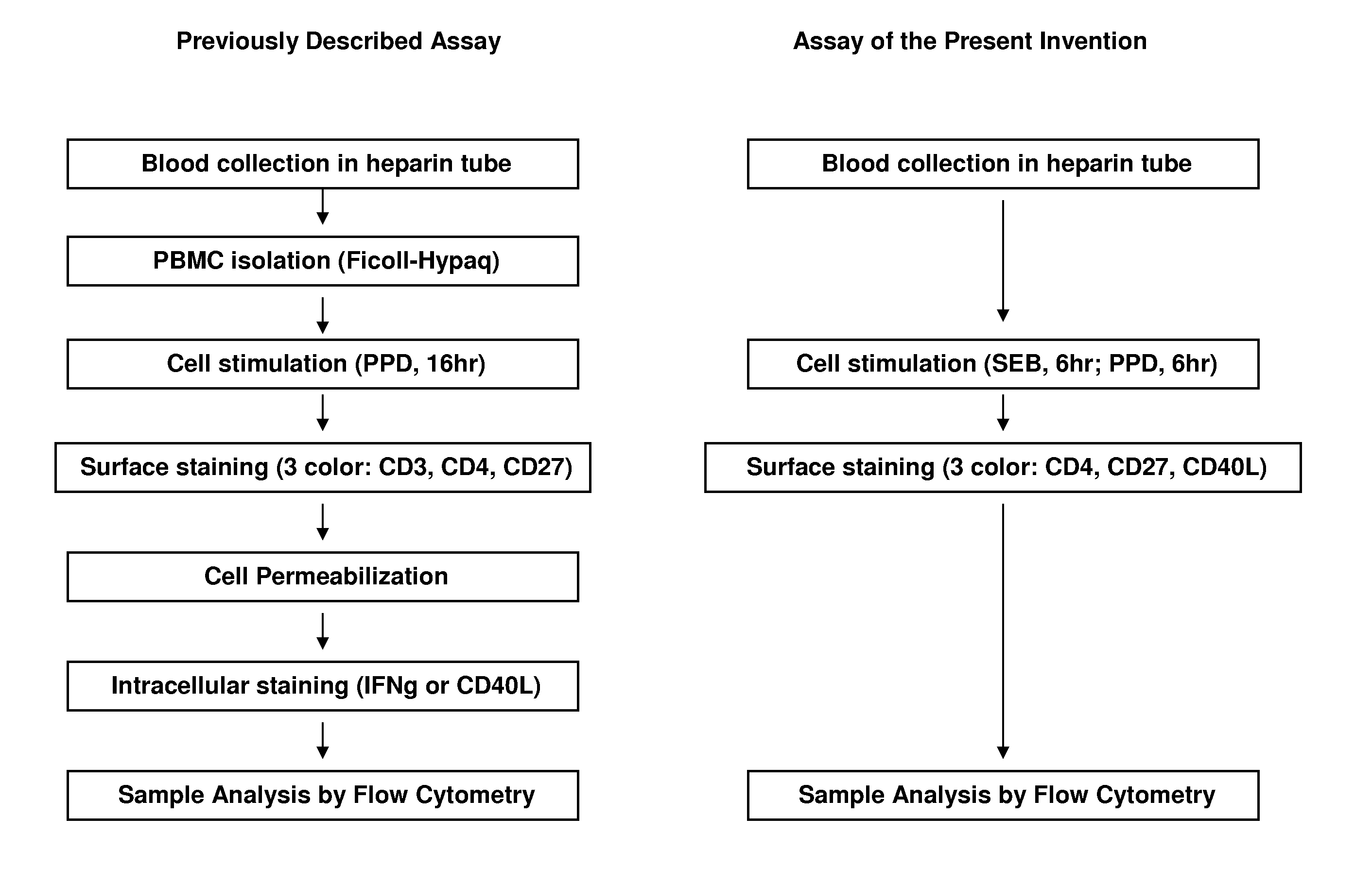
I. Whole blood sampleDraw blood into a heparinized Vacutainer™ tube, and hold at room temperature until use (no more than ˜4 hrs).
II. Activation
The protocol described below is designed for duplicates of each test condition; amounts of sample may be adjusted as long as reagent proportions remain identical. Use of more than 1 ml of blood per tube is not recommended.1. For each patient sample, prepare one appropriately labeled round-bottom polypropylene tube (e.g. 15 ml Falcon #352096, or 14 ml Falcon #352059; BD Biosciences, San Jose, Calif.) for each sample treatment: (1) an unstimulated negative control, (2) a PPD-stimulated test sample, and (3) an SEB (Streptococcal enterotoxin B)-stimulated positive control.2. Add 500 μl of whole blood into each tube.3. Add 500 ng of CD40-specific mAb into each tube. A preferred C...
example 2
Analysis by Flow Cytometry
The samples prepared as described in example 1 are analyzed using a flow cytometer. For use with the preferred choice of dyes, described above, a flow cytometer having three excitation lasers, a blue, a red, and a violet, is used. Examples of usable flow cytometers include the BD FACSCanto™ Flow Cytometer, the BD™ LSR II System, and the BD FACSAria™ II Cell Sorter, all available from BD Biosciences (San Jose, Calif.).
The flow cytometer is set up according to the manufacturer's instructions. The prepared samples are analyzed, and the cell populations of interest are identified and counted by appropriate gating using well-known methods.
CD4+ T-cells are identified by the expression of CD3 and CD4. Other combinations of markers that identify CD4+ T-cells, which are well known in the art, may be used to identify this cell subpopulation.
Activation of CD4+ T-cells is identified by the cell-surface expression of CD40L (also known as CD154) on the identified CD4+ T-...
example 3
Comparison of the Present Method with the Prior Art Method
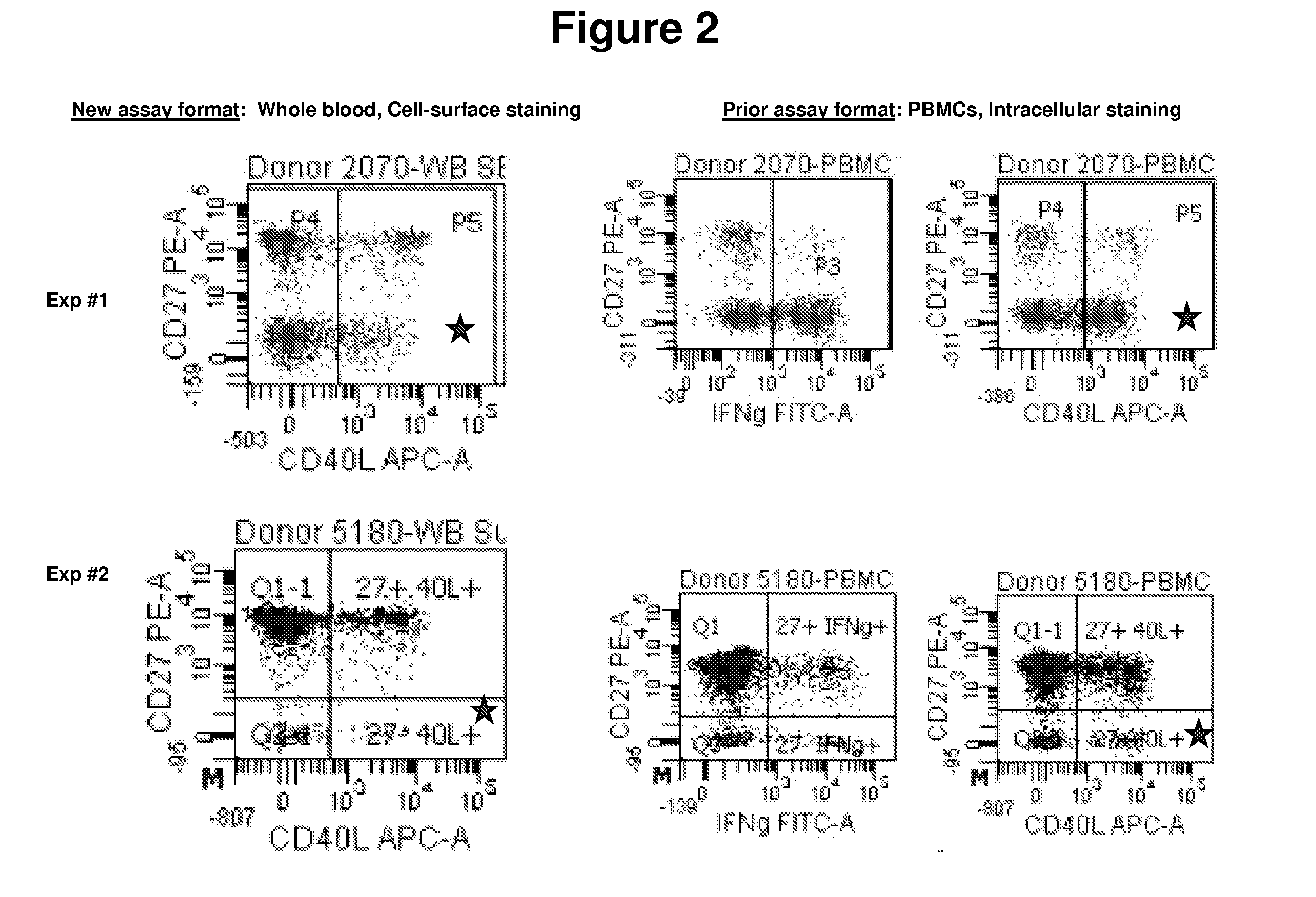
The ability of the present methods to detect CD27 positive and negative populations among activated CD4+ T-cells was compared with the prior art methods. Positive-control samples from two healthy donors were analyzed using each method.The present method was carried out essentially as described above using whole blood activated with SEB for 6 hours.The prior art methods were essentially as described in Streitz et al., 2007, supra.The data are shown in FIGS. 2 and 3. In FIG. 2, the CD27− populations are indicated with a star.The data demonstrate that the methods of the present invention, which work with whole blood and with only cell-surface staining, provide results that are comparable to the results obtained using the prior art methods.
PUM
| Property | Measurement | Unit |
|---|---|---|
| incubation time | aaaaa | aaaaa |
| incubation time | aaaaa | aaaaa |
| length | aaaaa | aaaaa |
Abstract
Description
Claims
Application Information
 - R&D
- Intellectual Property
- Life Sciences
- Materials
- Tech Scout
- Unparalleled Data Quality
- Higher Quality Content
- 60% Fewer Hallucinations
Browse by: Latest US Patents, China's latest patents, Technical Efficacy Thesaurus, Application Domain, Technology Topic, Popular Technical Reports.
© 2025 PatSnap. All rights reserved.Legal|Privacy policy|Modern Slavery Act Transparency Statement|Sitemap|About US| Contact US: help@patsnap.com