
Mechanically-actuated endotracheal tube cleaning device

Active Publication Date: 2011-02-03
AVENT INC
View PDF35 Cites 72 Cited by
  • Summary
  • Abstract
  • Description
  • Claims
  • Application Information

AI Technical Summary

Benefits of technology

There remains a need for systems, methods and devices for the cleaning of endotracheal tubes that are effective and efficient so that the tube cleanings can be reasonably carried out on a regular and preventative basis, rather than only when a particular problem arises. There also remains a need for systems, methods and devices for the cleaning of endotracheal tubes that prevent the build up of materials, perform the cleaning quickly, and that permit sufficient airflow through the endotracheal tube during use.
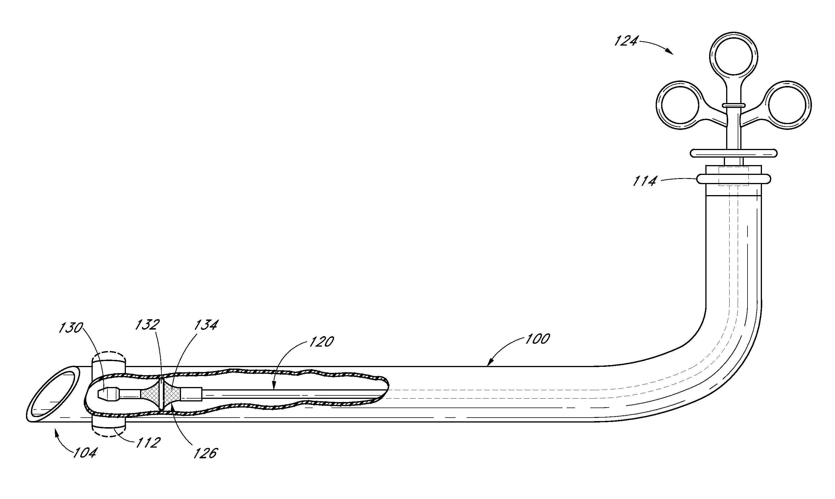
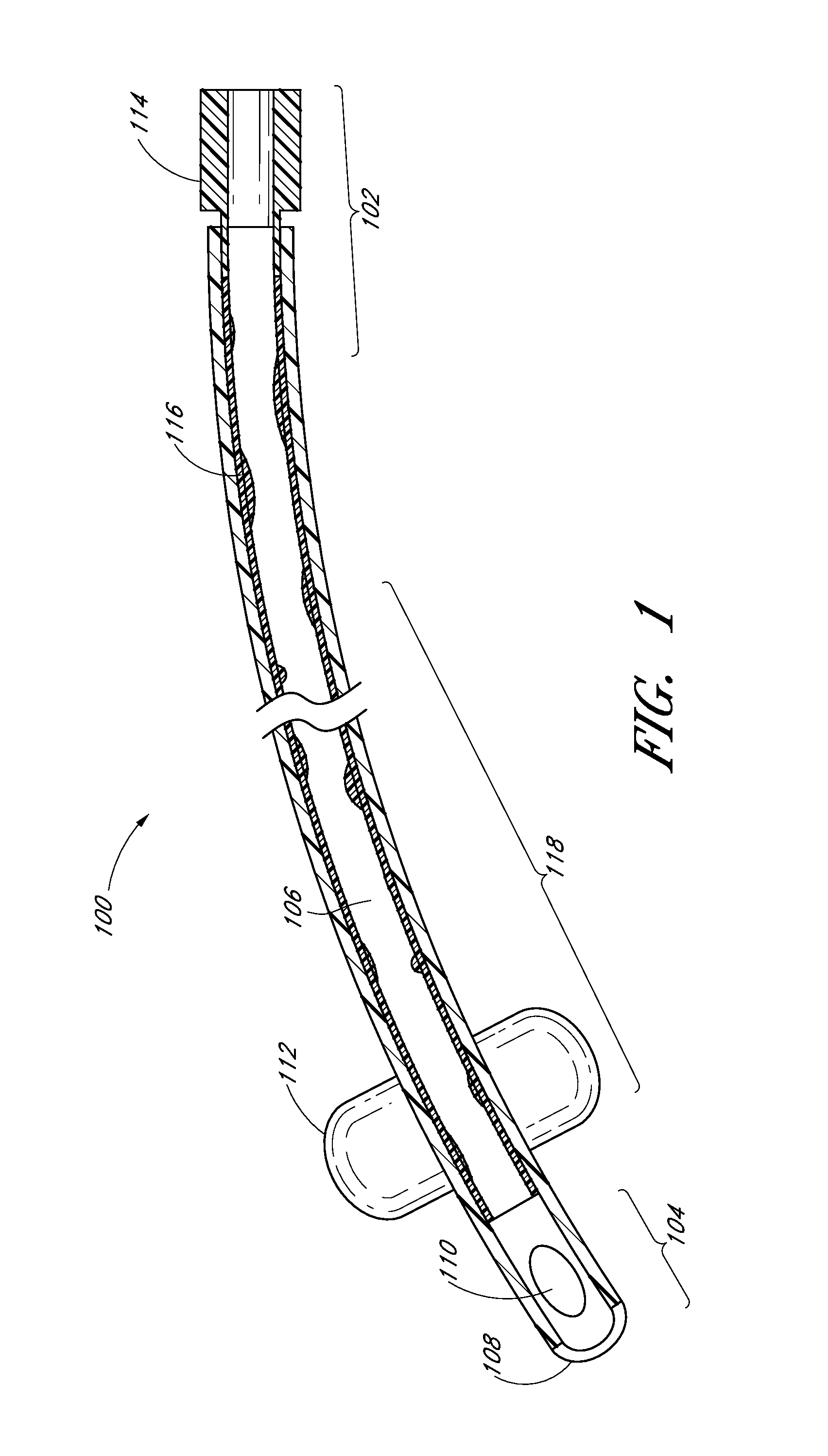
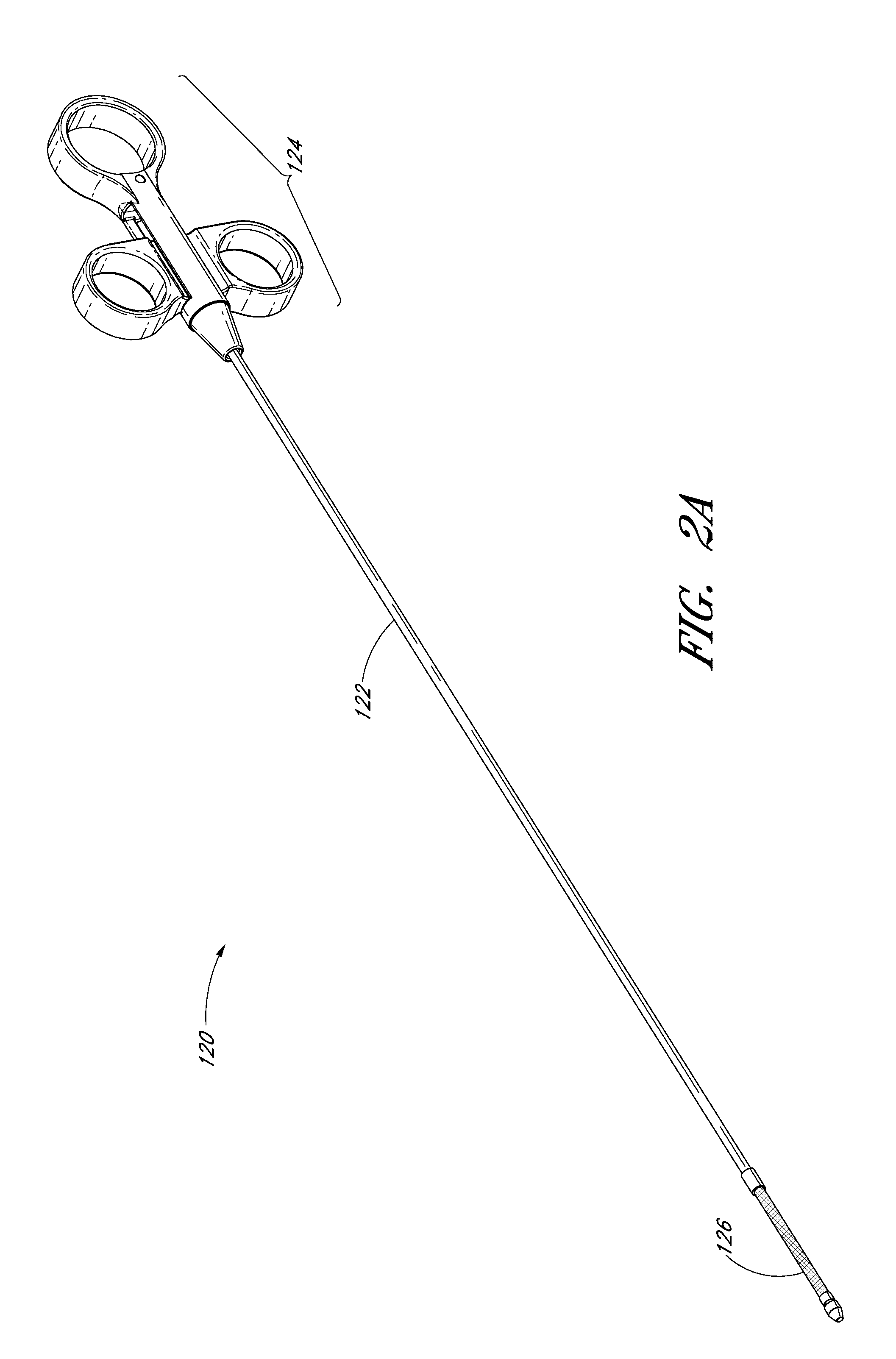
According to some embodiments, a non-inflatable, mechanically-actuated cleaning device for removing biofilm from an interior wall of an endotracheal tube or other conduit comprises an elongate body having a distal end, a proximal end, a longitudinal axis and a diameter in the range of about 1 mm to about 5 mm. The cleaning device further comprises a scaffold (e.g., mesh scaffold) positioned at the distal end of the elongate body. In some embodiments, the scaffold is positioned near the proximal end of the elongate body or at any other location along the elongate body. In one embodiment, the cleaning device comprises one or more O-rings or other removal members coupled to the scaffold (e.g., mesh scaffold) and a non-inflatable actuation assembly coupled to the proximal end of the elongate body for mechanically-actuating the expansion of the scaffold. In some embodiments, the scaffold is radially expandable between a collapsed position and an expanded position by manipulation of the actuation assembly. In one embodiment, the level of expansion and / or collapse can be precisely controlled between fully-expanded and fully-collapsed positions. In several embodiments, the O-ring or other removal member is configured to engage an interior surface of an endotracheal tube when the scaffold is in the expanded position. In some embodiments, the O-ring is configured to remove biofilm collected on the interior surface of an endotracheal tube when said O-ring is moved along the longitudinal axis of the elongate body. In one embodiment, the scaffold comprises a porous architecture configured for facilitating the in-flow of said biofilm into an interior of the scaffold, thereby trapping at least a portion of the biofilm within the scaffold. In other embodiments, one or more portions of the scaffold are non-porous or substantially non-porous so as to prevent or reduce the likelihood of biofilm and / or other materials from passing therethrough. In several embodiments, the scaffold (e.g., mesh scaffold) is configured to allow airflow through the endotracheal tube in the collapsed position and the expanded position. In some embodiments, the actuation assembly is configured for one-handed manipulation of the cleaning device during a cleaning procedure.

Problems solved by technology

Frequently, it is not practical or clinically acceptable to change out the endotracheal tube when there is a buildup of biofilm in order to remove the endotracheal tube for cleaning.
Removal and reinsertion of the endotracheal tube can be uncomfortable for the patient, can cause injury to the native airway, and can put reliable control of the airway at risk.

Method used


Image

  • Mechanically-actuated endotracheal tube cleaning device
  • Mechanically-actuated endotracheal tube cleaning device
  • Mechanically-actuated endotracheal tube cleaning device

Examples


Embodiment Construction

The discussion and the figures illustrated and referenced herein describe various embodiments of a body-inserted tube cleaning system and device, as well as methods related thereto. A number of these embodiments of tube cleaning systems, devices and methods are particularly well suited to remove biofilm from an interior surface of an endotracheal tube. However, the various devices, systems, methods and other features of the embodiments disclosed herein may be utilized or applied to other types of apparatuses, systems, procedures, and / or methods, whether medically-related or not. For example, the embodiments disclosed herein can be utilized for, but are not limited to, cleaning bronchoscopes, chest drainage tubes, gastrostomy drainage tubes, abdominal drainage tubes, other body drainage tubes, feeding tubes, endoscopes, percutaneous dialysis catheters, and any other percutaneous or per os catheters or body-inserted tubes. In addition, as discussed in greater detail herein, the variou...


PUM


Abstract

Systems, devices, and methods are disclosed for the cleaning of an endotracheal tube while a patient is being supported by a ventilator connected to the endotracheal tube for the purpose of increasing the available space for airflow or to prevent the build up of materials that may constrict airflow or be a potential nidus for infection. In one embodiment, a mechanically-actuated endotracheal tube cleaning device is configured to removably receive a visualization member to provide cleaning of the endotracheal tube under direct visualization.

Description

BACKGROUND1. FieldThis application relates generally to the cleaning of body-inserted tubes and, more specifically, to devices, systems, and methods for removing fluids, secretions and / or other materials from a lumen of an endotracheal tube.2. Description of the Related ArtAn endotracheal tube is used in patient care to provide a clear airway through the mouth, pharynx, and trachea into the tracheobronchial tree. Use of an endotracheal tube is appropriate when the integrity of the airway is, or may become, challenged due to trauma or pathology, or if a patient cannot otherwise breathe unaided. Often the endotracheal tube is coupled to a mechanical ventilator to aid the patient's respiration, and can be expected to remain in situ for an extended time until the patient is once again able to breathe on his own.Endotracheal tubes are used in millions of patients around the world to support life after major surgery, trauma, or the development of certain severe medical conditions such as ...

Claims


Application Information

Patent Timeline
IPC IPC(8): A61M16/04
CPCA61B1/00135A61B1/00045A61B1/00165A61B1/05A61B1/0669A61B1/267A61B1/2676A61B17/320725A61B19/34A61B2019/343A61M16/04A61M16/0434A61M16/0463A61M16/0472A61M2202/203A61B1/00142A61M16/0833A61M2016/0413A61M16/0418A61M16/0427A61M16/0431A61M16/0486A61M16/0484A61B90/70A61B2090/701A61B1/04A61B1/0661A61B1/07
Inventor VAZALES, BRAD E.BERTOLERO, ARTHURWATSON, KENGRACY, JAMES M.COLWELL, CLIFTON P.BERKY, CRAIG B.DAVIDNER, ALAN
Owner AVENT INC
Learn More