
Use of focused ultrasound for vascular sealing

a vascular catheter and focused ultrasound technology, applied in the field of vascular catheter entry wound sealing, can solve the problems of ischemia and/or thrombosis, time-consuming procedure, and discomfort for patients, and achieve the effect of safe sealing of vascular catheter entry wounds and little operator training

Inactive Publication Date: 2011-01-27
OTSUKA MEDICAL DEVICES
View PDF104 Cites 27 Cited by
  • Summary
  • Abstract
  • Description
  • Claims
  • Application Information

AI Technical Summary

Benefits of technology

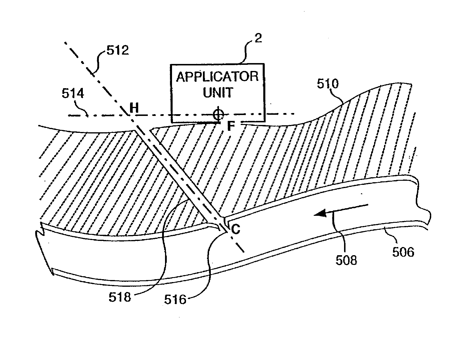
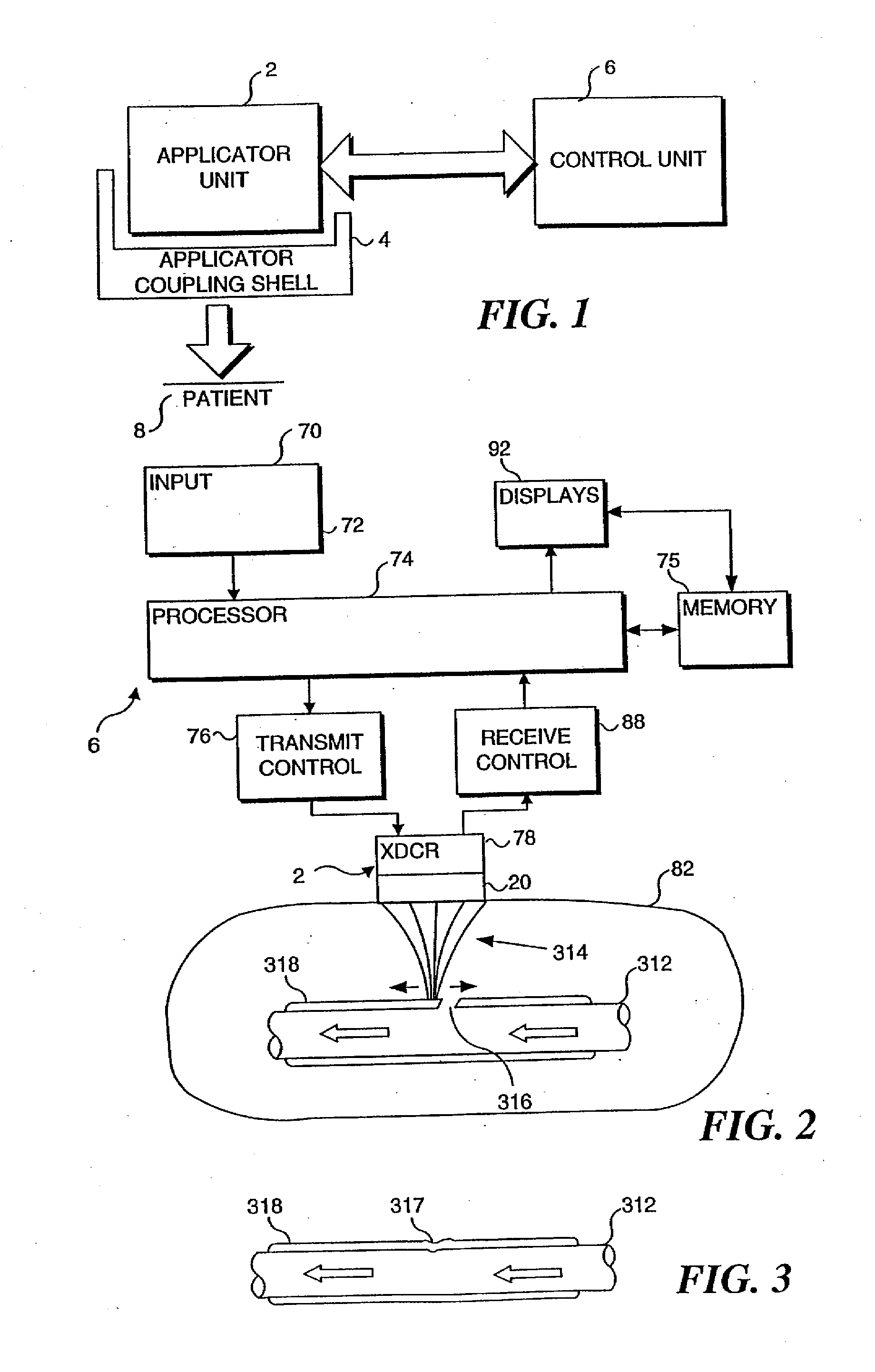
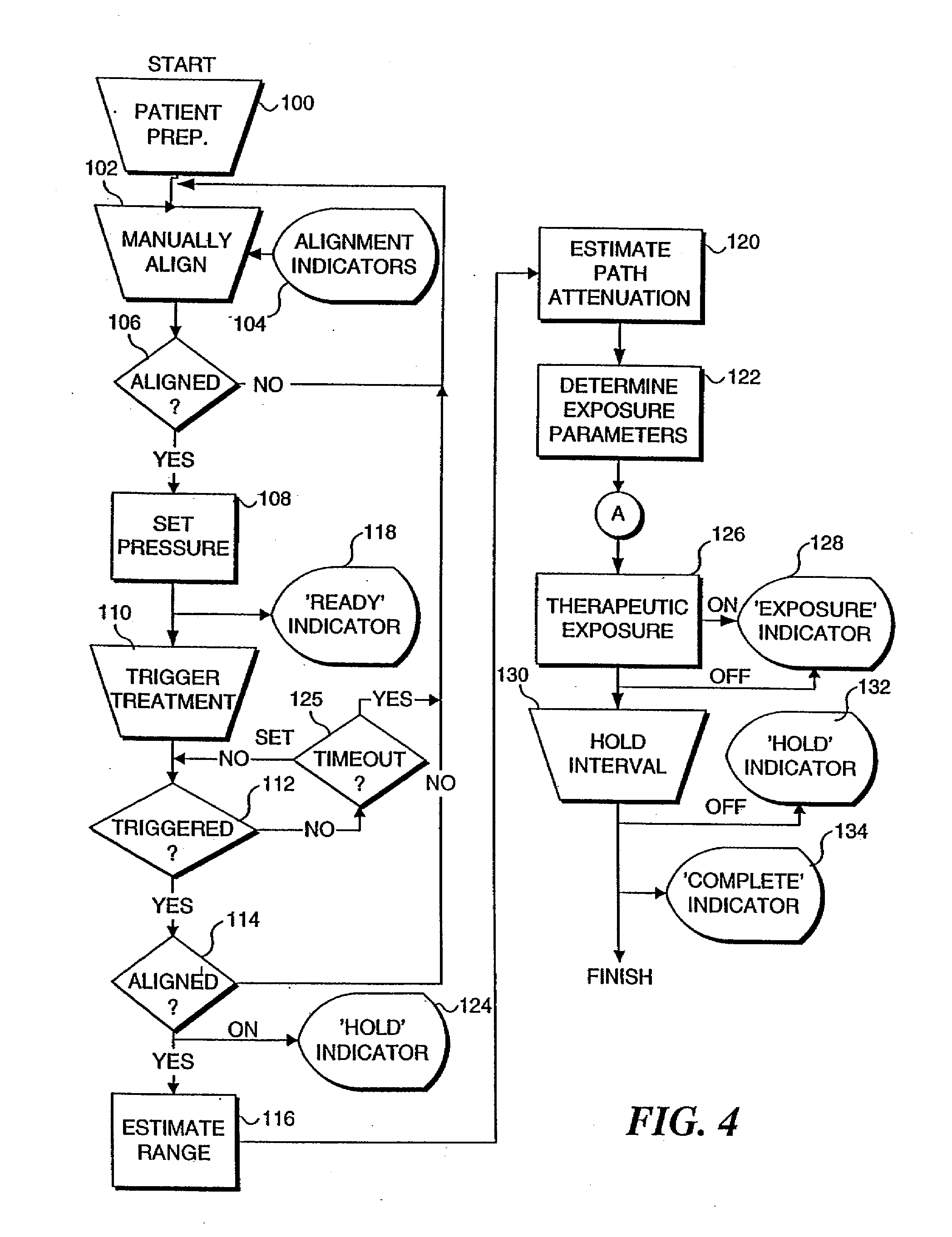
[0012]In accord with the present invention, a method and apparatus are defined that provide advantageous solutions to the problem of expeditiously and safely sealing vascular catheter entry wounds made in connection with medical procedures. The method includes the steps of determining a site of the puncture in the vascular vessel and positioning an ultrasonic transducer applicator at a position adjacent to the site. The ultrasonic transducer applicator is coupled to a control that includes a processor programmed to administer ultrasonic energy in a manner that efficaciously seals a puncture. A user is enabled to initiate a process that is controlled by the control, so that very little operator training is required. The control automatically controls the ultrasonic transducer applicator so that the ultrasonic energy produced by the ultrasonic transducer applicator is focused at the site and is administered at a sufficient intensity and duration to denature tissue at the puncture, closing and sealing the puncture.
[0013]To determine the site of the puncture, an imaging ultrasonic beam is generated with the ultrasonic transducer applicator and is transmitted into the patient, proximate an expected location for the site. A reflection of the imaging ultrasonic beam is then received from within the patient using the ultrasonic transducer applicator, producing a corresponding output signal. The output signal is processed with the processor included in the control to facilitate determining the site of the puncture.
[0014]For example, a visual indication of a location of the site of the puncture can be provided to enable an operator to position the ultrasonic transducer applicator so that the ultrasonic energy produced by the ultrasonic transducer applicator is directed at the site. Such a visual indication may be in the form of, for example, lighted display indicators. In one form of the present invention, the visual indication includes an image of the site in which an axis of the vascular vessel is visually evident, enabling the operator to position the ultrasonic transducer applicator longitudinally along the axis of the vascular vessel so that the ultrasonic energy is directed at the site of the puncture.
[0019]It is also contemplated that the administration of the ultrasonic energy be interrupted, to again generate the imaging ultrasound beam, thereby confirming whether the ultrasonic energy is being directed at the site of the puncture.

Problems solved by technology

When applied manually, the procedure is time-consuming and requires the presence of a medical professional for thirty minutes or more.
For both manual and mechanical pressure application, the procedure is uncomfortable for the patient and frequently requires the administration of analgesics to be tolerable.
Moreover, the application of excessive pressure can occlude the underlying artery, resulting in ischemia and / or thrombosis.
Renewed bleeding through the tissue tract is not uncommon and can result in hematoma, pseudoaneurisms, and arteriovenous fistulas.
The risk of these complications increases with the use of larger sheath sizes, which are frequently necessary in interventional procedures, and when the patient is anticoagulated with heparin or other drugs.
The large hemostasis plug stimulates blood coagulation in the vessel puncture site, but blocks the catheter entry tract, making catheter reentry more difficult, if required.
However, any device remaining in the vessel lumen increases the risk of thrombus formation.
Such a device also can detach and cause occlusion in a distal blood vessel, which would likely require major surgery to remove.
Suture material left in the vessel may cause tissue irritation that will prolong the healing process.
However, in some cases, the clotting agent may leak past the balloon into the vessel lumen and cause stenosis.

Method used


Image

  • Use of focused ultrasound for vascular sealing
  • Use of focused ultrasound for vascular sealing
  • Use of focused ultrasound for vascular sealing

Examples


Embodiment Construction

Use of Ultrasound for Sealing Punctures

[0043]Because of its unique properties in soft tissue, ultrasound can be brought to a tight focus at a distance from its source. If sufficient energy is radiated within the ultrasound beam, cells located in the focal volume can be rapidly heated, while intervening and surrounding tissues are spared. Surrounding tissues are unaffected in the unfocused portion of the ultrasound beam because the energy is spread over a correspondingly larger area and associated heating is minimized.

[0044]Whereas ultrasound intensities on the order of 0.1 Watts / cm2 are employed in diagnostic imaging applications, intensities in excess of 1,000 Watts / cm2 are typical in high-intensity focused ultrasound (HIFU) applications. At the focal point, these high intensities result in large, controlled temperature rises within a matter of seconds.

[0045]It has been demonstrated in numerous in vivo animal studies that HIFU can rapidly seal blood vessel punctures and lacerations...


PUM


Abstract

An ultrasonic applicator unit (2) is used diagnostically to locate a puncture wound (316) in an artery and then therapeutically to seal the puncture wound with high intensity focused ultrasound (HIFU). A control unit (6) coupled to the applicator unit includes a processor (74) that automates the procedure, controlling various parameters of the diagnostic and therapeutic modes, including the intensity and duration of the ultrasonic energy emitted by the applicator unit. A protective, sterile acoustic shell (4), which is intended to be used with a single patient and then discarded, is slipped over the applicator unit to protect against direct contact between the applicator unit and the patient and to maintain a sterile field at the site of the puncture. The apparatus and method are particularly applicable to sealing a puncture made when inserting a catheter into an artery or other vessel. Several different procedures are described for locating the puncture wound, including imaging the vessel in which the puncture is disposed and use of a locator rod to determine the disposition of the puncture along the longitudinal axis of the artery.

Description

RELATED APPLICATIONS[0001]This application is a continuation of co-pending U.S. application Ser. No. 11 / 619,996, filed Jan. 4, 2010, which is a continuation of co-pending U.S. application Ser. No. 10 / 616,831, filed Jul. 10, 2003, which is a continuation of U.S. application Ser. No. 09 / 696,076, filed Oct. 25, 2000, now U.S. Pat. No. 6,656,136, which claims the benefit of U.S. provisional patent application Ser. No. 60 / 163,466, filed Oct. 25, 1999, and U.S. provisional patent application Ser. No. 60 / 171,703, filed Dec. 23, 1999.FIELD OF THE INVENTION[0002]The present invention generally relates to methods and apparatus for sealing vascular punctures and wounds, and more particularly, to a device that may be used to deliver ultrasound energy to a vascular puncture site to arrest bleeding.BACKGROUND OF THE INVENTION[0003]Various surgical procedures are performed by medical specialists such as cardiologists and radiologists, utilizing percutaneous entry into a blood vessel. To facilitate...

Claims


Application Information

Patent Timeline
Patent Type & Authority Applications(United States)
IPC IPC(8): A61N7/00A61B8/00A61B17/00A61B17/12A61B18/00A61B19/00A61N7/02G10K11/32G10K11/34
CPCA61B8/4422A61B17/0057A61B2017/00504A61B2017/00641A61B2017/00907A61B2019/462G10K11/346A61B2019/5404A61B2019/5437A61B2562/164A61N7/02G10K11/32A61B2019/5276A61B2090/3904A61B2090/062A61B2090/378A61B2090/3937
Inventor WENG, LEEPEROZEK, DAVID M.ZHANG, JIMIN
Owner OTSUKA MEDICAL DEVICES
Learn More