Boundary conditions for the arrangement of cells and tissues
a technology applied in the field of boundary conditions for the arrangement of cells and tissues, can solve the problems of often failing to build biosynthetic materials or engineered tissues that recapitulate these structure-function relationships
- Summary
- Abstract
- Description
- Claims
- Application Information
AI Technical Summary
Benefits of technology
Problems solved by technology
Method used
Image
Examples
example 1
Sarcomere Alignment is Regulated by Myocyte Shape
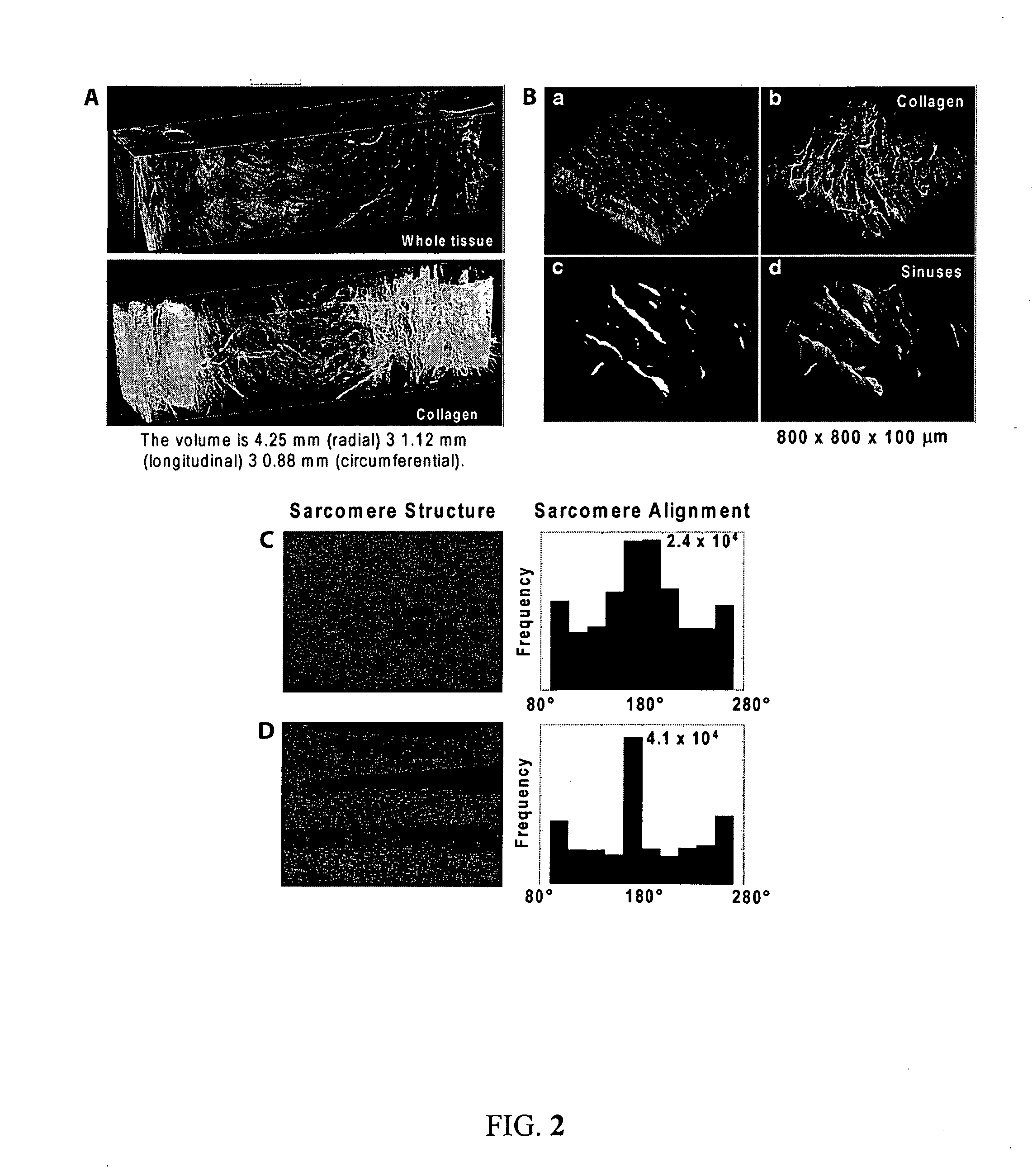
[0084]Cardiac organogenesis and pathogenesis are both characterized by changes in myocyte shape, cytoskeletal architecture, and the extracellular matrix (ECM). However, the mechanisms by which the cellular boundary conditions imposed by the ECM influence myocyte shape and myofibrillar patterning are unknown. Geometric cues in the ECM align sarcomeres by directing the actin network orientation.
[0085]The shape of cultured neonatal rat ventricular myocytes was altered by varying the cellular boundary conditions via soft lithography. Circular and 2500 μm2 rectangular ECM islands were microcontact printed on rigid substrates; adherent myocytes conformed to the ECM island shape after 48 hours in culture. Myocytes were immunostained against F-actin and sarcomeric α-actinin to visualize their cytoskeleton with fluorescent microscopy. Each immunofluorescence image was spatially registered, normalized and summed over all myocytes to obtain an a...
example 2
Ordered Processes in the Self-Organization of a Muscle Cell
[0120]Cellular form and function are the result of self-assembly and -organization of its molecular constituents into coupled networks. A specific example of this phenomenon is myofibrillogenesis, the formation and organization of myofibrils in striated muscle. Although several hypotheses have been proposed to describe maturation of myofibrils, the physical principles governing their development into a mature, functional structure were not well understood prior to the present invention Described herein is a mechanism of cytoskeletal and myofibril organization of cardiac myocytes. Computer simulations and in vitro assays to control myocyte shape demonstrated that distinct cytoskeletal architectures arise from two temporally-ordered, organizational processes: the interaction between stress fibers, premyofibrils and focal adhesions, as well as cooperative alignment and parallel bundling of myofibrils. The results identify a hie...
example 3
Modeling Dynamic Alignment of Premyofibrils
[0162]A 2D myocyte that fully spreads to a defined geometry Ω was evaluated by focusing on variables measured experimentally. A full description of the time-dependent organization of premyofibrils requires at least five field variables defined as a function of position {right arrow over (r)} and time t: (1, 2) the density of bound and unbound integrin, ρ and ρ*, respectively; (3) the local traction exerted on the bound integrin, {right arrow over (T)}; (4) the local density and orientation of the stress fiber network, {right arrow over (S)}′; and (5) the premyofibril, {right arrow over (u)}, in which the directional component of {right arrow over (u)} denotes the dominant orientation of premyofibrils, and the magnitude of {right arrow over (u)} represents the co-localization of premyofibrils into a bundle. Prior studies suggest that FAC formation takes place on a fast time scale (on the order of seconds), followed by the assembly / disassembl...
PUM
 Abstract
Description
Claims
Application Information
 - R&D
- Intellectual Property
- Life Sciences
- Materials
- Tech Scout
- Unparalleled Data Quality
- Higher Quality Content
- 60% Fewer Hallucinations
Browse by: Latest US Patents, China's latest patents, Technical Efficacy Thesaurus, Application Domain, Technology Topic, Popular Technical Reports.
© 2025 PatSnap. All rights reserved.Legal|Privacy policy|Modern Slavery Act Transparency Statement|Sitemap|About US| Contact US: help@patsnap.com


