
Imaging of myelin basic protein

a technology of myelin and basic protein, which is applied in the field of imaging of myelin basic protein, can solve the problems of spinal nerve roots being damaged, long time required for retrograde transport may not be clinically feasible, and signal is typically presen

Inactive Publication Date: 2010-12-09
GENERAL ELECTRIC CO
View PDF4 Cites 5 Cited by
  • Summary
  • Abstract
  • Description
  • Claims
  • Application Information

AI Technical Summary

Problems solved by technology

A challenge in the first approach is that the signal is typically ambiguous
Although this strategy may work, there are many inherent problems.
The long times required for retrograde transport may not be clinically feasible.
The spinal nerve roots can be damaged as they traverse the spinal canal, but are especially vulnerable in the intervertebral foramen, where the spinal nerve roots join to form the spinal nerves.
However, except for a few dyes such as bis-styrene-arylene dyes such as 1,4-bis(p-aminostyryl)-2-methoxy benzene (BMB), and (E,E)-1,4-bis(4′-aminostyryl)-2-dimethoxy-benzene (BDB), most of the publicly-disclosed dyes are unable to cross the blood nerve or blood brain barrier.

Method used


Image

  • Imaging of myelin basic protein
  • Imaging of myelin basic protein
  • Imaging of myelin basic protein

Examples


example 1

Preparation of Nerve Tissue Sections

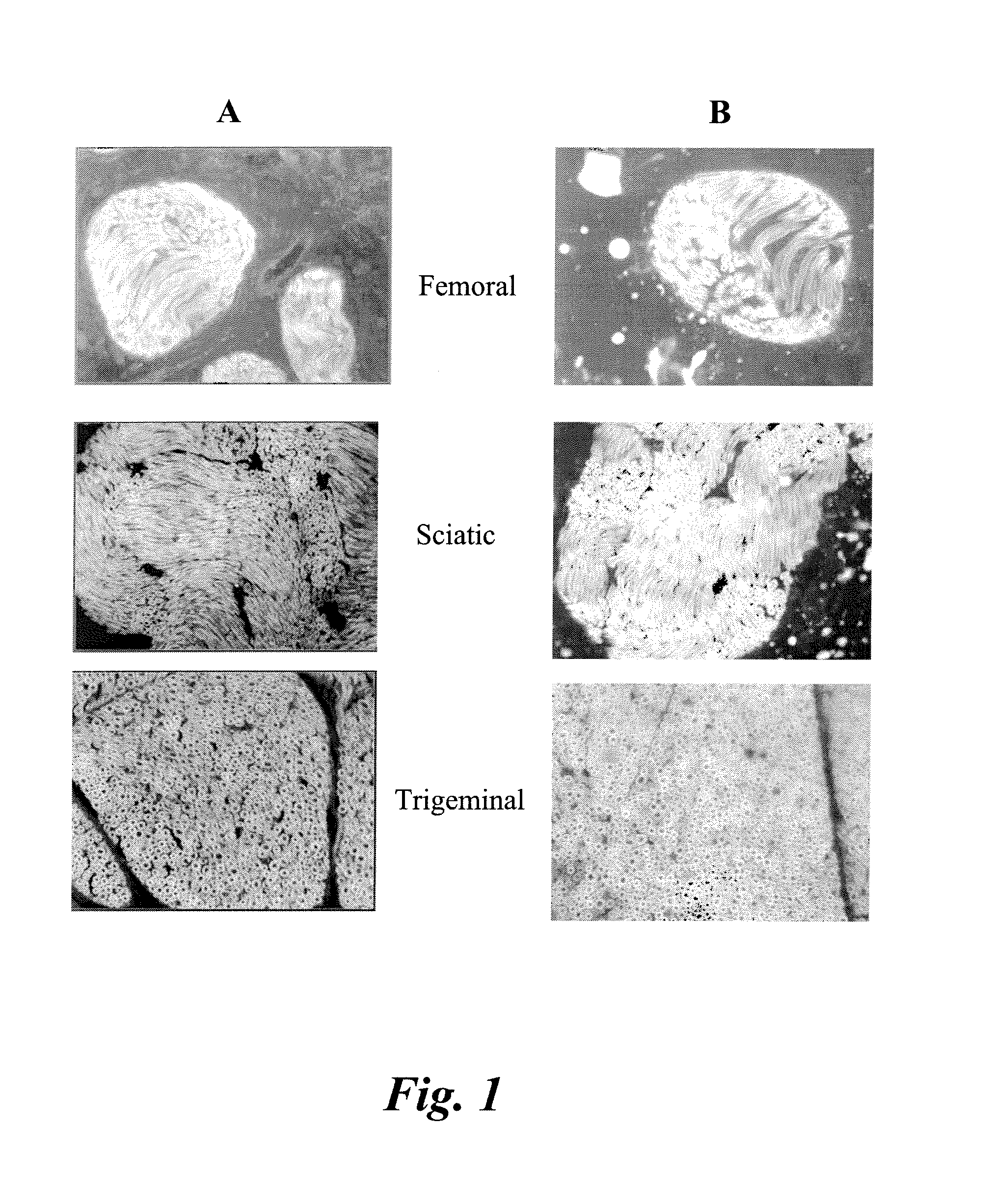
[0066]Various nerves including sciatic, femoral, brachial plexus, trigeminal, optic, and penile were harvested from male Sprague Dawley rats or male CD-1 mice. Tissue was fixed by perfusion and / or post-fixation with formalin. Following post-fixation, tissue was cryoprotected in a 20% sucrose solution made in phosphate buffered saline (PBS). Nerves were then flash-frozen using methanol and dry ice in OCT media. In some cases, PVDF membranes were used to help keep the nerves vertical in the OCT media. Thin sections (5-10 um) were sliced on a Leica microtome and stored in a −80° C. freezer prior to staining with antibodies or small molecule compounds.

example 2

Histological Evaluation of Nerve Tissue Sections by Antibody

[0067]Some nerves were stained for hematoxylin and eosin in order to identify basic nerve morphology. Serial sections of the nerves were stained for a panel of myelin proteins; including myelin basic protein (MBP), myelin protein zero (MPZ), myelin associated glycoprotein (MAG), and peripheral myelin protein 22 (PMP22), and Schwann cell proteins 2′,3′-Cyclic Nucleotide 3′-Phosphodiesterase (CNPase) and S100. Antibody vendor, catalog number and dilutions are shown in Table I. The nerves were stained on an automated Ventana Discovery XT immunostainer (Roche). Non-paraffin tissues were pre-treated in Cell Conditioning Solution, CC1, (Ventana). The slides were then blocked in 10% serum (species determined by host of secondary antibody). The primary and secondary antibodies were applied via manual application and incubated with heat (37° C.) on the immunostainer for one hour with rinses in between. The slides were then removed f...

example 3

Measurement of Optical Properties of the Small Molecule Fluorophores

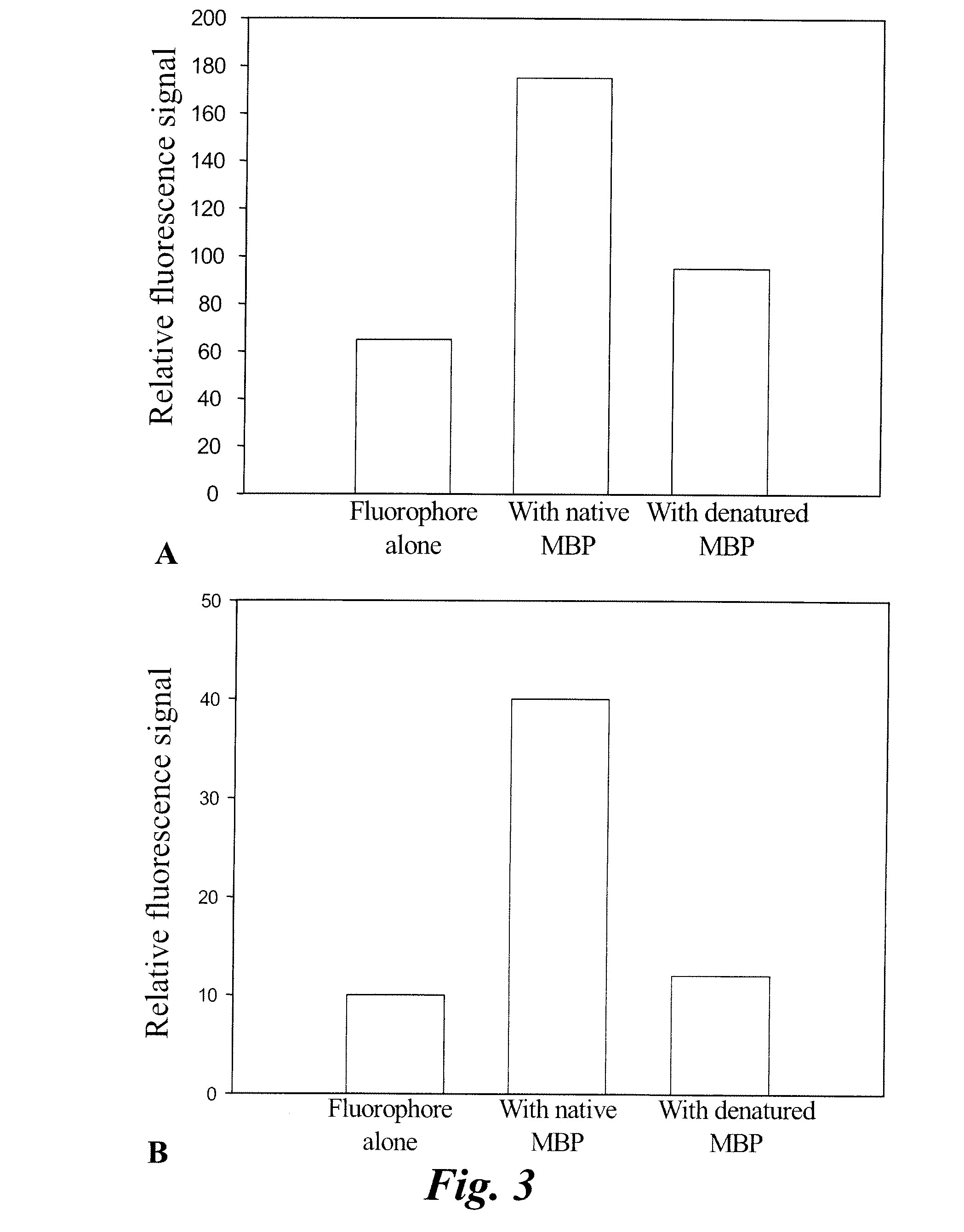
[0068]The fluorophores agents were dissolved in dimethylsulfoxide (DMSO) to make a 10 mM stock solution. An aliquot was taken to prepare a 10 nm-1 uM fluorophore solution in methanol, water, or DMSO. Optical measurements from the three solvents were taken. Absorbance spectra were measured using a Perkin Elmer Lambda 20 UV / VIS spectrometer. Emission spectra were generated using a PTI steady state fluorimeter.


PUM


Abstract

The present invention relates to methods for myelin basic protein detection comprises identifying a subject at risk of or diagnosed with a myelin-associated neuropathy, parenterally administering to the subject the agent, and determining myelination in the subject by detecting binding to myelin basic protein. Methods for the detection of myelin and a quantitative measurement of its local concentration in a sample using an agent with specific binding to myelin basic protein are also provided as is a kit containing the agent or its derivatives for use in detecting myelin basic protein

Description

CROSS-REFERENCE TO RELATED APPLICATIONS[0001]This application is a continuation-in-part to U.S. patent application Ser. No. 12 / 478,300 filed Jun. 4, 2009; the disclosure of which is incorporated herein by reference in its entirety.BACKGROUND[0002]Information flow within the nervous system requires the perpetuation of ionic gradients along neurons. In many neurons, effective and efficient perpetuation of such gradients along axons requires electrical insulation. Myelin, a lipid-rich, dielectric substance that ensheathes axons, serves this insulating function. The nervous system contains high levels of myelin, which is especially enriched where many myelinated axons are bundled together, such as in tracts of the spinal cord and spinal nerve roots, nerves in the peripheral nervous system, and fiber tracts in the brain, collectively called “white matter”, as opposed to “grey matter”. Because non-nervous system tissue lacks myelin, the presence of myelin can distinguish nerve tissue from...

Claims


Application Information

Patent Timeline
IPC IPC(8): A61K51/00A61K49/00
CPCA61K49/10
Inventor SICLOVAN, TIBERIU MIRCEAJOHNSON, BRUCE FLETCHERZHANG, RONGTAN HEHIR, CRISTINA ABUCAY
Owner GENERAL ELECTRIC CO
Learn More