
Device with which a histological section generated on a blade of a microtome can be applied to a slide

Active Publication Date: 2010-08-26
LEICA BIOSYST NUSSLOCH
View PDF5 Cites 13 Cited by
  • Summary
  • Abstract
  • Description
  • Claims
  • Application Information

AI Technical Summary

Benefits of technology

[0007]The present invention is thus based on the object to reduce the manual expense to be rendered by an operator during the application of a histological section to a slide and to preferably achieve a high quality standard during the application operation.
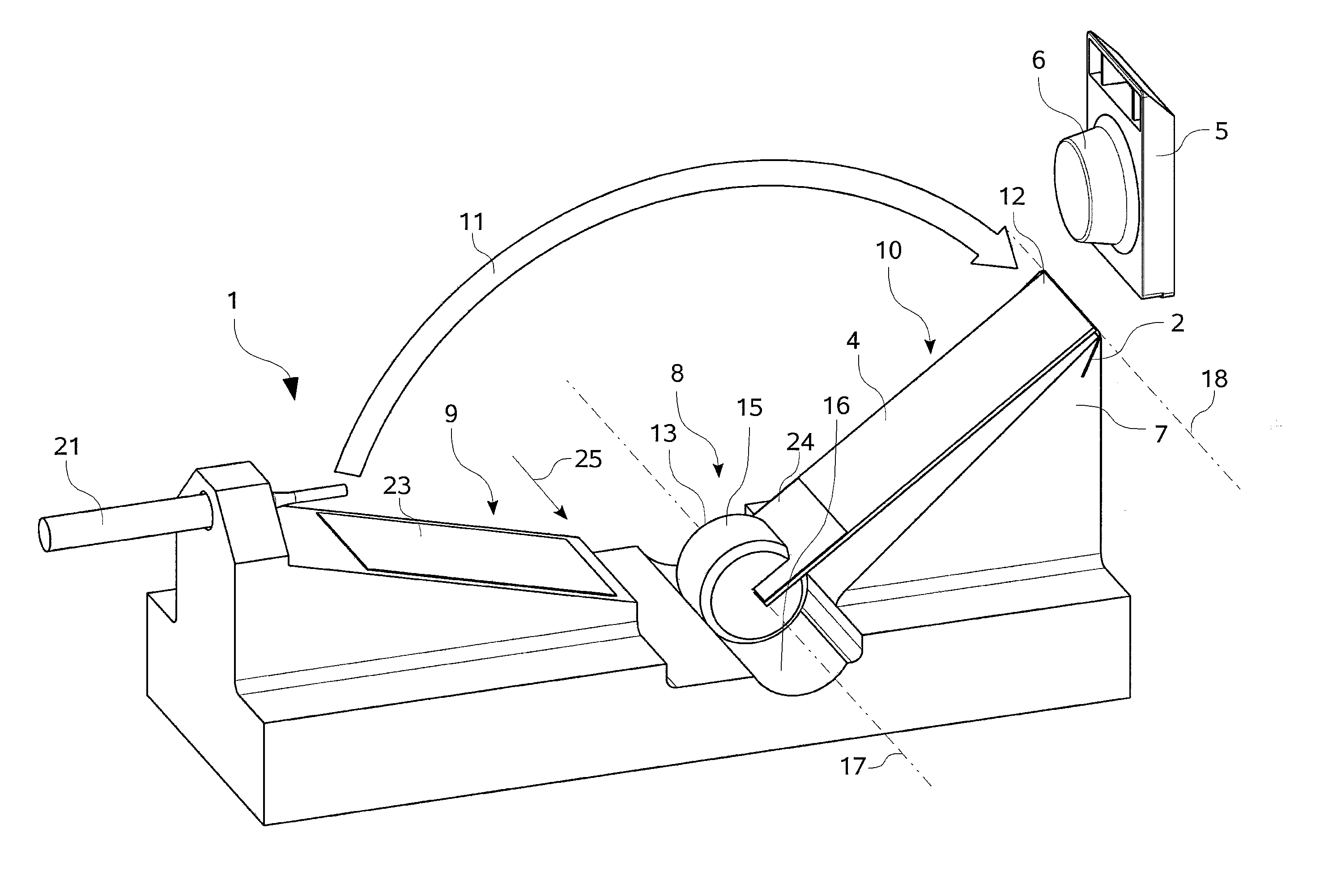
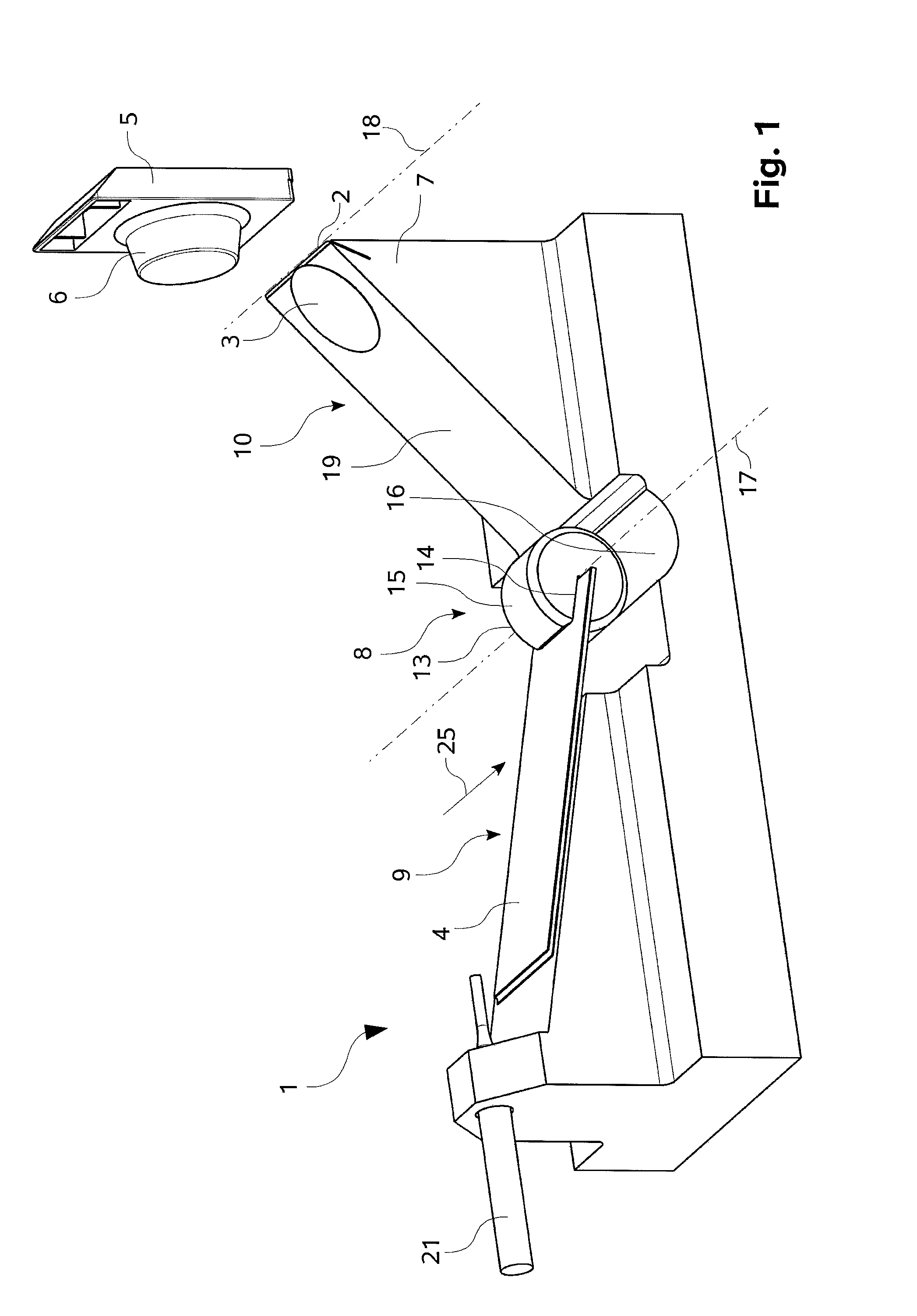
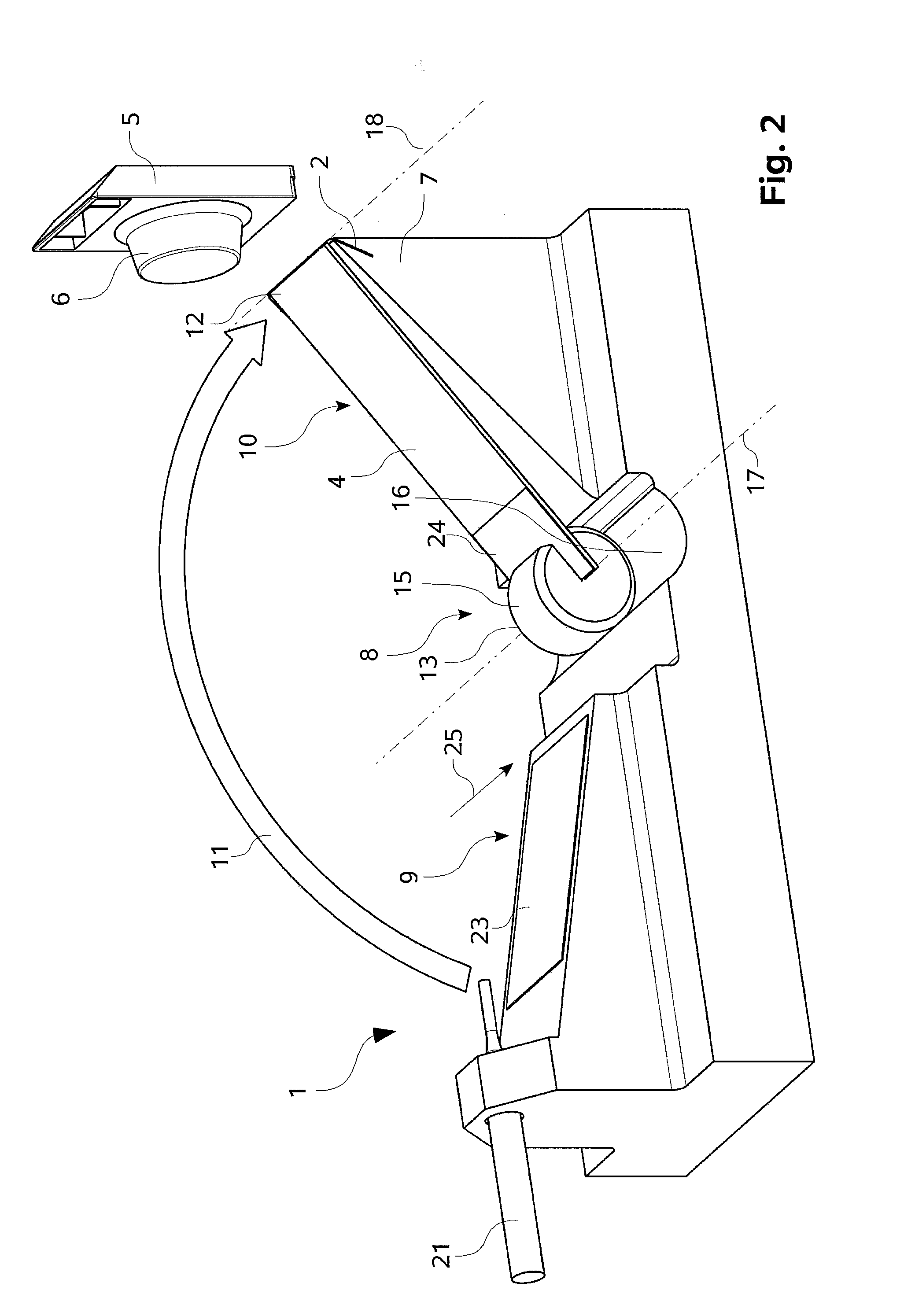
[0009]According to the invention, it has at first been recognized that the application operation of a histological section to a slide can be considerably simplified when, in a manner according to the invention, a positioning device is provided with which the slide can be suitably positioned so that the histological section is brought into contact with the surface of the slide. Due to the adhesive force between the histological section and the slide, the histological section adheres to the slide, thus is applied to the slide as a result thereof and can then be fed to the further histological treatment. Therefore, with the positioning device the slide can be positioned in a simple and reproducible manner at the histological section or, respectively, at the blade or, respectively, at the blade holder of the microtome. This can take place manually or automatically. In any case, according to the invention the histological section is applied to or on the slide immediately after preparing the histological section with the microtome.
[0015]According to a preferred embodiment, the axis of rotation of the rotatably mounted component is oriented substantially parallel to the blade's cutting edge. When the slide is rotated or swiveled towards the blade holder with the aid of the rotatably mounted component about the axis of rotation which is oriented substantially parallel to the blade's cutting edge, the slide approaches—at least with respect to a direction of the surface of the slide which corresponds to the direction of the axis of rotation—the blade holder in a uniform manner and can be effectively brought into contact with the histological section positioned on the blade holder. The rotatably mounted component could be arranged in front of the microtome and below the knife blade or, respectively, the knife holder, for example between the microtome and an operator actuating the microtome. Alternatively and in particular additionally thereto, the axis of rotation of the rotatably mounted component could lie in a plane which is oriented substantially identically or parallel to the surface of the blade holder. Such an orientation or arrangement of the axis of rotation of the rotatably mounted component can likewise allow a uniform approach of the slide to the blade holder and thus to the histological section positioned on the blade holder. As a result thereof, in particular the side of the slide facing the histological section, in an area near the application position, can be brought closer to the surface of the blade holder in a substantially parallel manner. According to this alternative, for example, the rotatably mounted component could be arranged to the left or the right of the knife holder and thus not directly between the microtome and the operator.
[0025]In a comparable manner, a take-away device could be provided for taking away slides on which one histological section each has been applied. The take-away device could be designed in the form of a transport belt as well. Insofar, the application operation of the histological section onto a slide after a cutting operation with the microtome manually initiated by an operator can be largely automated in an advantageous manner. Preferably, it is provided that the histological section can be automatically prepared with the microtome. When in this case both a feed device and a take-away device are provided with which slides can each time be fed to the inventive device or, respectively, taken away from the device according to the invention, the application operation of histological sections to slides on the microtome can be fully automated. For monitoring the automatic application operation at least one sensor and / or at least one camera can be provided with which the cutting operation with the microtome on the one hand and the positioning of the slide with the positioning device on the other hand can be monitored.

Problems solved by technology

When proceeding like this, there is the risk of a contamination of the water bath and thus of the various histological sections which swim in the water bath one after the other.
This application operation, too, involves high manual expense and requires a post-treatment by UV light.

Method used


Image

  • Device with which a histological section generated on a blade of a microtome can be applied to a slide
  • Device with which a histological section generated on a blade of a microtome can be applied to a slide
  • Device with which a histological section generated on a blade of a microtome can be applied to a slide

Examples


Embodiment Construction

[0009]According to the invention, it has at first been recognized that the application operation of a histological section to a slide can be considerably simplified when, in a manner according to the invention, a positioning device is provided with which the slide can be suitably positioned so that the histological section is brought into contact with the surface of the slide. Due to the adhesive force between the histological section and the slide, the histological section adheres to the slide, thus is applied to the slide as a result thereof and can then be fed to the further histological treatment. Therefore, with the positioning device the slide can be positioned in a simple and reproducible manner at the histological section or, respectively, at the blade or, respectively, at the blade holder of the microtome. This can take place manually or automatically. In any case, according to the invention the histological section is applied to or on the slide immediately after preparing ...


PUM

PropertyMeasurementUnit
Temperatureaaaaaaaaaa
Forceaaaaaaaaaa
Friction coefficientaaaaaaaaaa

Abstract

A device for applying a histological section to a slide is described. The histological section is generated by a cutting action performed by a blade of a microtome. The device comprises a positioning device having a component that is rotatably mounted to a bearing and has a receptacle for receiving and holding the slide, wherein the positioning device is designed such that the slide received in the receptacle can be rotated about an axis of rotation of the rotatably mounted component.

Description

CROSS-REFERENCE TO RELATED APPLICATIONS[0001]This application is a national phase entry based on the International Patent Application PCT / EP2008 / 065535 filed on Nov. 14, 2008 that claims the priority of the German patent application DE 102007047797.1 that was filed on Nov. 15, 2007. The entire content of this prior German patent application is herewith incorporated by reference.BACKGROUND OF THE INVENTION[0002]The present invention relates to a device for applying a histological section which can be generated on a blade of a microtome to a slide.[0003]Microtomes have been known for a long time from the prior art. Thin histological sections of tissue samples embedded in paraffin are produced with them. One histological section each is applied to a slide. The slides with the histological sections are usually subjected to a further treatment, the histological sections being stained by means of a staining method in order to be finally observed with the microscope.[0004]When a thin histo...

Claims


Application Information

Patent Timeline
IPC IPC(8): C12M1/00
CPCG01N1/06G01N1/2813G01N1/312
Inventor WALTER, ROLANDLAUDAT, ANDREASSCHMITT, CHRISTOPH
Owner LEICA BIOSYST NUSSLOCH
Learn More