
Diagnostic markers of wound infection

a diagnostic marker and wound infection technology, applied in the field of monitoring patients, can solve the problems of poor correlation between clinical signs of infection and actual measured bioburden in the wounds that were studied, and cannot mount an efficient immune response to microbial contamination, and achieve the effect of low level of relevant cytokine and low cytokine level

Inactive Publication Date: 2010-07-01
WOUNDCHEK LAB US
View PDF2 Cites 9 Cited by
  • Summary
  • Abstract
  • Description
  • Claims
  • Application Information

AI Technical Summary

Benefits of technology

[0037]The wound dressing material may be in the form of a solid, or a semi-solid ointment or gel. Suitably, the wound dressing material comprises only up to 20% by weight, suitably less than 10% by weight of water. The relatively low water content improves the stability of the material and makes it possible to sterilize by heat or irradiation without loss of activity. The material may also contain 0-40% by weight, suitably 0-25% by weight of a plasticiser, suitably a polyhydric alcohol such as glycerol. All of the above percentages are on a dry weight basis.
[0095]In certain embodiments of the present invention, the device may further comprise a control moiety which can interact with a component of the wound sample to improve the accuracy of the device. The control moiety may be adapted to reduce false positive or false negative results. Devices of this type adaptable for use in accordance with the present invention are described in patent publications GB-A-2415039, GB-A-2426335, GB-A-2430029, GB-A-2430030 and GB-A-2430031.

Problems solved by technology

It is hypothesised that the underlying pathology of diabetic foot ulcers, may be a contributing factor, with these ulcers unable to mount an efficient immune response to microbial contamination.
The present inventors have also found that the correlation between clinical signs of infection and actual measured bioburden in the wounds that were studied is poor, with the clinical diagnosis giving a number of false positives and false negatives for wound infection as measured by bacterial bioburden.

Method used


Image

  • Diagnostic markers of wound infection
  • Diagnostic markers of wound infection
  • Diagnostic markers of wound infection

Examples


reference example 1

Comparison of Venous Leg Ulcers and Diabetic Foot Ulcers

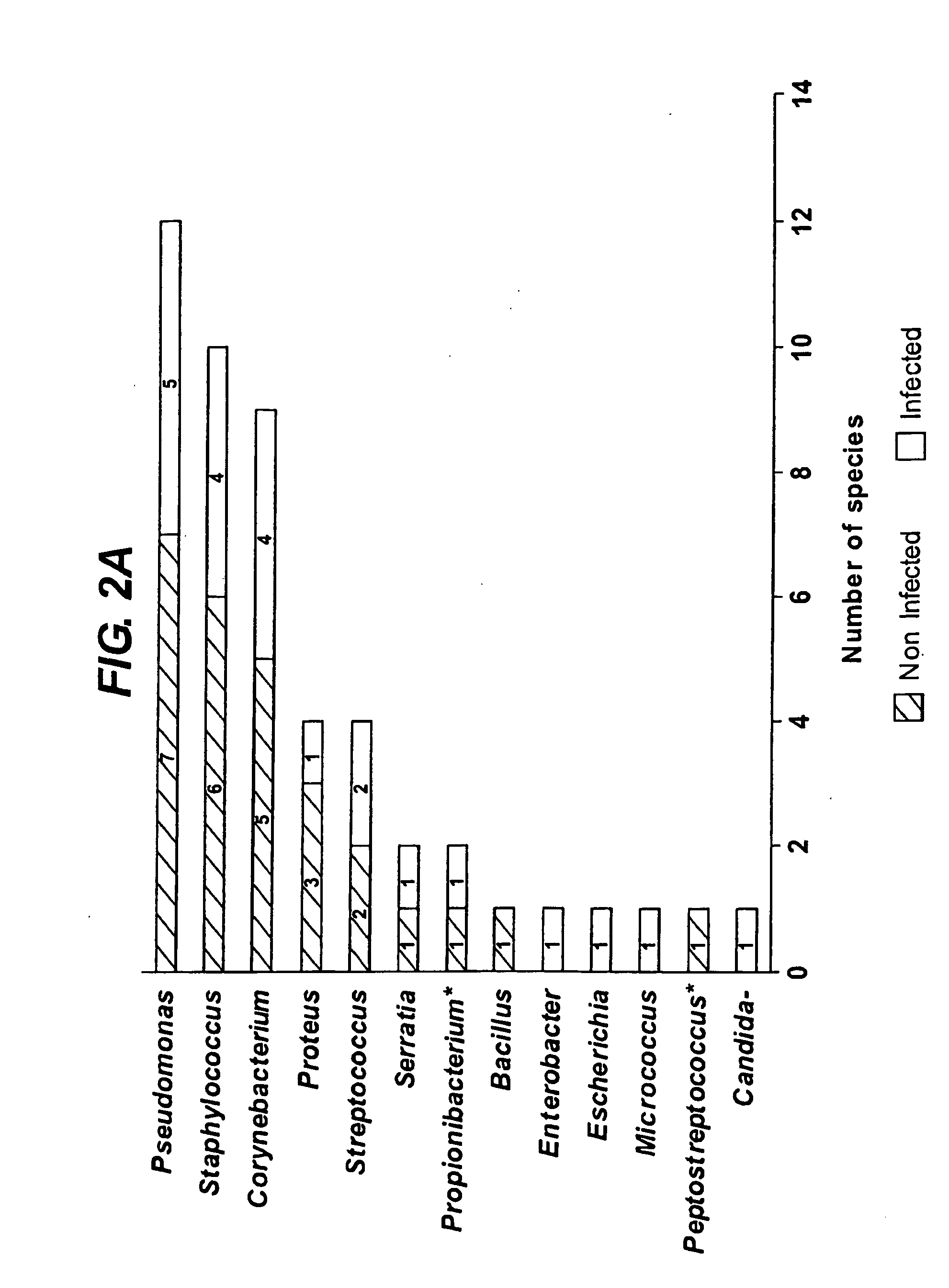
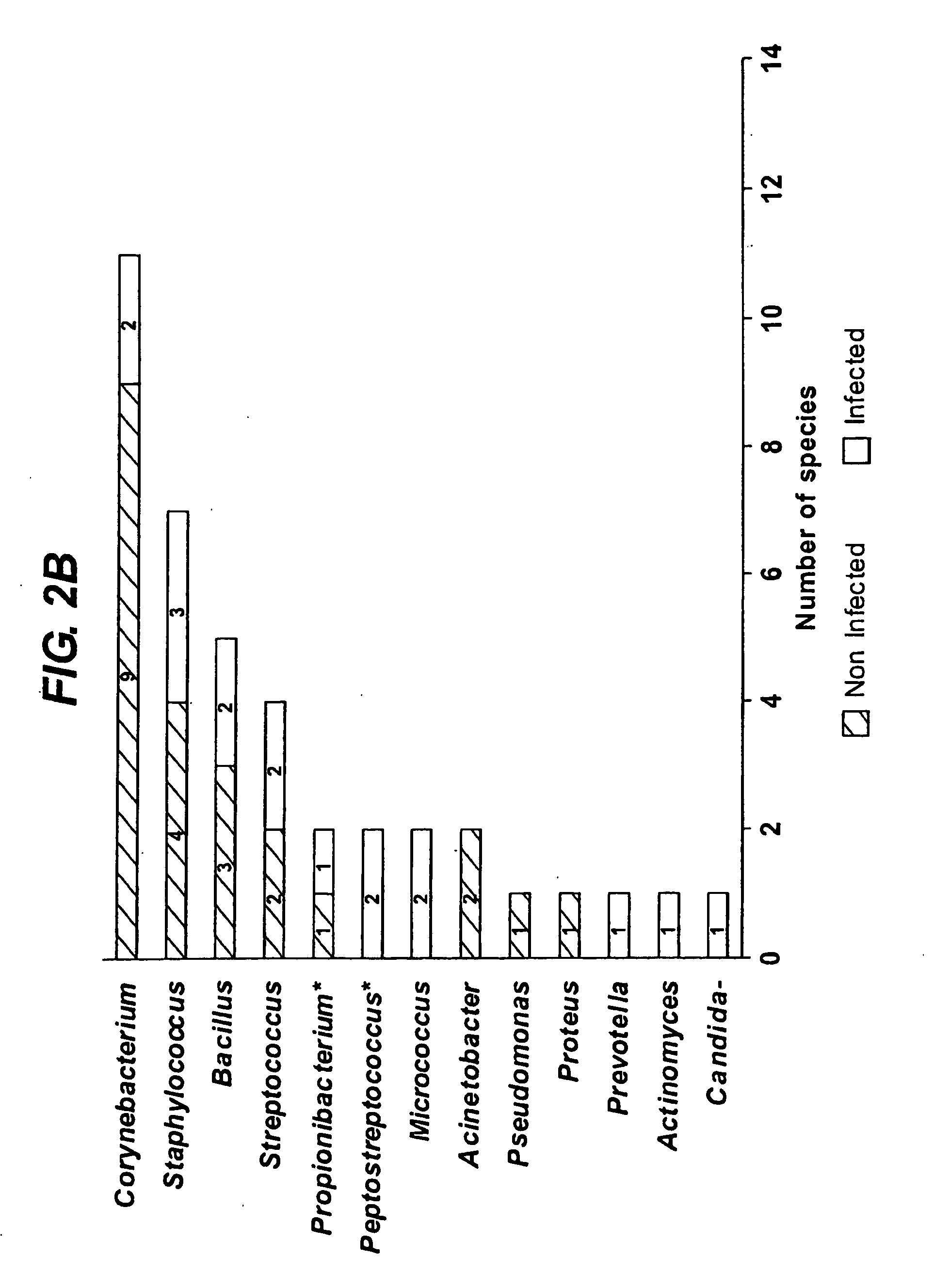
[0122]The inventors' previous work revealed that levels of certain cytokines in wound fluid correlate with the microbial bioburden in venous leg ulcers, and thus that increased cytokine levels can be used in the diagnosis and prognosis of venous leg ulcer infection (e.g. see the references listed above under Background to the Invention). In contrast, the inventors have now found that levels of certain cytokines in diabetic ulcer wound fluid inversely correlate with the microbial bioburden in diabetic ulcers, in particular diabetic foot ulcers, and thus that decreased cytokine levels can be used in the diagnosis and prognosis of diabetic ulcer infection, including diabetic foot ulcer infection.

[0123]FIG. 1 shows a comparison of the microbial bioburden (measured in CFU / ml) in infected and non-infected venous leg ulcers and diabetic foot ulcers.

[0124]All of the wounds tested contained less than 1×10⁹ CFU / ml (represented as 5 CFU / m...

example 2

Analysis of Diabetic Foot Ulcer Wound Fluid Samples

[0130]The inventors have investigated cytokine levels in diabetic foot ulcer wound fluids, and have found that levels of certain cytokines in wound fluid inversely correlate with the microbial bioburden in diabetic ulcers, in particular diabetic foot ulcers. The experimental results are summarised in the table below, which shows the amount of cytokine in microbiologically defined non-infected and infected diabetic foot ulcer wound fluid. The values are median values (in picogram / ml) for each cytokine in diabetic foot ulcer wound fluid that had a significant relationship with the microbial bioburden (ns=not significant).

[0131]It was found that within the diabetic foot ulcer population tested, some of the cytokines with significant differences at a bioburden level of 106 CFU / ml did not all display significant differences at a level of 107 CFU / ml. This change in significance was due to the type of statistical analysis employed, compoun...

reference example 3

Analysis of Venous Leg Ulcer Wound Fluid Samples

[0132]Table 3 shows the measured levels of cytokine in microbiologically defined non-infected and infected venous leg ulcer wound fluids. In contrast to the data for diabetic foot ulcer wound fluids in Example 2, median values for a number of cytokines in venous leg ulcer wound fluids exhibited a significant positive correlation with the levels of bacterial bioburden. Namely, the pro-inflammatory cytokines, IL-1β, IL-6 (FIG. 4.12a), the angiogenic cytokines, angiogenin and VEGF (FIG. 4.12b), cell surface receptors, ICAM and TNFr2, and TNF-α, and the T-cell differentiating cytokine, IL-4.

[0133]The most significant relationships discriminating venous leg ulcers wound fluids with bacterial bioburden level of 1×10⁶ CFU / ml and at a level of 1×10⁷ CFU / ml, were IL-1β (p=0.004), and IL-4 (p=0.001), respectively.

TABLE 3Total BioburdenTotal Bioburden(CFU / ml)Significance(CFU / ml)SignificanceCytokine>106 vrs 6(p values)>107 vrs 7(p values)IL-1β1250...


PUM

PropertyMeasurementUnit
Timeaaaaaaaaaa
Timeaaaaaaaaaa
Contentaaaaaaaaaa

Abstract

The present invention relates to a method of determining the microbial bioburden in a wound (in particular a diabetic ulcer) in a test subject, the method comprising the step of measuring the level of a cytokine in a wound sample, wherein a cytokine level lower than a reference level indicates a significant microbial bioburden in the wound (or a cytokine level higher than a reference level indicates an insignificant microbial bioburden in the wound). The invention provides methods of diagnosis, prognosis and treatment of wound infection, and devices and kits for use in such methods.

Description

[0001]All documents cited herein are incorporated by reference in their entirety.TECHNICAL FIELD[0002]The present invention relates to monitoring patients for the onset or development of wound infection, by determining the microbial bioburden in a wound in a test subject. The invention provides methods of diagnosis, prognosis and treatment of wound infection. The invention also provides devices and kits for use in such methods.BACKGROUND ART[0003]In mammals, injury triggers a complex cascade of cellular and biochemical events that result in a healed wound. Wound healing is a complex dynamic process that results in the restoration of anatomic continuity and function; an ideally healed wound is one that has returned to normal anatomic structure, function and appearance.[0004]Chronically contaminated wounds all contain a microbial flora. These microbes may be indigenous to the patient or might be exogenous to the wound. Closure, or eventual healing of the wound is often based on a phys...

Claims


Application Information

Patent Timeline
IPC IPC(8): A61K31/79C07K16/24A61K31/65A61K31/43A61K31/7048A61K38/12A61K31/655A61K33/38A61P17/02G01N33/68C12Q1/02G01N33/53G01N30/00
CPCC12Q1/04G01N33/54366G01N33/569G01N33/6863G01N2800/042A61P17/02A61L15/16A61L15/44C07K14/52C07K16/24G01N33/53
Inventor STEPHENS, PHILIPMOSELEY, RYANCLARK, RACHAELCULLEN, BREDA MARY
Owner WOUNDCHEK LAB US
Learn More