Photographing apparatus of composition-image for dental diagnosis

Inactive Publication Date: 2010-05-27
CHA YOUNG JIN
View PDF14 Cites 28 Cited by
  • Summary
  • Abstract
  • Description
  • Claims
  • Application Information

AI Technical Summary

Benefits of technology

[0013]As clearly understood in the above explanation, the composite imaging apparatus for dental diagnosis in accordance with the present invention has the cephalometric image detector, which is provided as a unitary part on the rotary arm, laterally spaced apart from and positioned lower than the CT and panoramic image detector on the rotary arm. This structure can resolve the cumbersome of equipment usage of the prior art, in which the cephalometric image detector was installed due to its nature on the elevating frame, separate from the rotary arm, and thereby maximize cost saving and the efficiency of equipment usage for radiography.
[0014]Further, the position of the patient's head can be automatically changed by moving the object moving device, so that various kinds of radiography can be easily done without taking some annoying actions, such as the patient needs to change his / her pose according to an instruction from a nurse or a surgeon while standing on the base of the conventional X-ray imaging apparatus. The object moving device can be elevated up and down, moved forward and backward, or rotated for 360 degrees in a direction opposite the movement of the rotary arm, so that the radiography time can be dramatically reduced and the X-ray exposure harmful to the human body can be minimized at the same time.

Problems solved by technology

Since radiographies including the teeth, the alveolar bone and the skull's jawbone of an object (patient) are being processed only by the simple rotation of the rotary arm 940 in the leftward and rightward direction while the patient is positioned on the base 910, there can be some problems such as an inefficient image-taking process, for example, in which the patient has to change his / her pose according to the body portion, the image of which is being taken, and resultant time delay.
In addition, the cephalometric image detector 950 is not rotatable as mentioned above and is installed apart from the X-ray generator 942 with a certain interval, and there has been a cumbersome job of removing the panoramic image detector 944 for the cephalometric image taking process.

Method used


Image

  • Photographing apparatus of composition-image for dental diagnosis
  • Photographing apparatus of composition-image for dental diagnosis
  • Photographing apparatus of composition-image for dental diagnosis

Examples


Embodiment Construction

[0036]Exemplary embodiments of the present invention will be described below in detail with reference to the accompanying drawings so as to be easily implemented by those skilled in the art.

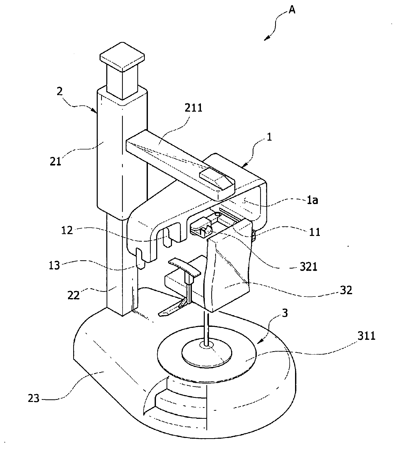
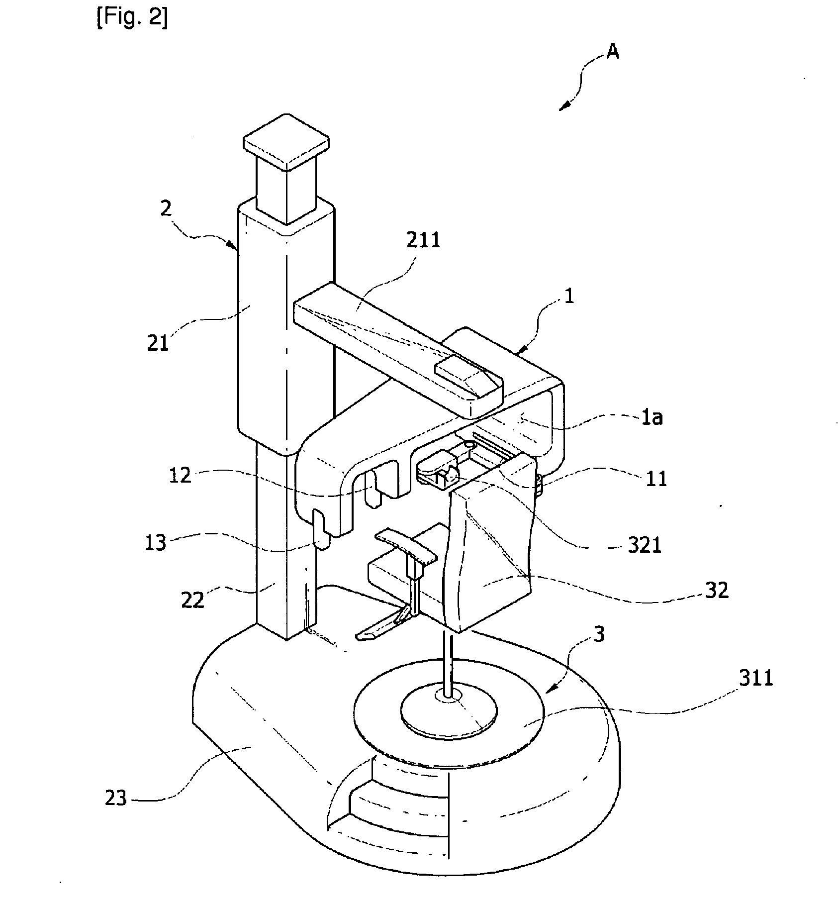
[0037]FIG. 2 is an overall perspective view showing the structure of a composite imaging apparatus for dental diagnosis in accordance with the present invention, FIG. 3 and FIG. 4 are cross-sectional views showing the structure of the base portion of the composite imaging apparatus for dental diagnosis in accordance with the present invention, taken from the side thereof, FIG. 5 is a front elevational view showing the structure of a rotary arm as an important part of the composite imaging apparatus for dental diagnosis in accordance with the present invention, FIG. 6 is an exploded diagram illustrating the structure of an object moving device in the composite imaging apparatus for dental diagnosis in accordance with the present invention, and FIG. 7 and FIG. 8 are exemplary diagrams illustrating ...


PUM


Abstract

A composite imaging apparatus for dental diagnosis, wherein dental diagnosis for teeth / periodontal diseases and orthodontics can be simply done even with an imaging apparatus, and a patient's head portion can be automatically rotated according to image taking directions so as to simplify radiography, save radiography time, and minimize X-ray exposure. The apparatus includes a rotary arm horizontally rotating in left and right directions in order to take an X-ray image of teeth, jawbone and alveolar bone of a patient; a support frame vertically moving with a proper range according to a height of the patient, supporting and enabling the rotary arm to be fixed and horizontally rotate; and an object moving device formed on the base so as to move up and down a for certain range, reciprocate in forward and backward directions, horizontally rotate in forward and reverse directions while the patient is sitting thereon, and facilitate radiography.

Description

TECHNICAL FIELD[0001]The present invention relates to a composite imaging apparatus for dental diagnosis, and more particularly, to a composite imaging apparatus for dental diagnosis, wherein a dental diagnosis for teeth / periodontal diseases and orthodontics can be simply done even with an imaging apparatus, and a patient's head portion can be automatically rotated according to image taking directions so as to simplify radiography, save radiography time, and minimize X-ray exposure.BACKGROUND ART[0002]In the dentistry, an imaging apparatus is generally provided to diagnose the conditions of the alveolar bone including the teeth for the cure of the teeth / periodontal disease and the orthodontics.[0003]The main objective of the X-ray imaging apparatus is for the dentist to consider any existing diseased factors in the patient's jawbone and information affecting the medical treatment together with the recognition of the size / shape of the jawbone and the anatomical changes of the skull a...

Claims


Application Information

Patent Timeline
IPC IPC(8): A61B6/14H05G1/60
CPCA61B6/14A61B6/51A61B6/00
Inventor CHA, YOUNG-JIN
Owner CHA YOUNG JIN
Why Eureka
Try Eureka