
Assessment of breast density and related cancer risk

Inactive Publication Date: 2010-05-20
CARESTREAM HEALTH INC
View PDF2 Cites 42 Cited by
  • Summary
  • Abstract
  • Description
  • Claims
  • Application Information

AI Technical Summary

Benefits of technology

[0012]It is an advantage of the present invention that it is relatively insensitive to differences in image contrast or other quality characteristics or to differences due to the specific type of radiology system used for obtaining the image.

Problems solved by technology

It is known that mammographic imaging techniques are less successful with denser breast tissue than with predominantly fat tissue.
Fibroglandular tissue in the breast tends to attenuate x-rays to a greater degree than does fat tissue, leading to increased difficulty in detection of cancer sites for denser breasts.
In addition, some studies indicate that lesions in higher density areas are themselves more difficult to detect from the mammogram than are lesions in fatty regions, somewhat compounding the problem.
Moreover, although tissue density has been recognized as a significant factor for risk assessment, conventional mammography CAD systems have not utilized this information to help obtain improved results from diagnostic tools.

Method used


Image

  • Assessment of breast density and related cancer risk
  • Assessment of breast density and related cancer risk
  • Assessment of breast density and related cancer risk

Examples


Embodiment Construction

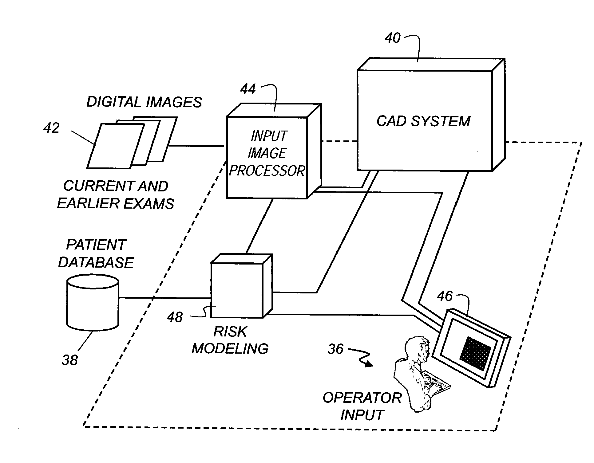
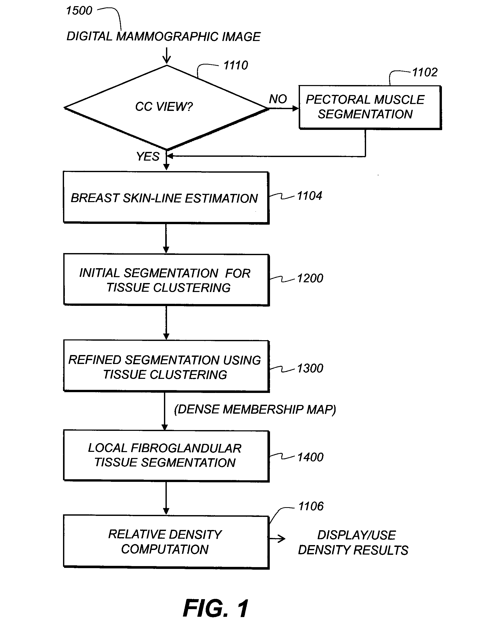
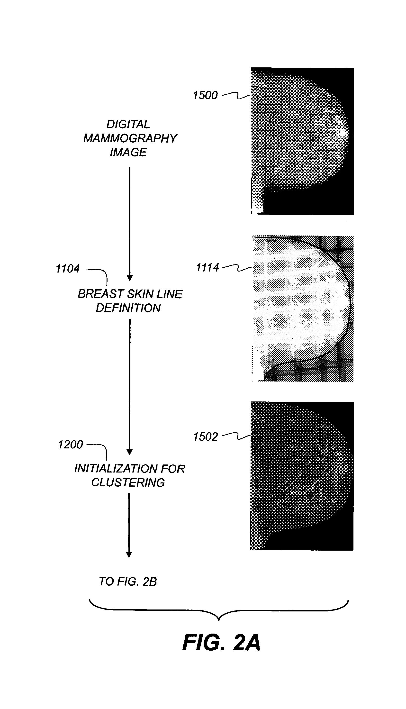
[0029]The following is a detailed description of the preferred embodiments of the invention, reference being made to the drawings in which the same reference numerals identify the same elements of structure in each of the several figures.

[0030]Reference is also made to commonly assigned U.S. patent application Ser. No. 11 / 616,953 filed 28 Dec. 2006 and entitled “Method for Classifying Breast Tissue Density” by Luo et al.

[0031]For the detailed description that follows, the mammographic image is defined as f(X), where X denotes the pixel array and f(x) denotes the intensity value for pixel x in X.

[0032]In the context of the present disclosure, the term “dense tissue” is generally considered synonomous with fibroglandular tissue of the breast. Within the mammography image, this dense tissue is readily distinguishable from fatty tissue to those skilled in breast cancer diagnosis.

[0033]The logic flow diagram of FIG. 1 and graphical sequence of FIGS. 2A, 2B, and 2C show a basic sequence f...


PUM


Abstract

A method for assessing breast density executed at least in part by a computer system, identifies breast tissue from the electronic image data for at least one mammographic image, then performs an initial segmentation of fibroglandular tissue within the breast tissue according to at least one of gradient and uniformity data that is derived from the image data. The initial segmentation is refined using a pixel clustering process. A localized segmentation is obtained from the refined segmentation by generating and combining a density probability mapping and a homogeneity mapping from the image data. A percent density value for the at least one image is calculated and stored in a memory.

Description

CROSS REFERENCE TO RELATED APPLICATIONS[0001]Reference is made to, and priority is claimed from, U.S. Ser. No. 61 / 116,047, filed as a provisional patent application on Nov. 19, 2008, entitled “Assessment Of Breast Density And Related Cancer Risk”, in the names of Zhimin Huo et al., and which is commonly assigned.FIELD OF THE INVENTION[0002]The invention generally relates to image processing and analysis and computer-aided diagnosis (CAD) and more particularly relates to methods that assess and use data related to the density of breast tissue as a risk factor in breast cancer diagnosis.BACKGROUND OF THE INVENTION[0003]In a number of studies, breast density has been found to be a factor for assessing cancer risk. Among factors that determine density is the relative proportion of dense to fatty tissues, sometimes expressed as mammographic percent density, or MPD. The average breast generally has about 50% fibroglandular tissue, a mixture of fibrous connective tissue and the glandular e...

Claims


Application Information

Patent Timeline
IPC IPC(8): G06K9/00
CPCG06T7/0012G06T7/0081G06T2207/30068G06T2207/20221G06T7/0087G06T7/11G06T7/143
Inventor HUO, ZHIMINLAO, ZHIQIANG
Owner CARESTREAM HEALTH INC
Learn More