Methods and devices for sonographic imaging
a sonographic imaging and sonography technology, applied in tomography, ultrasonic/sonic/infrasonic diagnostics, catheters, etc., can solve the problems of limited clinical use, no contrast agent indicated for ultrasound contrast enhancement, and limited use in cardiac and vascular applications
- Summary
- Abstract
- Description
- Claims
- Application Information
AI Technical Summary
Benefits of technology
Problems solved by technology
Method used
Image
Examples
example 1
[0064]Preparation of Contrast Medium with Dual Syringe Pump
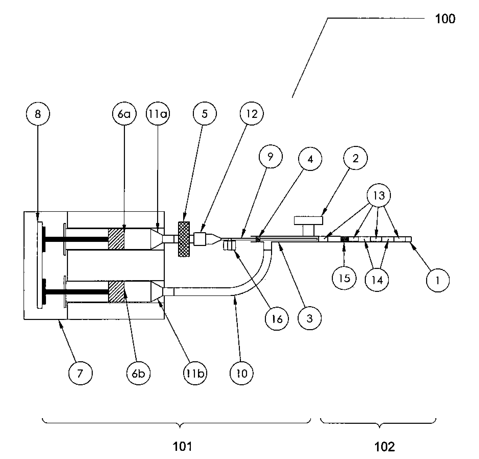
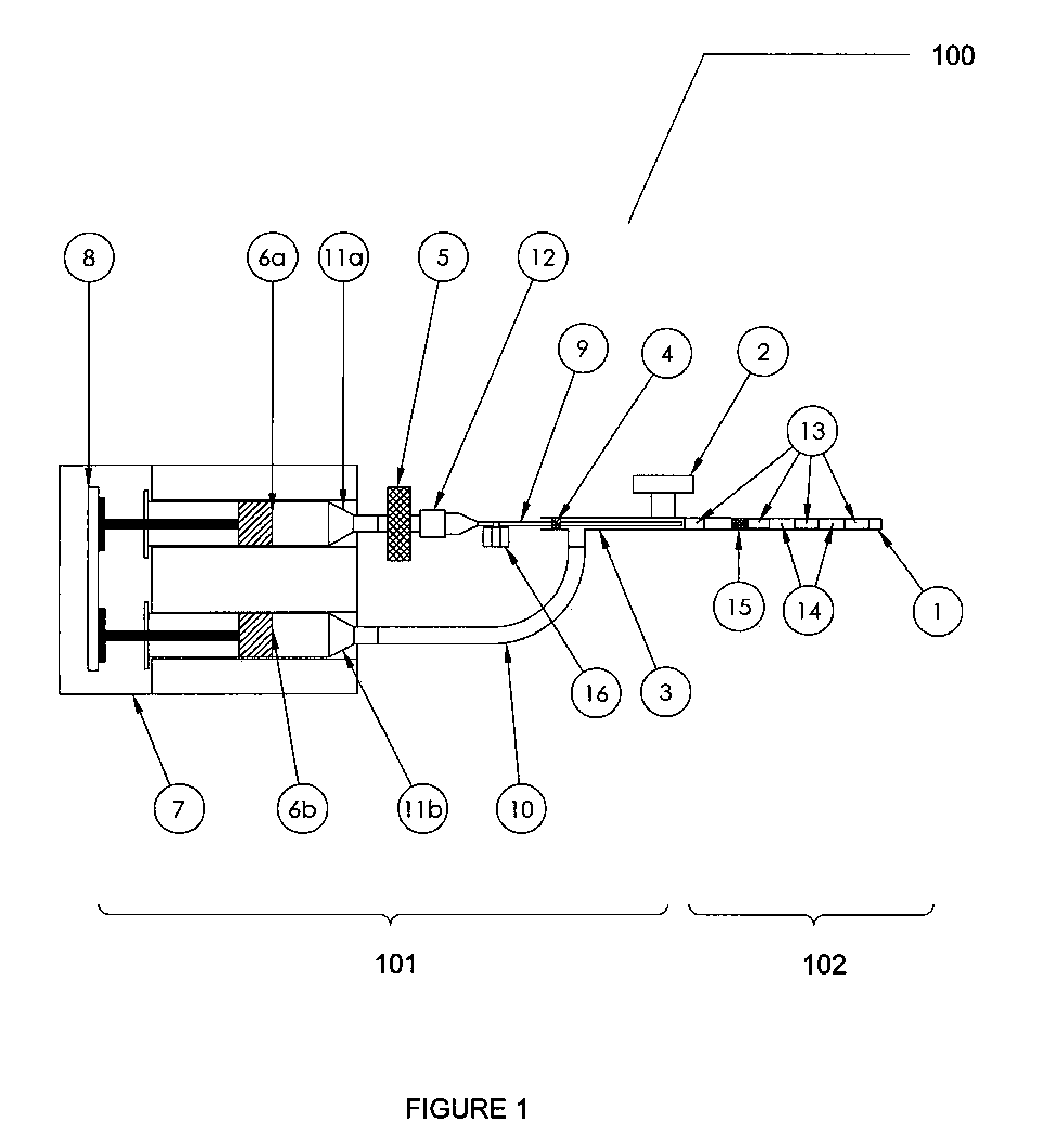
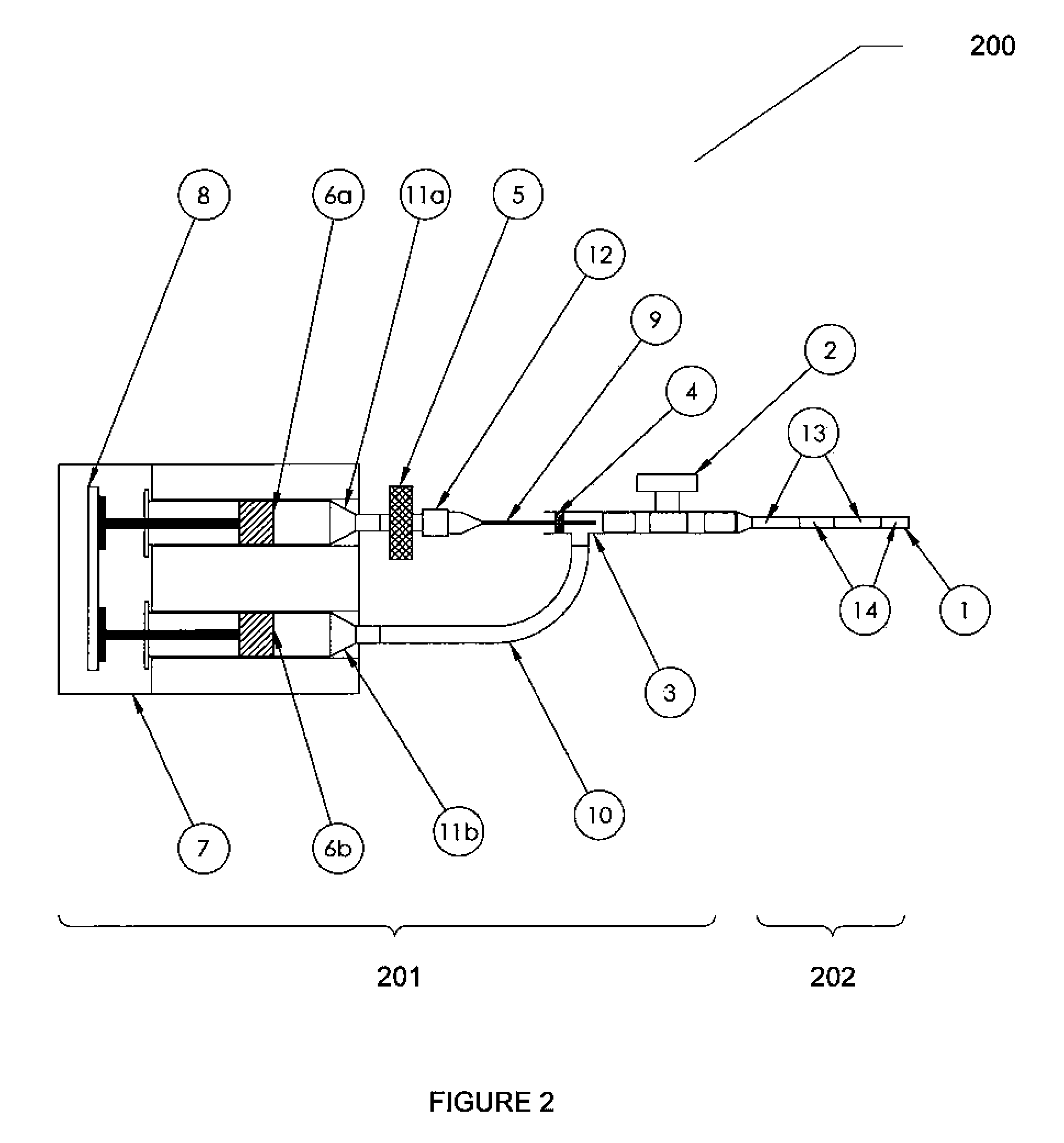
[0065]A container assembly comprising a dual pump was made, as generally depicted in FIG. 1, with two syringes, one 6 cc and the other 20 cc in volume. The 6 cc syringe was completely filled with saline and the 20 cc was filled with air. Sterile 0.2 μm filters (Sartorius Minisart or Whatman Syrfil-MF) were attached to the syringes, as sterile technique was desired. A 27 gauge, 3.5″ length spinal needle was used to inject a gas phase into a fluid phase in the contrast pattern generating chamber to create the alternating air and liquid phase interfaces. A PICC-Nate catheter T-port extension and two lengths of extension tubing were utilized in the set-up.
[0066]Variations of syringe ID, pump volume, pump rate and pump delay settings were evaluated and yielded an acceptable contrast medium, as visualized in a catheter assembly forward of the container assembly. The contrast medium was delivered into clear PVC tubing that simulate...
example 2
[0067]Preparation of Contrast Medium with Handheld Dual Syringes
[0068]The assembly of Example 1 was followed using a housing to support the dual syringes. A block was placed behind the plunger of the 6 cc syringe containing saline to align with the plunger distance of the 20 cc syringe containing air. The creation of the contrast medium and its delivery to a catheter were controlled and manipulated by hand force on the plungers of the dual syringes as necessary to deliver the contrast medium into the catheter. When the two plungers of the syringes were pushed simultaneously, the pattern of the contrast medium was uniform, with substantially equal amounts of air and saline phases, alternating in the catheter. When one plunger was pushed, followed by pushing of the plunger of the other syringe, the pattern was sometimes regular and sometimes irregular, depending on the activation of the individual plungers. Although the sizes of the individual seqments of air and saline phases were no...
example 3
[0069]Preparation of Contrast Medium with Syringe Containing Porous Substance
[0070]A sterile Optipore scrubbing sponge was cut lengthwise in two equal parts. The plunger from a 60 cc syringe was removed and the sponge halves were inserted, one behind the other. The plunger was reinserted in the syringe and depressed to the 15 cc mark. The syringe tip was submerged into a sterile container of saline and the plunger was withdrawn to the 30 cc mark. The container assembly was now assembled and loaded. The container assembly was attached to a catheter assembly and the plunger was depressed to create an air and saline composition, a contrast medium composition, for sonographic visualization. The contrast medium was delivered into clear PVC tubing that simulated the dimensions of the Fallopian tube. An irregular pattern or random pattern was visualized as the user controled the delivery of the contrast medium. Although the sizes of the individual seqments of air and saline phases were not...
PUM
 Abstract
Description
Claims
Application Information
 - R&D
- Intellectual Property
- Life Sciences
- Materials
- Tech Scout
- Unparalleled Data Quality
- Higher Quality Content
- 60% Fewer Hallucinations
Browse by: Latest US Patents, China's latest patents, Technical Efficacy Thesaurus, Application Domain, Technology Topic, Popular Technical Reports.
© 2025 PatSnap. All rights reserved.Legal|Privacy policy|Modern Slavery Act Transparency Statement|Sitemap|About US| Contact US: help@patsnap.com


