
Methods for promoting neovascularization

Inactive Publication Date: 2010-02-18
CHILDRENS MEDICAL CENT CORP
View PDF8 Cites 23 Cited by
  • Summary
  • Abstract
  • Description
  • Claims
  • Application Information

AI Technical Summary

Benefits of technology

[0008]In one embodiment, the method of promoting neovascularization occurs in a tissue that is ischemic. Such neovascularization occurs in therapeutic vasculogenesis. Therapeutic vasculogenesis is useful for promoting tissue repair and wound healing. Promoting neovascularization at the site of injury or damage can help speed the repair. Ischemic tissues and organs having reduced blood flow can also benefit from therapeutic vasculogenesis using the invention. In one embodiment, the ischemic tissue includes, for example, the heart, skin, adipose tissue, muscle, brain, bone, liver, lungs, intestines, legs, limbs and kidneys. The composition containing the progenitor cells is contacted by direct injection to the ischemic tissue or to healthy tissue adjacent to the ischemic tissue. The composition containing endothelial progenitor cells can be delivered alone or mixed with mesenchymal progenitor cells prior to delivery. Alternately, the composition containing mesenchymal progenitor cells can be delivered alone or mixed with endothelial progenitor cells prior to delivery.
[0013]In one embodiment, the endothelial progenitor cells and mesenchymal progenitor cells are autologous to a recipient. Endothelial progenitor cells and mesenchymal progenitor cells are isolated from a sample of peripheral blood of a patient and expanded in vitro. The same autologous endothelial progenitor cells and mesenchymal progenitor cells are then used in tissue engineered constructs which are then implanted into the same donor patient. In another embodiment, the same endothelial and mesenchymal progenitor cells are used in tissue repair and / or wound healing in the donor patient. This greatly reduces the immune rejection of the engineered tissue and implanted progenitor cells, and further eliminates the need for life-long immune suppression therapy.

Problems solved by technology

Despite advances in this field, TE still faces major constraints.
Currently, there are no TE constructs presently available that have an inherent microvascular bed ready to be connected to the host vascular system.
Consequently, tissues implanted with a volume greater than 2 to 3 mm cannot obtain appropriate provision of nutrients, gas exchange, and elimination of waste products since all these mechanisms are limited by the diffusion distance.
Without the development of the microvascular network, the engineered tissue is not sustainable and dies with time.
Therefore, neovascularization of engineered tissues and organs is a major challenge of TE.

Method used


Image

  • Methods for promoting neovascularization
  • Methods for promoting neovascularization
  • Methods for promoting neovascularization

Examples


example 1

[0142]In Vivo Vasculogenic Potential of Human Blood-Derived Endothelial Progenitor Cells (EPC).

[0143]Materials and Methods.

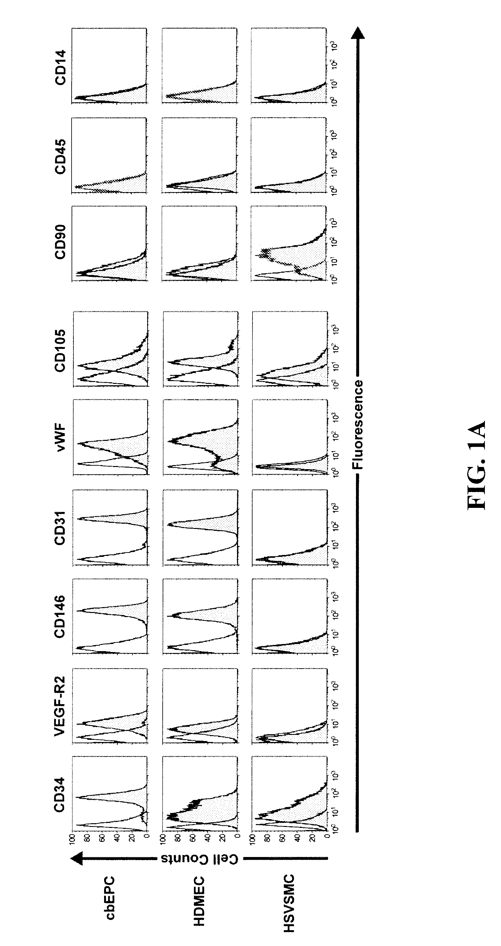
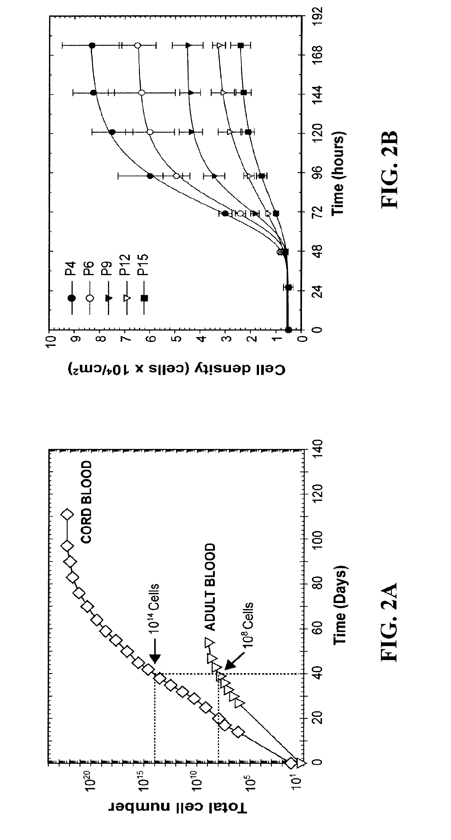
[0144]Isolation and culture of blood-derived EPCs—Human umbilical cord blood was obtained from the Brigham and Women's Hospital in accordance with an Institutional Review Board-approved protocol. Adult peripheral blood was collected from volunteer donors in accordance with a protocol approved by Children's Hospital Boston Committee on Clinical Investigation. Both cord blood-derived EPCs (cbEPCs) and adult peripheral blood-derived EPCs were obtained from the mononuclear cell (MNC) fractions similarly to other authors (Ingram D A, et. al., Blood. 2004,104:2752-60; Lin Y, et. al., J Clin Invest. 2000,105:71-77; Yoder M C, et. al., Blood. 2006, 109:1801-9). MNCs were seeded on 1% gelatin-coated tissue culture plates using Endothelial Basal Medium (EBM-2) supplemented with SingleQuots (except for hydrocortisone) (Cambrex BioScience, Walkersville, Md.), 20% FBS (Hyclo...

example 2

[0171]Engineering Vascular Networks In Vivo with Human Postnatal Progenitor Cells Isolated From Blood and Bone Marrow.

[0172]Materials and Methods

[0173]Isolation and culture of EPCs—EPCs from human umbilical cord blood and adult peripheral blood were isolated and cultured as described above.

[0174]Isolation and culture of MPCs—bmMPCs were isolated from the MNC fractions of a 25 mL human bone marrow sample (Cambrex Bio Science, Walkersville, Md.). MNCs were seeded on 1% gelatin-coated tissue culture plates using EGM-2 (except for hydrocortisone, VEGF, bFGF, and heparin), 20% FBS, 1× GPS and 15% autologous plasma. Unbound cells were removed at 48 hours, and the bound cell fraction maintained in culture until 70% confluence using MPC-medium: EGM-2 (except for hydrocortisone, VEGF, bFGF, and heparin), 20% FBS, and 1× GPS. Commercially available bmMPCs (Cambrex) were used as control to those isolated in our laboratory. Similarly, cbMPCs were isolated from the MNC fractions of 25 mL human c...


PUM


Abstract

The success of tissue engineering and therapeutic neovascularization depends on the development of a microvascular network. The present invention provides methods for promoting neovascularization in tissue engineering constructs, tissue repair, and wound healing comprising endothelial and mesenchymal progenitor cells.

Description

CROSS REFERENCE TO RELATED APPLICATIONS[0001]This application claims benefit of U.S. Provisional Patent application No. 60 / 875,737 filed Dec. 19, 2006, the contents of which are incorporated herein by reference in its entirety.GOVERNMENT SUPPORT[0002]This invention was made with Government support under Grant No.: W81XWH-05-1-0115 awarded by the Department of Defense. The Government has certain rights in the invention.BACKGROUND OF THE INVENTION[0003]Tissue engineering (TE) holds a great promise as a new approach for creating replacement tissue to repair congenital defects or diseased tissue. One strategy is to seed the appropriate cells on a biodegradable scaffold engineered with the desired mechanical properties, followed by stimulation of cell growth and differentiation in vitro, such that, on implantation in vivo, the engineered construct undergoes remodeling and maturation into functional tissue. Examples of this approach include blood vessels, cardiovascular substitutes, bladd...

Claims


Application Information

Patent Timeline
IPC IPC(8): A61K35/12A61P9/00A61K35/28A61K35/44
CPCA61K35/28A61L27/3886A61L27/3804A61K35/44A61P9/00A61P9/10
Inventor MELERO-MARTIN, JUAN M.BISCHOFF, JOYCE E.
Owner CHILDRENS MEDICAL CENT CORP
Learn More