Differential gene expression in physiological and pathological angiogenesis
- Summary
- Abstract
- Description
- Claims
- Application Information
AI Technical Summary
Benefits of technology
Problems solved by technology
Method used
Image
Examples
example 1
Materials and Methods
[0258]Cell lines and animal studies. EMT6 cells were a kind gift of Dr. Robert S. Kerbel, KM12SM cells were a kind gift of Isaiah J. Fidler, HCT116 cells were from the DCT tumor repository (NCI, Frederick) and LS174T, SW620, CT26 and LLC were from the American Type Culture Collection (Manassas, Va.). Tumor cell lines were maintained in DMEM containing 10% fetal bovine serum. Tumors were made by inoculating 5×10⁵−1×10⁶ cells subcutaneously or intrasplenically. To produce liver metastasis by intrasplenic injection, the spleen was exteriorized through a left lateral incision prior to tumor cell injection. The tumor cell suspension was allowed to enter the portal circulation over a period of five minutes, after which the spleen was removed and the skin sutured. For partial hepatectomy, the liver was exposed through a midline abdominal incision and the two anterior lobes were exteriorized and the suspensory ligaments severed. The left lateral and caudal lobes were ge...
example 2
Purification of Endothelial Cells from Normal and Malignant Tissues
[0267]This example describes methods used to immunopurify endothelial cells (ECs) from various tissue types.
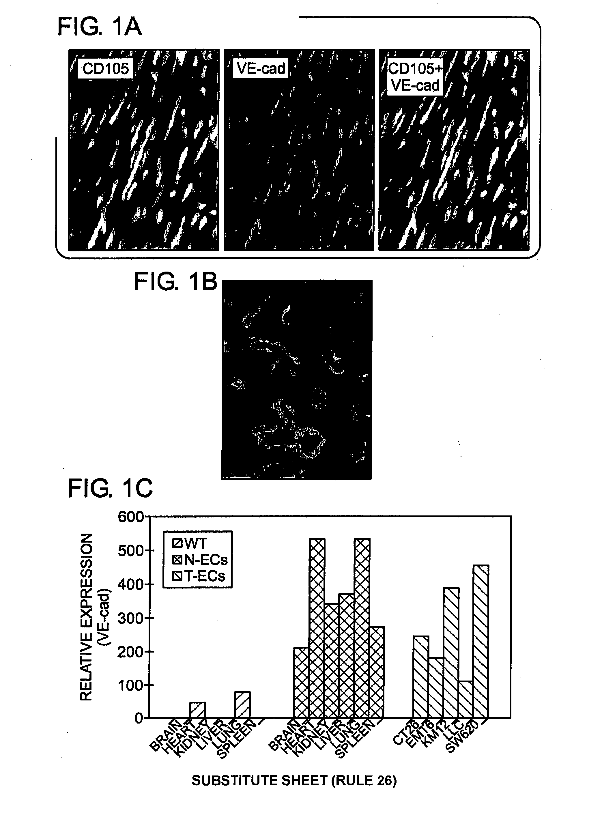
[0268]Initial attempts to purify ECs involved antibody recognition of CD31, the conventional cell surface marker used for affinity purification of mouse ECs, were difficult because of its cross reactivity with hematopoietic cells. CD105 (endoglin) and / or VE-cadherin were found to be specifically localized to the ECs of normal and tumor tissues. For example, as illustrated in FIG. 1A, immunofluorescence staining of heart tissue demonstrated co-localization of CD105 (green) with VE-cadherin (red) in the heart vessels. Further, FIG. 1B demonstrates immunofluorscence staining of liver tissue with CD105 (green). CD105 was determined to be a preferred marker in liver because CD105 stained all the endothelium including sinusoidal ECs whereas VE-cadherin did not.
[0269]The cell isolation involved tissue dissociation, th...
example 3
Gene Expression in Resting Normal ECs, Regenerating Liver ECs and Tumor ECs
[0277]This example illustrates the expression of various markers in resting normal ECs, regenerating liver ECs and tumor ECs.
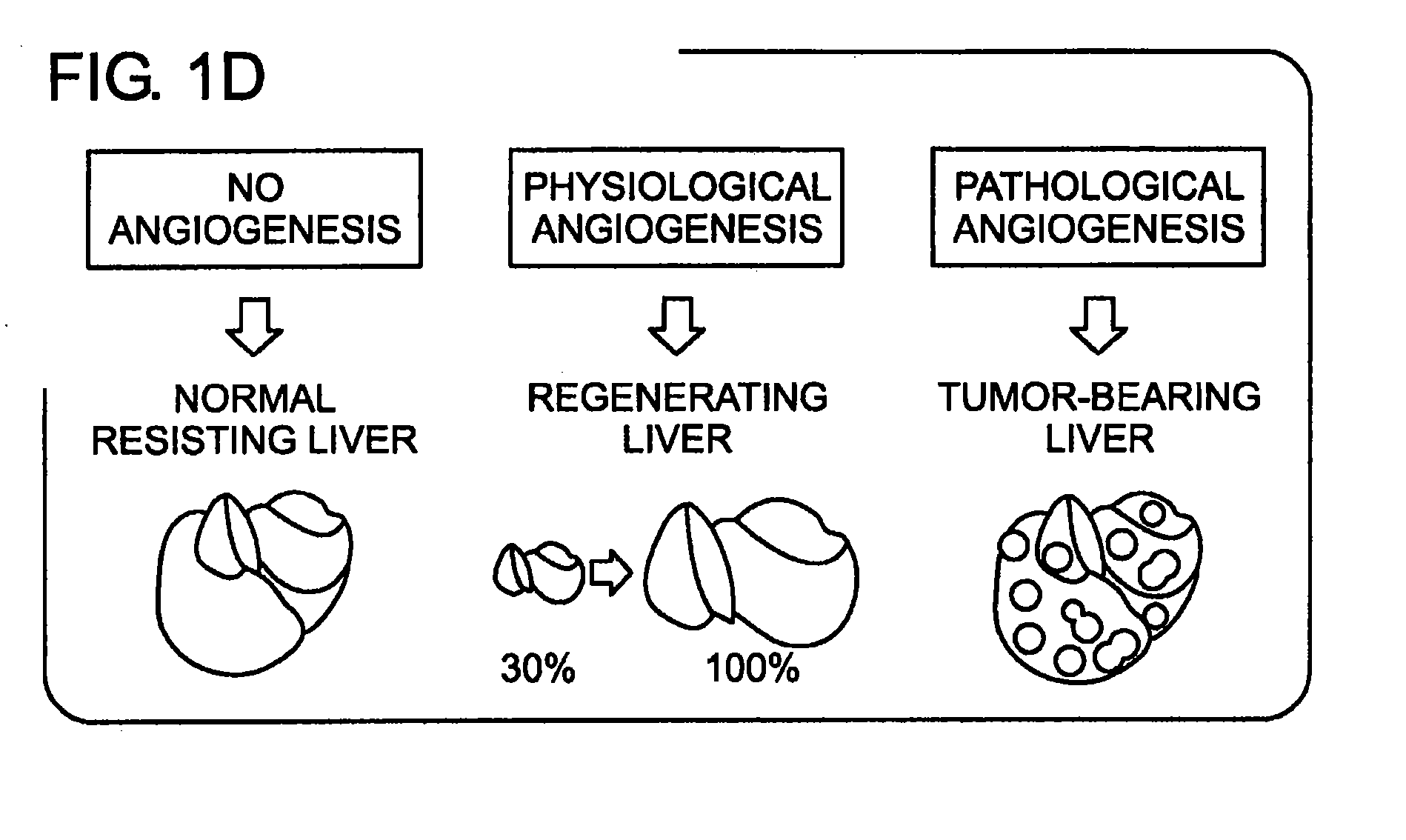
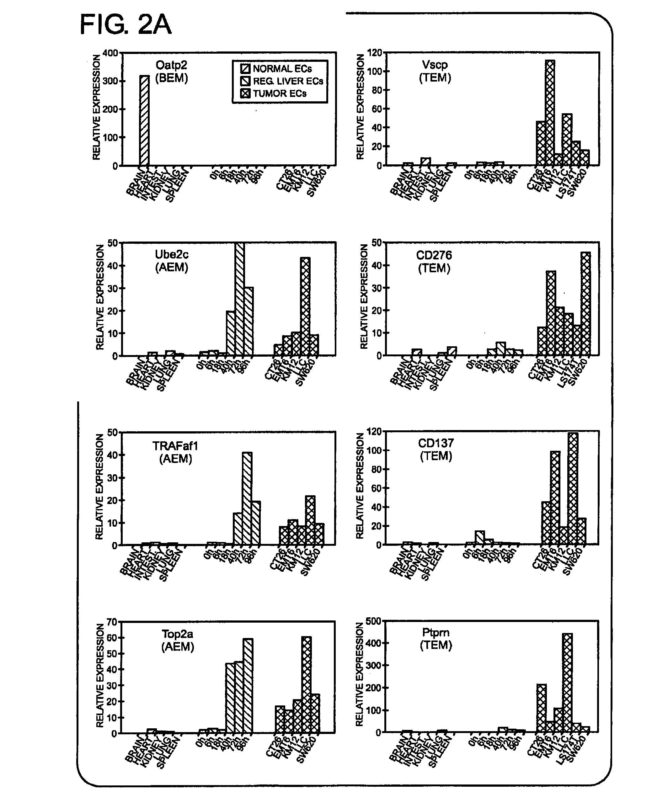
[0278]In order to identify genes that were elevated during physiological angiogenesis, ECs were isolated from liver at 24-, 48- or 72-hours following partial hepatectomy, the period during which endothelial growth is thought to occur (Michalopoulos & DeFrances. Science 276:60-66, 1997). In total, 395,234 SAGE tags were isolated from regenerating liver (See Table 6). Gene expression patterns of regenerating liver ECs were compared with a combined set of EC libraries derived from all non-proliferating normal organs including resting liver (see FIG. 1D). This comparison revealed 12 genes that were overexpressed in regenerating liver ECs compared to non-angiogenic ECs (Table 9), which were referred to as physiological angiogenesis endothelial markers.
[0279]At least seven of these genes may ...
PUM
 Abstract
Description
Claims
Application Information
 - R&D
- Intellectual Property
- Life Sciences
- Materials
- Tech Scout
- Unparalleled Data Quality
- Higher Quality Content
- 60% Fewer Hallucinations
Browse by: Latest US Patents, China's latest patents, Technical Efficacy Thesaurus, Application Domain, Technology Topic, Popular Technical Reports.
© 2025 PatSnap. All rights reserved.Legal|Privacy policy|Modern Slavery Act Transparency Statement|Sitemap|About US| Contact US: help@patsnap.com


