
In Vivo Measurement of Trace Elements in Bone by X-Ray Fluorescence

a trace element and fluorescence technology, applied in the direction of material analysis using wave/particle radiation, instruments, applications, etc., can solve the problems of time-consuming and burdensome, toxicity is a serious environmental and societal problem affecting intelligence, behavior and health, and the standard technique has significant problems associated with i

Active Publication Date: 2009-11-05
THERMO NITON ANALYZERS
View PDF9 Cites 9 Cited by
  • Summary
  • Abstract
  • Description
  • Claims
  • Application Information

AI Technical Summary

Benefits of technology

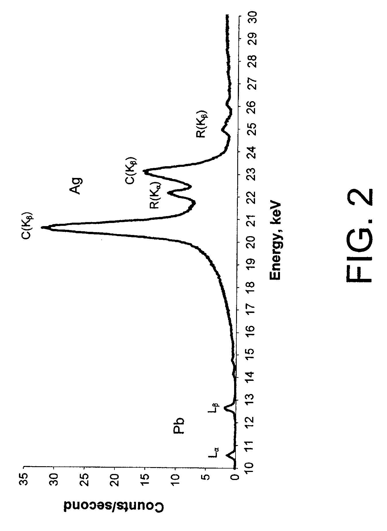
[0009]Embodiments of the present invention provide XRF-based methods and apparatuses for in vivo measurement of the concentration in bone of a specified trace element, such as lead, which provide reliable results without requiring independent experimental determination of the thickness of tissue overlying the irradiated bone. In a first embodiment, a portion of a bone with overlying tissue is irradiated with x-rays having energies suitable for causing the specified trace element to fluoresce, producing first and second fluoresced emission peaks characteristic of the trace element (e.g., the Lα and Lβ x-ray peaks of lead). The concentration of the trace element is then calculated based on the measured intensities of the first emission peak and a function having as an argument the ratio of the intensities of the first and second fluoresced emission peaks, the function having at least one empirically determined parameter and being representative of the attenuation of the overlying tissue thickness.

Problems solved by technology

Lead toxicity is a serious environmental and societal problem affecting intelligence, behavior and health.
A test for lead in blood, however, reflects only the exposure during the few months prior to the test and may not reflect the whole-body burden.
These time-consuming and burdensome tests require the analysis of the urine following the administering of a chelating agent that causes lead to be removed from fluids and bone.
While offering excellent sensitivity, accuracy and precision, the standard technique has significant problems associated therewith.
Utilization of a radioisotope source has inherent disadvantages: it cannot be turned off, requires licensing and periodic replacement, and poses onerous problems with disposal.
Furthermore, testing conducted by the standard K x-ray technique requires about thirty minutes (during which the subject must be immobilized) to gather sufficient counts for statistically significant measures of the lead concentration.
That long duration, with its concomitant costs, precludes the test being used for routine clinical evaluations.
The disadvantages of the standard K x-ray technique have prompted exploration of the potential of measuring the lead concentration using the L x-rays of lead, fluoresced responsive to irradiation by relatively low-power x-rays that can be generated by x-ray tubes.
1399-1419 (2002)), a significant limitation on the accuracy and precision of lead concentration measurements based on detection of L x-rays arises from the sensitivity of such measurements on the thickness of tissue overlying the irradiated bone.
While various direct and indirect techniques are currently available (e.g., ultrasound gauges) for determination of overlying tissue thickness, such techniques may yield results having relatively large inherent variability, thereby compromising the accuracy and precision of lead concentration measurements that rely on determination of tissue thickness.
It is believed by the present inventor, however, that this proposed method, which is based upon a thin-layer approximation of the lead-containing bone, may not produce reliable results for bones of clinical interest, i.e., bone having a thickness in excess of 1 mm and thus being “infinitely” thick for the L x-rays of lead.

Method used


Image

  • In Vivo Measurement of Trace Elements in Bone by X-Ray Fluorescence
  • In Vivo Measurement of Trace Elements in Bone by X-Ray Fluorescence
  • In Vivo Measurement of Trace Elements in Bone by X-Ray Fluorescence

Examples


Embodiment Construction

[0022]Embodiments of the present invention are described in the context of their use for in vivo measurement of the concentration in bone of a particular trace element, namely lead. However, it should be understood that the techniques described and depicted herein are useful for measurement of a variety of trace elements of interest, including but not limited to strontium, mercury, cadmium and selenium. Thus, unless specifically limited to lead, the claims should be construed as extending to the full range of elements suitable for measurement using the techniques of the invention. It should also be noted that embodiments of the invention are suitable for use in both human and animal test subjects.

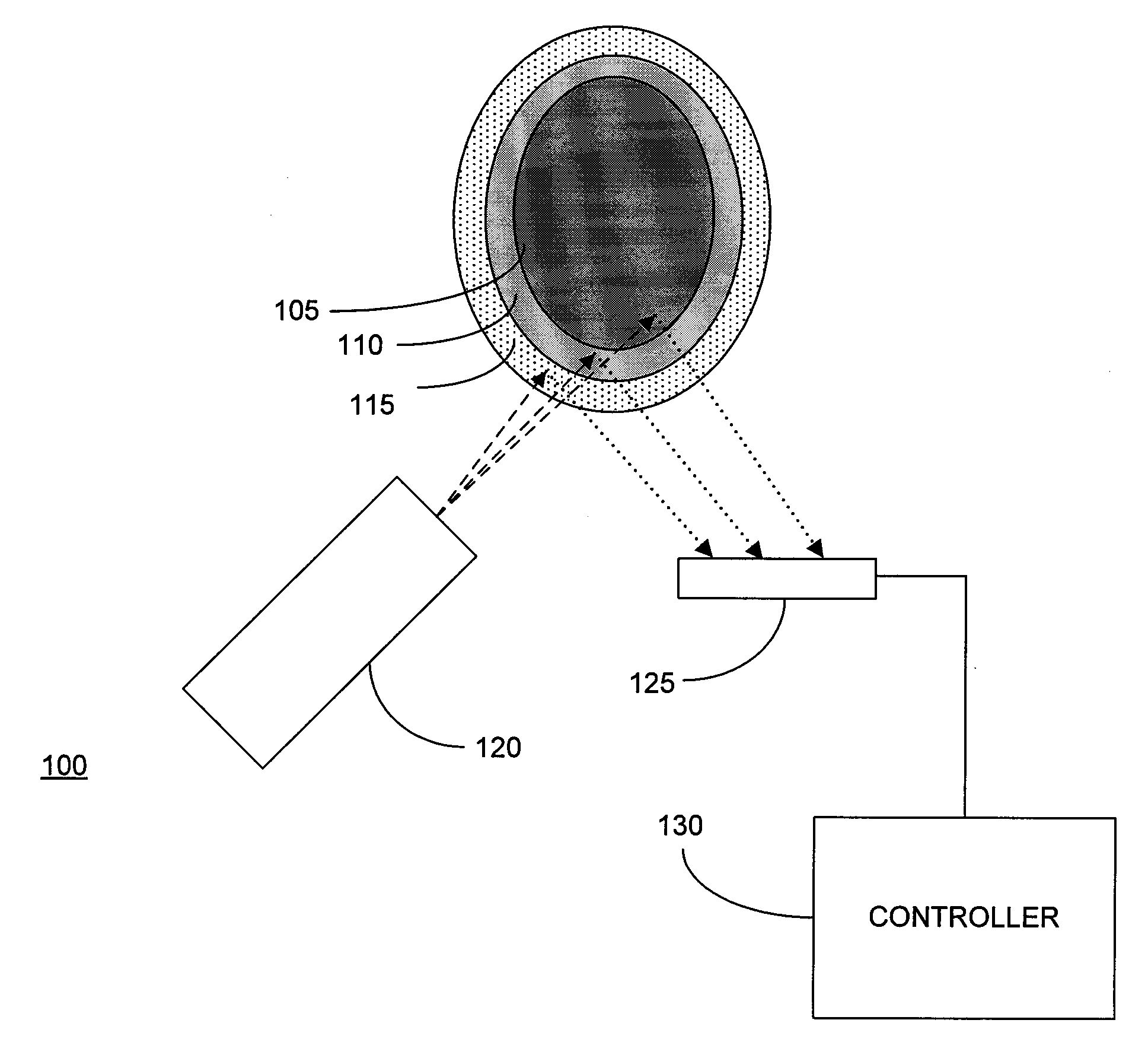
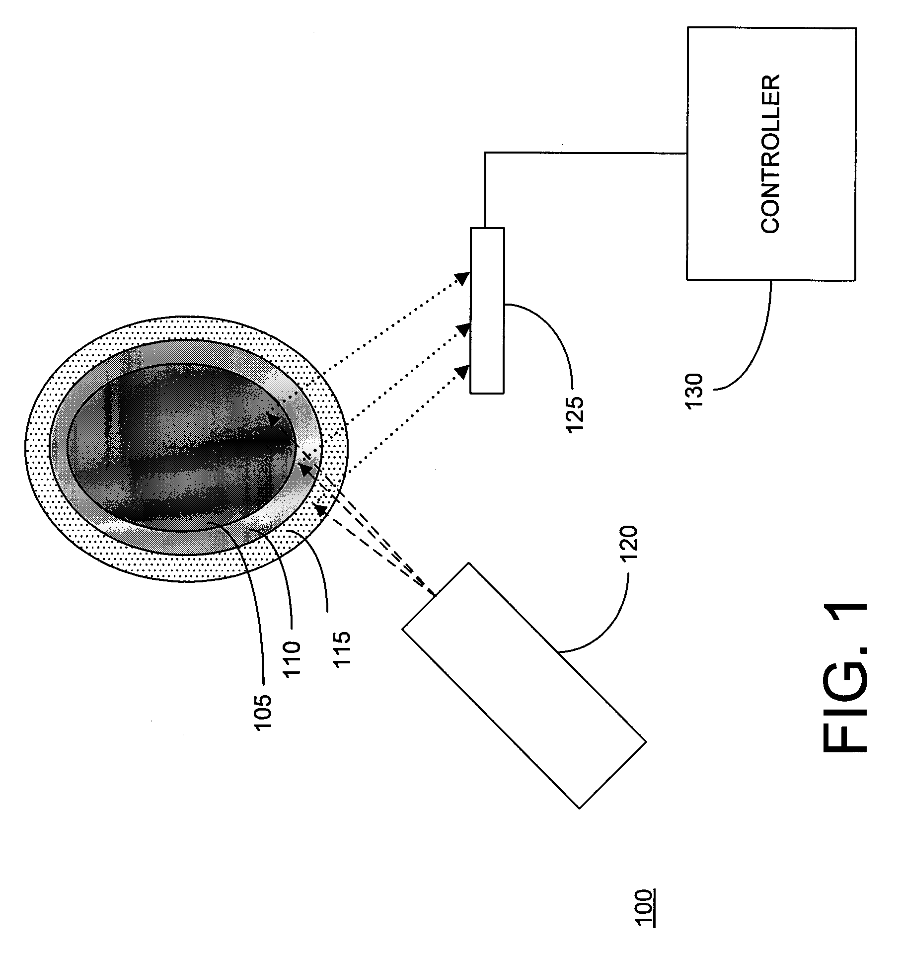
[0023]FIG. 1 depicts an XRF analyzer 100 disposed to measure the lead concentration of bone 105 covered by a relatively thin layer of tissue (typically 110 (subcutaneous fat) and skin 115 (typically ˜1 mm in thickness). In human test subjects, it is generally preferable to select the tibia,...


PUM


Abstract

Methods for in vivo measurement of lead or other trace elements in bone by x-ray fluorescence (XRF) without independent measurement of underlying tissue thickness are disclosed. In one method, the lead concentration is calculated based on the intensity of a first characteristic fluoresced peak and a function having as an argument the intensity ratio of first and second characteristic fluoresced peaks, with at least one parameter of the function being empirically determined by measurements of calibration phantoms having differing thicknesses of tissue surrogate material. In another method, the lead concentration is measured by estimating tissue thickness based on the intensity of the Compton scattering peak, or ratio of Compton / Rayleigh intensities, and the intensity of a characteristic fluoresced x-ray peak corrected for attenuation by tissue of the estimated thickness. Also disclosed is a method for determining the calcium concentration and density of bone based on XRF spectrum data.

Description

CROSS-REFERENCE TO RELATED APPLICATION[0001]This application claims the priority benefit under 35 U.S.C. § 119(e)(1) of U.S. Provisional Patent Application No. 61 / 049,373 by Grodzins entitled “In Vivo Measurement of Heavy Elements in Bone”, filed Apr. 30, 2008, the disclosure of which is herein incorporated by reference.FIELD OF THE INVENTION[0002]The present invention relates generally to methods and apparatuses for in vivo measurement of the concentration in bone of trace elements, such as lead, and more particularly to in vivo measurement of the concentration in bone of trace elements utilizing x-ray fluorescence.BACKGROUND OF THE INVENTION[0003]Lead toxicity is a serious environmental and societal problem affecting intelligence, behavior and health. Lead toxicity is defined by the U.S. Center for Disease Control in terms of the lead concentration in blood: a lead content in blood above 25 micrograms per deciliter of whole blood, associated with an erythrocyte proptoporphyrin con...

Claims


Application Information

Patent Timeline
IPC IPC(8): G01N23/223
CPCA61B6/482A61B6/485G01N2223/6123A61B6/583G01N2223/076A61B6/505
Inventor GRODZINS, LEE
Owner THERMO NITON ANALYZERS
Learn More