
Methods for Treating Hematopoietic Neoplasms

a hematopoietic neoplasm and neoplasm technology, applied in the direction of biocide, drug composition, active ingredients of phosphorous compounds, etc., can solve the problems of reducing affecting the survival rate of hematopoietic neoplasms, and vla4 expression by leukemic cells, etc., to achieve the effect of reducing

Inactive Publication Date: 2009-10-22
CHAPLIN DAVID +1
View PDF5 Cites 6 Cited by
  • Summary
  • Abstract
  • Description
  • Claims
  • Application Information

AI Technical Summary

Benefits of technology

[0002]Although chemotherapy induces remission in the majority of adult patients with acute myeloid leukemia (AML), only small percent are cured with conventional chemotherapy. Relapse of leukemias is in part due to the persistence of minimal residual leukemias that remain viable within specialized niches, such as vascular niches. Hence, novel treatment strategies are urgently needed to block the interaction of hematopoietic neoplasms with activated vasculature, interfering with the establishment of pro-leukemic niches in various organs and to eradicate resistant disease.
[0003]Adhesion of leukemic cells to stromal cells has been shown to confer increased resistance to chemotherapeutic agents and diminish the rate of apoptosis of the leukemic cells. This process, named cell adhesion-mediated drug resistance (CAM-DR), depends on the interaction of integrins with their ligands. Adhesion of VLA4 (very late antigen 4, α4β1) integrin-positive myeloid cells, to VCAM-1+ stromal cells is an important mediator of CAM-DR. Indeed, expression of VLA4 by leukemic cells portends a poor prognosis and a decreased five-year survival rate. Therefore, identification of novel anti-leukemic agents that inhibit interaction of leukemic cells with vascular cells provides novel strategies to target organ-infiltrating, angiogenesis-dependent leukemias.
[0004]Within the marrow or in circulation, hematopoietic neoplasms are closely associated with endothelium, supporting establishment of neo-vessels by elaboration of angiogenic factors. In addition, leukemic cells may activate endothelial cells by releasing pro-inflammatory factors, including interleukin-1 (IL-1), facilitating invasion into tissues and formation of infiltrative organ disease or subcutaneous tumors, namely chloromas, thereby establishing chemotherapy-refractory leukemic minimal residual disease.
[0005]One approach to destabilize interactions of hematopoietic neoplasms with endothelium is through disruption of the cytoskeletal organization of the neoplastic cells. Indeed, disruption of cytoskeletal stability of hematopoietic neoplasms may not only promote cell death directly, but also diminish the cellular interaction of the hematopoietic neoplasms with vascular cells, thereby increasing sensitivity to chemotherapy.
[0006]Combretastatin-A4, a novel tubulin-destabilizing agent, was isolated from the South African tree Combreturn caffrum. Combretastatin-A4 binds to tubulin at the same site as colchicine does, but with even higher affinity. Its pro-drug, combretastatin-A4 phosphate (CA4P) induces rapid microtubule depolymerization and vascular shutdown in subcutaneous solid tumors causing tumor necrosis at concentrations well below the maximum tolerated dose. CA4P also can induce apoptosis of the endothelial cells by disengaging VE-cadherin interaction. Thus, CA4P may not only target rapidly proliferating leukemic cells directly, but also diminish interaction of the leukemic cells with activated endothelial cells, thereby preventing establishment of a perivascular nidus for leukemic chloromas.
[0007]The inventors demonstrate that CA4P at low, non-toxic doses, surprisingly induces rapid cell death of non-adherent leukemic cells through caspase activation, mitochondria destabilization and accumulation of reactive oxygen species (ROS), accompanied by the release of pro-apoptotic mitochondrial membrane proteins. Additionally single-agent CA4P treatment is effective in eradicating both circulating, and vascular-adherent leukemic cells in subcutaneous and systemic mouse models of AML, without affecting normal hematopoiesis. CA4P-treated mice had significantly prolonged survival and showed a drastic reduction of detectable leukemic cells in the marrow and peripheral circulation, and significantly decreased leukemic organ infiltration. In addition, CA4P decreases expression of VCAM-1 on endothelial cells both in vitro and in vivo, thereby decreasing leukemic cell adhesion to the vascular cells, thereby reversing drug resistance. Therefore, CA4P delivered in combination with chemotherapeutic agents represents a promising novel therapeutic approach to treat hematopoietic neoplasms.III. SUMMARY OF THE INVENTION

Problems solved by technology

Indeed, expression of VLA4 by leukemic cells portends a poor prognosis and a decreased five-year survival rate.
Indeed, disruption of cytoskeletal stability of hematopoietic neoplasms may not only promote cell death directly, but also diminish the cellular interaction of the hematopoietic neoplasms with vascular cells, thereby increasing sensitivity to chemotherapy.

Method used


Image

  • Methods for Treating Hematopoietic Neoplasms
  • Methods for Treating Hematopoietic Neoplasms
  • Methods for Treating Hematopoietic Neoplasms

Examples


example 1

A. Example 1

Combretastatin A4 Phosphate (CA4P) Inhibits Leukemic Cell Proliferation

[0065]Leukemic cells were seeded at 1×10⁵ cells / ml in X-vivo medium (Bio-Whittaker, MA) with 5% FBS and CA4P or pre-incubated with the PARP-inhibitor DPQ or caspase-inhibitors Z-VAD-fmk and Q-VD-OPh (R&D, Minneapolis, Minn.). A panel of leukemic cells was incubated with CA4P at the different concentrations indicated in FIG. 1, and viable cells were counted after 48 hours using trypan blue exclusion. Results of four experiments in duplicate are expressed as the ratio of the percentage of viable cells / control±SEM (*p50 ranging from 2.5 to 5 nM (FIG. 1). The majority of the AML cell lines tested was sensitive to CA4P at a concentration of 2.5 nM or less. All leukemic cell lines, as well as a recently established primary leukemic cell line (R81) 16 were sensitive at low doses of CA4P (<10 nM).

example 2

B. Example 2

CA4P Causes Cell Cycle Arrest in G2 / M Phase

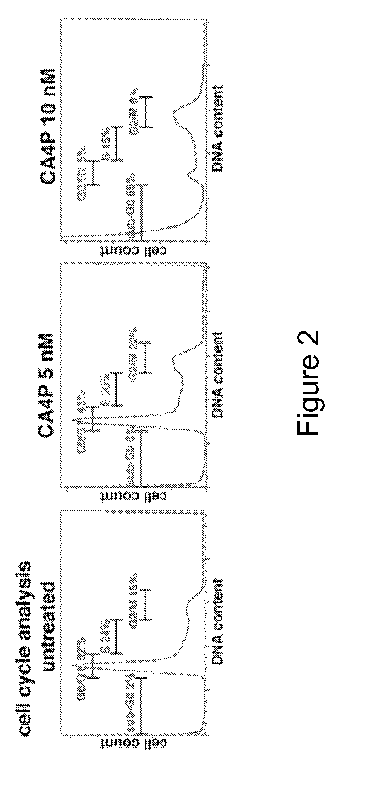
[0066]CA4P induces G2 / M arrest and cell death, as evidenced by increase in the sub-G0 / G1 peak (FIG. 2). Leukemic cells were seeded at 105 per ml in X-vivo supplemented with 5% FBS and then incubated with CA4P. KG1a leukemic cells were treated with CA4P at 0, 5 and 10 nM concentrations. After incubation for 48 hours, apoptotic cells were quantified by ApoAlert Annexin-V-fluorescine isothiocyanate (FITC) propidium iodide (PI) Apoptosis Kit (BD) using a Coulter Elite flow cytometer. In CA4P-treated cells, cell cycle analysis with propidium iodine (PI) showed G2 / M arrest and evidence of DNA fragmentation (sub G0-phase) at 48 hours.

example 3

C. Example 3

Combretastatin A-4 Phosphate Causes DNA Fragmentation and Morphological Evidence of Mitotic Catastrophe

[0067]DNA damage in CA4P-incubated leukemic cells was assessed by comet assay. The concept behind this assay is based upon the ability of denatured, cleaved DNA fragments to migrate out of the cell under the influence of an electric field, whereas undamaged DNA migrates more slowly and remains within the confines of the nucleus. Evaluation of the DNA “comet” tail shape and migration pattern allows for assessment of DNA damage. Results were expressed as the percentage of cells with a comet tail in 100 randomly selected, non-overlapping cells visualized by standard light microscopy. Quantification of the number of leukemic cells displaying a comet tail strongly increased after CA4P treatment (FIG. 3), consistent with CA4P-induced DNA damage. Results of three experiments in duplicate are expressed as the mean of the number of cells with a comet tail (%)±SEM (*p<0.05 as com...


PUM

PropertyMeasurementUnit
concentrationsaaaaaaaaaa
refractoryaaaaaaaaaa
Adhesionaaaaaaaaaa

Abstract

This invention relates to methods for treating, preventing and / or managing hematopoietic neoplasm in a subject by administering to the subject combretastatin A-4 phosphate, or a pharmaceutically acceptable salt thereof. The method may further comprise co-administering a chemotherapeutic agent.

Description

I. CROSS REFERENCE TO RELATED APPLICATIONS[0001]This application claims the benefit of U.S. provisional patent application No. 60 / 989,786, filed 21 Nov. 2007.II. INTRODUCTION[0002]Although chemotherapy induces remission in the majority of adult patients with acute myeloid leukemia (AML), only small percent are cured with conventional chemotherapy. Relapse of leukemias is in part due to the persistence of minimal residual leukemias that remain viable within specialized niches, such as vascular niches. Hence, novel treatment strategies are urgently needed to block the interaction of hematopoietic neoplasms with activated vasculature, interfering with the establishment of pro-leukemic niches in various organs and to eradicate resistant disease.[0003]Adhesion of leukemic cells to stromal cells has been shown to confer increased resistance to chemotherapeutic agents and diminish the rate of apoptosis of the leukemic cells. This process, named cell adhesion-mediated drug resistance (CAM-D...

Claims


Application Information

Patent Timeline
Patent Type & Authority Applications(United States)
IPC IPC(8): A61K31/7076A61K31/661A61K31/7064A61P35/00
CPCA61K31/52A61K31/661A61K31/675A61K31/7048A61K31/7068A61K2300/00A61K31/6615A61P35/00A61P35/02A61P43/00A61K45/06
Inventor CHAPLIN, DAVIDRAFII, SHAHIN
Owner CHAPLIN DAVID
Learn More