Method of modulating cellular activity and agents for use therein
- Summary
- Abstract
- Description
- Claims
- Application Information
AI Technical Summary
Benefits of technology
Problems solved by technology
Method used
Image
Examples
example 1
Tissue Section Staining using Immunofluorescence
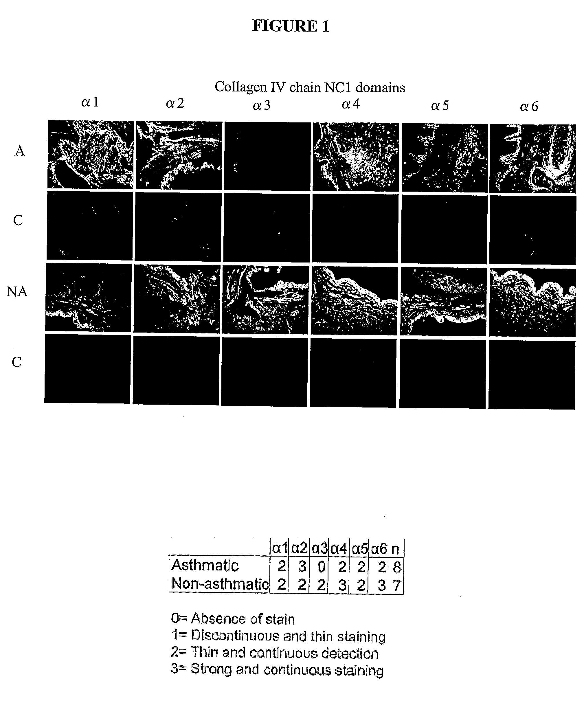
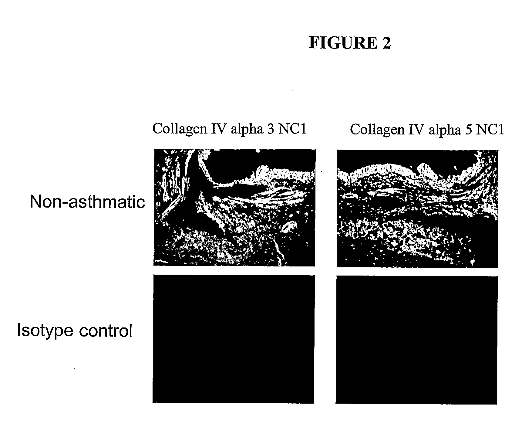
[0225]Lung tissue sections from a total of 15 asthmatics, 9 non-asthmatics and 10 volunteers with Lymphangioleiomyomatosis (LAM) were examined using immunohistochemistry. For 7 asthmatic patients, diagnosed according to the Global Initiative for Asthma (GINA) guidelines, bronchial biopsies were taken. In addition, biopsies were also taken from two volunteers who were shown not to have asthma according to GINA guidelines. For the remaining asthmatic and non-asthmatic individuals tissue was sourced from a tissue bank. In the case of asthmatic samples, many of these were derived from individuals who had died of asthma (status asthmaticus). Diagnosis of LAM was made by respiratory physician with the aid of a CT scan.
[0226]The presence of the non-collagenous 1 (NC1) domain of the collagen IV α3 chain (tumstatin) was examined in all tissue sections. The presence of the non-collagenous 1 (NC1) domain of the collagen IV α1,2,3,4,5 and 6 chains...
example 1a
[0227]Paraffin embedded tissue sections of bronchial rings from 8 asthmatics, 7 non-asthmatics and 10 LAM patients were deparaffinised and rehydrated through graded alcohol. Blocking serum was then added to the sections (10% non-immune horse serum) for 20 minutes at room temperature. Without rinsing, either primary antibodies (collagen IV α1-6 NC1 Shigei Medical Research Institute, Okayama, Japan [1 ng / ml]) or isotype control antibodies (Goat IgG, Jackson ImmunoResearch PA, USA [1 ng / ml]) were added to the sections and incubated for 1 hour at room temperature. Sections were then washed with phosphate buffered saline (PBS) and a rat anti-goat FITC-conjugated secondary antibody (MP Biomedicals Ohio, USA [1 ng / ml]) was added, and incubated for 30 minutes at room temperature. Sections were then rinsed with PBS (to wash away unbound secondary antibody) and mounted using vestashield mounting media (Vecta Laboratories USA). Images were taken on an Olympus fluorescence microscope and captur...
example 1b
Colorimetric Analysis of Stained Tissue Sections
[0233]To further validate the above findings (Example 1A) and further investigate the pattern of tissue expression of collagen IV α3, it was investigated whether the 7 s domain of collagen IV α3 was present in tissue sections from asthmatic and non-asthmatic fresh frozen bronchial biopsies as well as paraffin sections from LAM and non-asthmatic patients. In addition it was investigated whether the NC1 domain of collagen IV α3 was present in a further asthmatic volunteer. In contrast to the staining protocol described in Example 1A, the inventors here used an automated quantitative colorimetric analysis of the staining patterns.
[0234]Frozen sections were thawed and placed into PBS buffer for 5 minutes. Paraffin sections were de-paraffinised and placed in water for 5 minutes. Sections were blocked using a pre-made peroxidase block (DakoCytomation, Denmark) for 5 minutes. Primary antibodies were then added, Mouse anti-human collagen IV α3...
PUM
| Property | Measurement | Unit |
|---|---|---|
| Fraction | aaaaa | aaaaa |
| Electrical conductance | aaaaa | aaaaa |
| Level | aaaaa | aaaaa |
Abstract
Description
Claims
Application Information
 - R&D
- Intellectual Property
- Life Sciences
- Materials
- Tech Scout
- Unparalleled Data Quality
- Higher Quality Content
- 60% Fewer Hallucinations
Browse by: Latest US Patents, China's latest patents, Technical Efficacy Thesaurus, Application Domain, Technology Topic, Popular Technical Reports.
© 2025 PatSnap. All rights reserved.Legal|Privacy policy|Modern Slavery Act Transparency Statement|Sitemap|About US| Contact US: help@patsnap.com


