
Ink-jet printing of tissues

a tissue and inkjet printing technology, applied in the field of inkjet printing, can solve the problem that the possibility of simultaneously printing multiple cell types to build viable heterogeneous cellular constructs has not been demonstrated to da

Inactive Publication Date: 2009-08-20
WAKE FOREST UNIV HEALTH SCI INC
View PDF11 Cites 112 Cited by
  • Summary
  • Abstract
  • Description
  • Claims
  • Application Information

AI Technical Summary

Benefits of technology

[0007]Although the capability of inkjet printing of viable single cells has been verified, the possibility of simultaneously printing multiple cell types to build viable heterogeneous cellular constructs has not been demonstrated to date. It has been found that distinct cell types can be mixed with support compounds (collagen gels) and printed into the target areas to form 3-dimensional tissue structures. Further, basic physiological functions and properties of each cell type within the structure can be maintained.

Problems solved by technology

Although the capability of inkjet printing of viable single cells has been verified, the possibility of simultaneously printing multiple cell types to build viable heterogeneous cellular constructs has not been demonstrated to date.

Method used


Image

  • Ink-jet printing of tissues
  • Ink-jet printing of tissues
  • Ink-jet printing of tissues

Examples


example 1

Printing of Multiple Cell Types

[0081]Materials and Methods. Three distinct cell types were used in this study: human amniotic fluid-derived stem cells (hAFSC) transfected with lacZ, bladder smooth muscle cells (BSMC), and GFP labeled MS1 (mouse pancreatic islet endothelial cell line). Each cell type was grown separately, trypsinized, collected and resuspended in Type I collagen solution. Different mixtures of collagen and cells were loaded into different ink cartridges. Each cell-collagen mixture was printed layer-by-layer into the pre-designed target locations using a modified HP 550 printer. A solution containing NaOH was subsequently printed in order to neutralize the pH. The printed constructs were placed in the incubator for 3-5 hours. Once the collagen gel was set, 3-D viable multi-cellular constructs with a specific shape were formed. After 2 days of culture, the printed multi-cellular constructs were fixed and characterized using cell specific markers (α-actin, X-gal).

[0082]...

example 2

In vivo Generation of Tissues with Ink-Jet Printing

[0086]In this example we investigated whether the printed multi-cell derived tissue constructs could maintain their structural and spatial orientation in vivo. We examined whether these tissues are able to survive and mature into functional tissues when implanted in vivo.

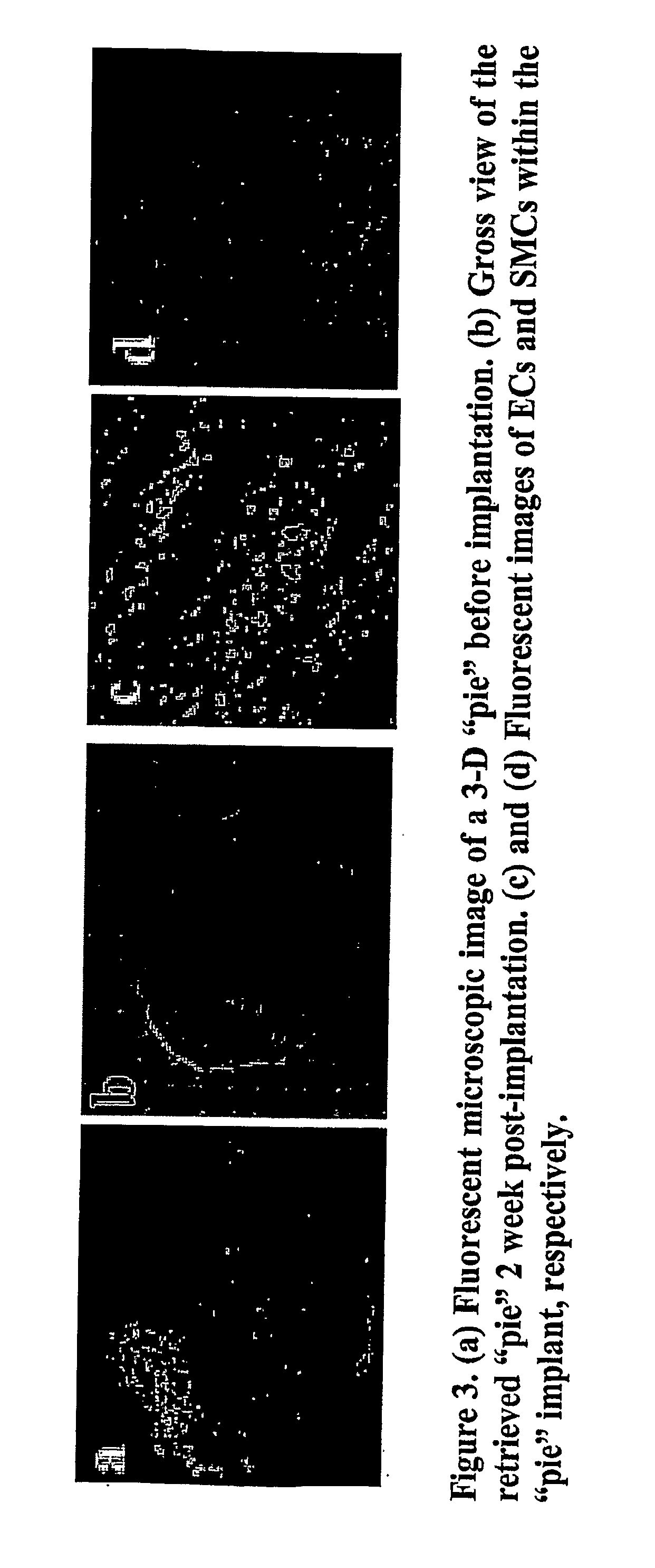
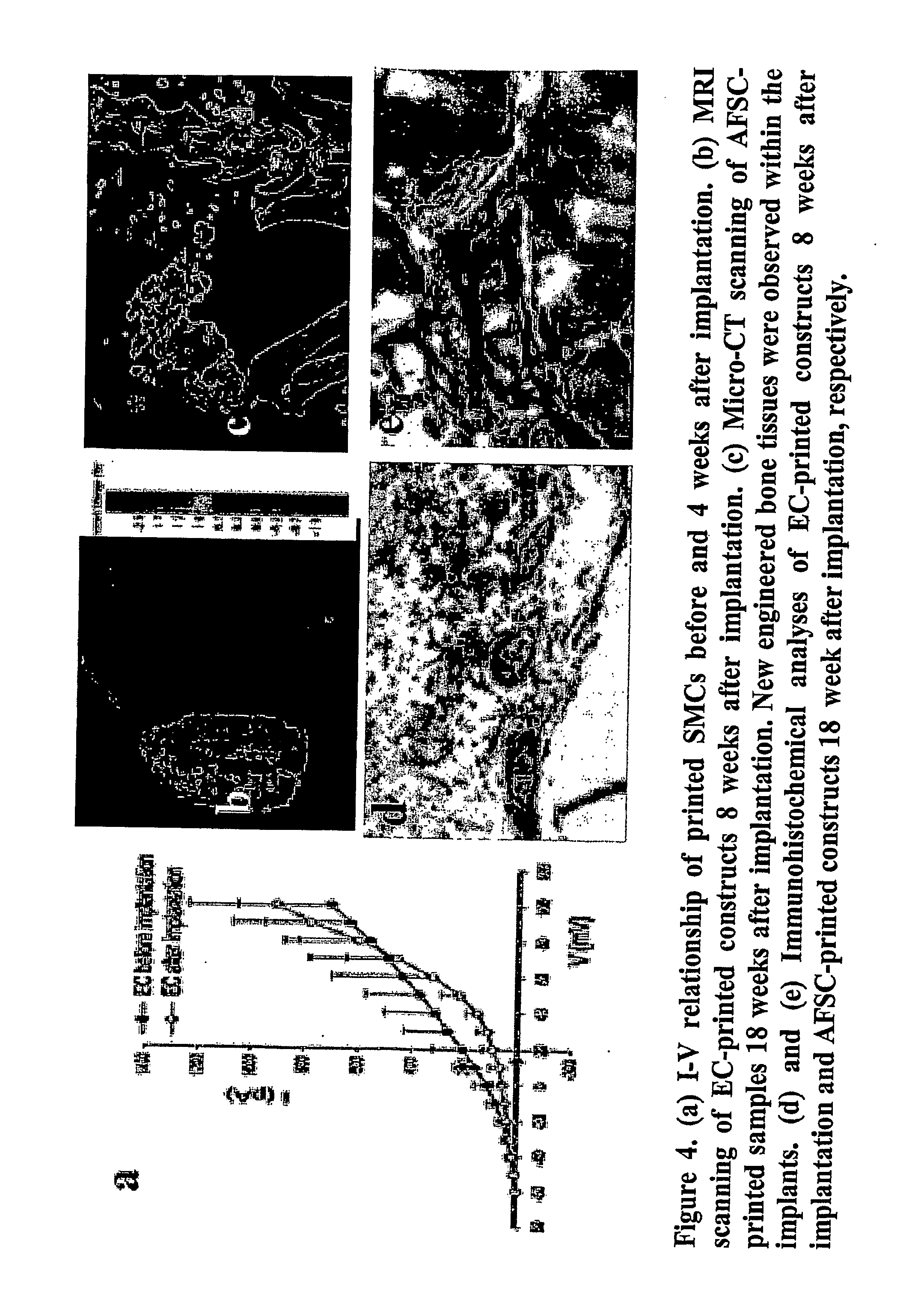
[0087]Materials and Methods: Three-dimensional multi-cell constructs with a “pie” configuration were fabricated by simultaneously printing 3 different cell types [canine bladder smooth muscle cells (SMC), bovine aortal endothelial cells (EC), and human amniotic fluid-derived stem cells (AFSC)] into collagen / alginate gel. The cells were labeled with 3 different membrane bound tracers, which include [PKH67 (red), PKH26 (green), and CMHC (blue)], respectively, prior to printing. Individual cells were also printed separately for additional testing. The printed 3D constructs were subcutaneously implanted into athymic mice. AFSC-printed constructs were cultured in osteoge...


PUM

PropertyMeasurementUnit
viscositiesaaaaaaaaaa

Abstract

A method of forming an array of viable cells is carried out by ink-jet printing a cellular composition containing said cells on a substrate. At least two different types of viable mammalian cells are printed on the substrate, the at least two different types of viable mammalian cells selected to together form a tissue. In some embodiments at least three or four different viable mammalian cells are printed on the substrate, the cells selected to together form a tissue. In some embodiments one of the viable mammalian cell types is a stem cell. In some embodiments the method further comprises printing at least one support compound on the substrate, the support compound selected to form a tissue together with said cells. In some embodiments the method further comprises printing at least one growth factor on the substrate, the growth factor selected to cause the cells to form a tissue.

Description

FIELD OF THE INVENTION[0001]The present invention concerns ink-jet printing of viable cells and arrays of cells so produced.BACKGROUND OF THE INVENTION[0002]Living tissues maintain an inherent multi-cellular heterogeneous structure. Rebuilding of such complex structure requires subtle arrangements of different types of cells and extracellular matrices (ECM) at their specific anatomical target sites. To achieve tissue reconstitution, an effective method for a precise delivery of cells and biomaterials is needed. The inkjet printing technology has been applied to address this endeavor.[0003]The following references are noted herein:[0004]T. Boland et al., Ink-jet printing of viable cells, U.S. Pat. No. 7,051,654;[0005]W. Warren et al., Architecture tool and methods of use, U.S. Pat. No. 6,986,739; and[0006]J. Barron et al., Biological laser printing via indirect photon-biomaterial interactions, US Patent Application Publication No. 2005 / 0018036.SUMMARY OF THE INVENTION[0007]Although t...

Claims


Application Information

Patent Timeline
Patent Type & Authority Applications(United States)
IPC IPC(8): A61K35/12C40B50/12A61P35/00
CPCC12N5/0062B01L3/0268C12N2533/54A61P35/00B29C70/76B33Y10/00B33Y80/00C12N2533/74
Inventor YOO, JAMESXU, TAOATALA, ANTHONY
Owner WAKE FOREST UNIV HEALTH SCI INC
Learn More