
Coated hypodermic needle

a hypodermic needle and coating technology, applied in the field of coating hypodermic needles, can solve the problems of time-consuming epicardial procedures, fluoroscopy does not provide a clear image, and the use of fluoroscopy for pericardial access has several potential limitations

Active Publication Date: 2009-07-02
ST JUDE MEDICAL ATRIAL FIBRILLATION DIV
View PDF18 Cites 26 Cited by
  • Summary
  • Abstract
  • Description
  • Claims
  • Application Information

AI Technical Summary

Problems solved by technology

The use of fluoroscopy for achieving pericardial access has several potential limitations.
Furthermore, fluoroscopy does not provide a clear image.
Due to these limitations, epicardial procedures can be time consuming.
Also, there remains a risk of puncturing a coronary artery or puncturing a heart chamber.
In particular, there remains the risk of ventricular puncture (i.e., puncture in the ventricular wall) and bleeding (e.g., bleeding in the pericardial space).

Method used


Image

  • Coated hypodermic needle
  • Coated hypodermic needle
  • Coated hypodermic needle

Examples


Embodiment Construction

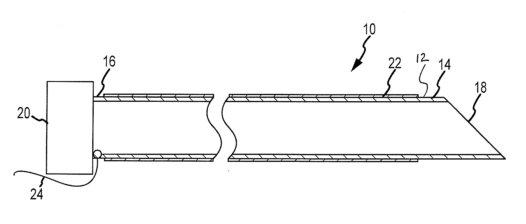
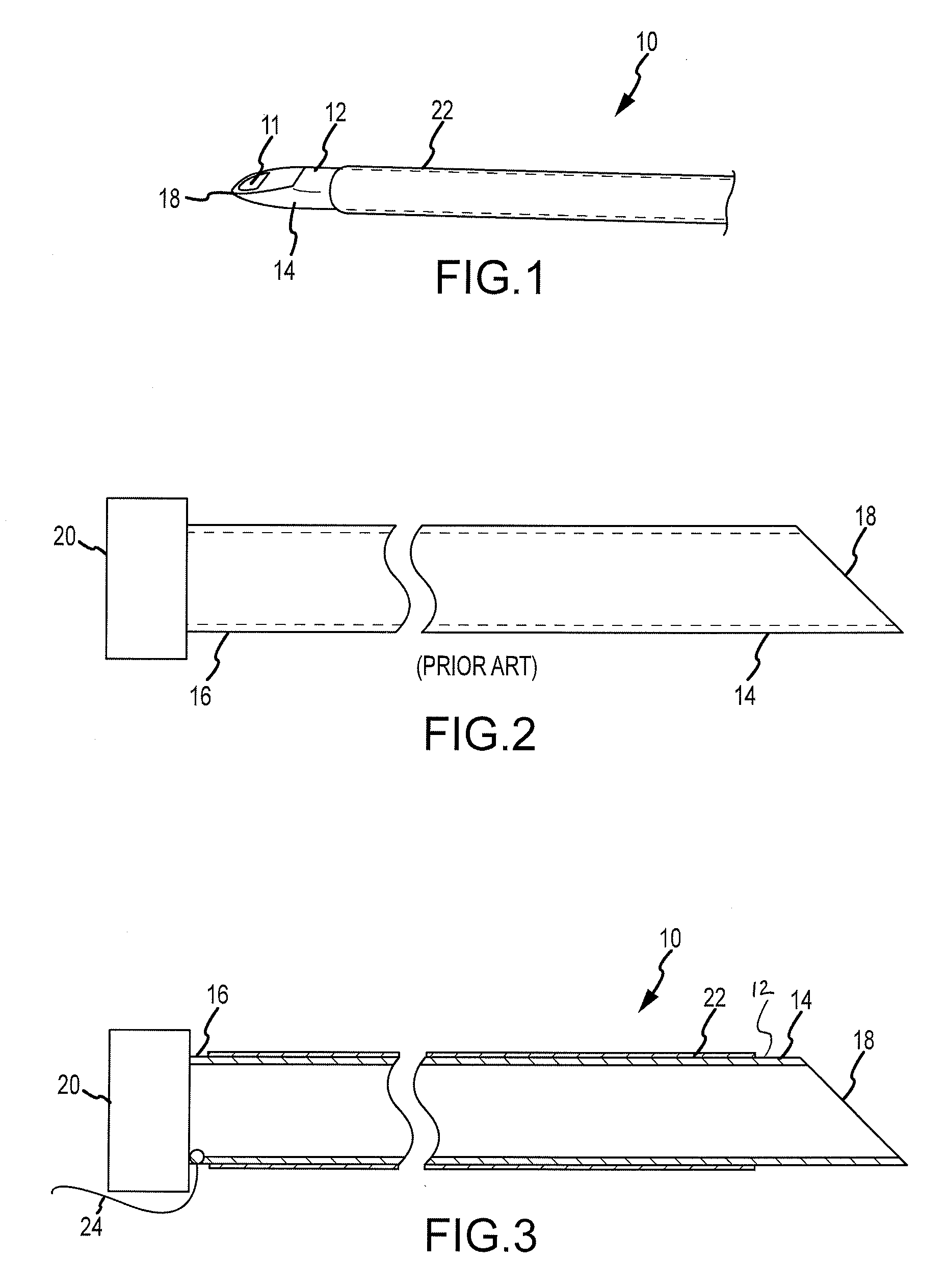
[0023]During an epicardial procedure, the needle used to enter the pericardial space may be the same needle that is used to enter the epidural space when administering epidural anesthesia. The needle conventionally used for pericardial access is a Tuohy needle. A Tuohy needle may have a shaft that is generally curved for at least a portion of its length and defines a lumen. It may have a stylet within the lumen and may be blunt-tipped. The shaft may comprise stainless steel. For some embodiments, the shaft may be between about 89 and 125 mm in length and approximately 1.5 mm in outer diameter.

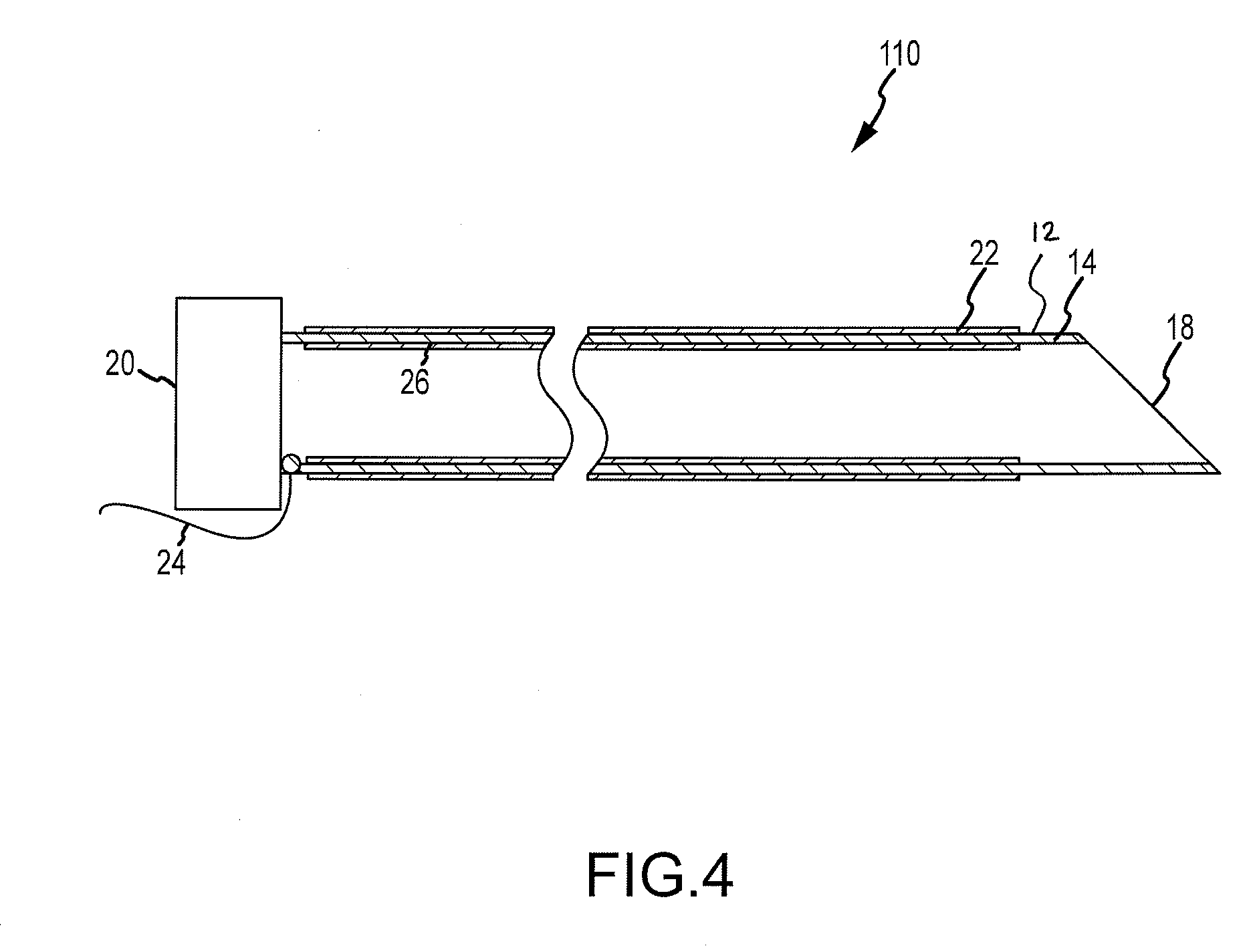
[0024]Referring now to FIG. 1, a first needle 10 in accordance with the present teachings may be similar to a Tuohy needle. The shaft of needle 10 may be hollow with an opening 11 at a distal end. The shaft of the needle may therefore define a lumen. A lumen may be provided because the needle 10 may be used to access the pericardial space, and various fluids (e.g., saline, contrast agents, and ...


PUM


Abstract

A needle for a medical procedure includes a shaft with an inner surface, an outer surface, a proximal section, and a distal section. The distal section has a conductive tip configured to be a first electrode for voltage measurement. The needle further includes a first electrically insulative outer layer over a portion of the outer surface of the shaft. The conductive tip is adapted for insertion through tissue into, for example, a pericardial space of a patient. A system for determining the location of a needle during a medical procedure includes the needle and an anatomical mapping and localization system electrically coupled to the needle and adapted to measure voltage at the conductive tip. A system for determining tissue thickness during a medical procedure includes the needle, an anatomical mapping and localization system electrically coupled to the needle, and an electrocardiograph operatively coupled to the needle and adapted to monitor electrical activity at the conductive tip and at the second electrode. The needle may include a second electrode.

Description

BACKGROUND OF THE INVENTION[0001]a. Field of the Invention[0002]The instant invention is directed toward a needle and a system suitable for use in a medical procedure. The instant invention includes a needle where an electrically insulative outer layer is disposed over a portion of an outer surface of the needle, and a distal section of the needle is exposed to allow for mapping of the needle during a medical procedure.[0003]b. Background Art[0004]In an electrophysiology (EP) procedure, electrode catheters may be guided into the chambers of the heart and to strategic places along the heart's conduction system. The electrodes may then be used to record the electrical impulses of the heart and may define the location of abnormal electrical activity. EP procedures may be used to diagnose and treat ventricular tachycardia (VT) or atrial fibrillation (Afib) ablation, for example. One EP procedure is catheter ablation in which a catheter is inserted through the vasculature and into the he...

Claims


Application Information

Patent Timeline
Patent Type & Authority Applications(United States)
IPC IPC(8): A61M5/32
CPCA61B17/3478A61B18/1477A61B2017/00247A61B2019/5251A61B2018/00392A61B2018/00839A61B2018/1425A61B2017/00256A61B2034/2051
Inventor CAO, HONGPAUL, SAURAVTHAO, CHOU
Owner ST JUDE MEDICAL ATRIAL FIBRILLATION DIV
Learn More