
System and method for urinary tract cell collection, diagnosis, and chemotherapy

a technology for urinary tract cells and urinary tracts, applied in the field of medical devices, can solve the problems of significant and potentially deadly medical conditions, available treatments carry the risk of recurrence, and still at risk for developing “bladder cancer

Inactive Publication Date: 2009-07-02
GARCIA MAURICE M
View PDF7 Cites 81 Cited by
  • Summary
  • Abstract
  • Description
  • Claims
  • Application Information

AI Technical Summary

Benefits of technology

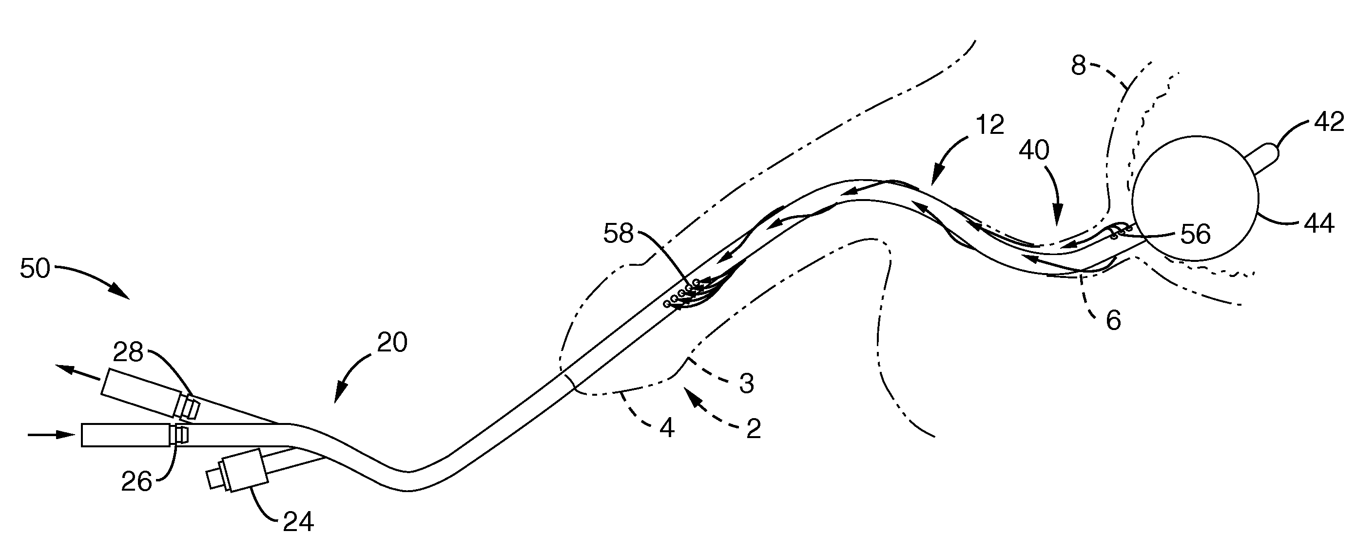
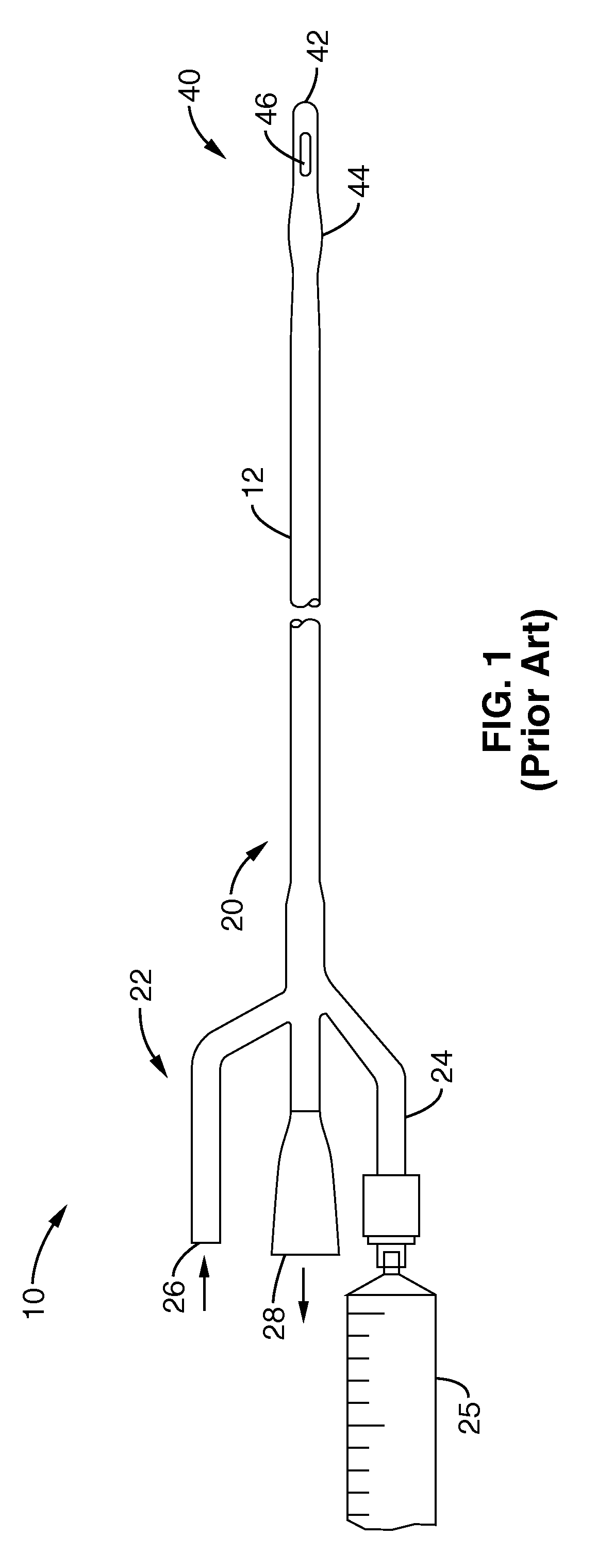
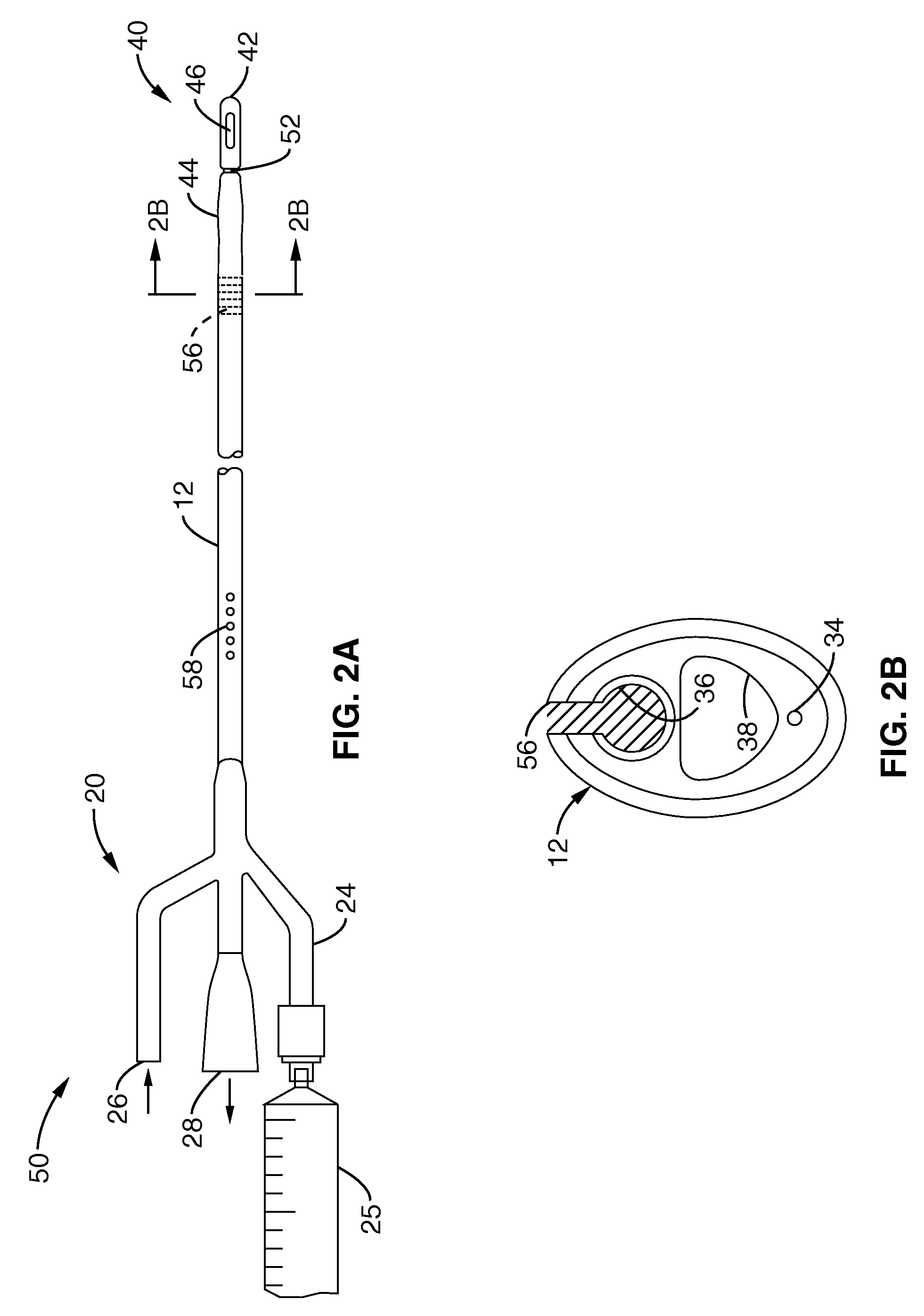
[0024]In another mode, the infusion lumen extends to a distal port located distally of the infusion port and also distally of the distal balloon. A valve that comprises a deflectable member is located along a portion of an internal wall of the catheter assembly that separates the infusion lumen and an inflation lumen of the distal balloon. The valve is positioned distally from the distal infusion port and proximally of the distal balloon. The valve remains open relative to the infusion lumen when the distal balloon is deflated. This allows fluid communication between the distal port and a proximal port located proximally of the balloon assembly and outside the body, such as for example to facilitate drainage of urine from the bladder through the infusion lumen when the distal port is located within the bladder. The valve is actuatable to a closed condition relative to the infusion lumen as follows. Pressurization of the balloon inflation lumen during inflation of the distal balloon deflects the deflectable member into the infusion lumen to thereby collapse across and substantially close the infusion lumen to fluid communication between the distal port and the distal infusion port. This closed configuration allows for pressurized fluid within the infusion lumen to be expelled from the infusion lumen through the distal infusion port proximally of the inflated distal balloon.
[0050]According to one further embodiment, the valve comprises a deflectable member located along a portion of an internal wall of the catheter assembly that separates the infusion lumen and an inflation lumen of the distal balloon. In further variations, the deflectable member is positioned distally from the distal inflation port, and the valve remains open relative to the infusion lumen when the distal balloon is deflated, thereby allowing fluid communication between the distal port and the proximal infusion port. The valve is actuatable to a closed condition relative to the infusion lumen upon pressurization of the balloon inflation lumen during inflation of the distal balloon, thereby deflecting the deflectable member into the infusion lumen to collapse across and substantially close the infusion lumen with respect to fluid communication between the distal port and the distal infusion port. The valve in the closed configuration allows for pressurized fluid within the infusion lumen to be expelled from the infusion lumen substantially through the distal infusion port proximally of the inflated distal balloon.
[0064]Another aspect is a method for performing a medical procedure in a lumen of a urinary tract in a patient, comprising: transurethrally delivering a distal end portion of a catheter assembly at least in part to the bladder; infusing a volume of wash medium from the distal end portion into a region of the lumen around the catheter; preventing the infused wash medium from flowing distally and proximally from the region; and collecting the infused wash medium from the region via a distal collection port located along the catheter in the region.

Problems solved by technology

Cancer as it relates to the urinary tract is a significant and potentially deadly medical condition.
Moreover, available treatments often carry the risk of recurrence, which also need early detection for most favorable re-treatment results.
Therefore, when a patient has bladder cancer, and ultimately has their diseased bladder removed (even when only the bladder contained cancer), they are still at risk for developing “bladder cancer” elsewhere.
However, a significant number of patients do not have any symptoms at all.
However, in particular, patients who have had their bladders removed pose unique challenges.
But, for the cytopathologists who have to examine their urine under a microscope to look for cancer cells, it is a problem.
A frustrated ability to recognize the difference, and the ability to find the samples for the determination in the first place, can have catastrophic consequences.
Accordingly, if a patient has had their bladder replaced with a new bladder made of intestine, conventional urine analysis is a relatively poor study to use to look for cancer cells.
This is because the mucus makes it very hard to see the cells clearly.
Also, neobladders get inflammatory reactions frequently.
An additional problem is that the mucus is already loaded with cellular debris, and this debris is what prevents a good view of any urothelial cells that might be cancerous.
While the conventional diagnostic techniques using voided urine sample is still of some value, this is mainly because there are no real alternatives.
Nonetheless, the “sensitivity” of such approach is low.
Notwithstanding these recognized shortcomings of current diagnostic techniques, the diagnostic quality of a urine specimen from a patient with a neobladder (e.g., made from the patient's own intestine) has not been appropriately studied or addressed in the past.
Moreover, the cytopathologists have also not conducted prolific study or debate about the poor quality of their specimens, possibly because it is intuitive to them—it is because of the mucus and cellular debris, etc., as a necessary evil of examining neobladder urine voids.
To the extent chemotherapy agents may be delivered locally to the area, the correct combination of drug and delivery system has yet to be optimized for optimal results.

Method used


Image

  • System and method for urinary tract cell collection, diagnosis, and chemotherapy
  • System and method for urinary tract cell collection, diagnosis, and chemotherapy
  • System and method for urinary tract cell collection, diagnosis, and chemotherapy

Examples


Embodiment Construction

[0092]Various aspects, modes, and embodiments of the present invention are further described and variously illustrated as follows.

[0093]By convention, the end furthest inside the patient (furthest from the outer skin surface) has been herein designated the “distal end”, while the “proximal end” of the catheter is the end that remains either outside of the body, or, the working end of the catheter, through-which fluids are irrigated into / out of the catheter. Medical conventions typically refer to proximal as “upstream” and distal as “downstream” when referring to anatomy, such as down the urological tract from “proximal” structures such as the kidney or ureters and “distally” toward the outside world such as via a urethra. For consistency of this disclosure, o the extent catheters devices and methods are herein described in retrograde delivery within such anatomies, “proximal” will hereunder mean outward from the patient and “distal” will hereunder mean further inward or upstream int...


PUM


Abstract

A system and method delivers and collects a wash medium into a lumen of the urinary tract system. Isolated saline delivery washes cells from a urethra that are collected for cancer diagnosis. BCG is delivered for local chemotherapy. The isolated delivery and collection is from a region bound by a distal occlusion and proximal occlusion. Inflatable balloons provide the occlusion and or a penis clamp may be used. The two balloons may telescope relative to each other. A balloon inflation lumen may collapse a valve membrane to occlude distal flow of infused fluid to only proximal of the balloon.

Description

CROSS-REFERENCE TO RELATED APPLICATIONS[0001]This application claims priority from, and is a 35 U.S.C. § 111(a) continuation of, co-pending PCT international application serial number PCT / US2007 / 063209, filed on Mar. 2, 2007, incorporated herein by reference in its entirety, which claims priority from U.S. Provisional Patent Application Ser. No. 60 / 778,750, filed on Mar. 3, 2006, incorporated herein by reference in its entirety, and also claims priority from U.S.[0002]Provisional Patent Application Ser. No. 60 / 802,316, filed on May 19, 2006, incorporated herein by reference in its entirety.STATEMENT REGARDING FEDERALLY SPONSORED RESEARCH OR DEVELOPMENT[0003]Not ApplicableBACKGROUND OF THE INVENTION[0004]1. Field of the Invention[0005]This invention relates to the field of medical devices. More specifically, it relates to a system, apparatus, and method for diagnosing and treating cancer in a patient, and still more particularly as it relates to the patient's urethra.[0006]2. Descrip...

Claims


Application Information

Patent Timeline
Patent Type & Authority Applications(United States)
IPC IPC(8): A61B10/02
CPCA61B10/0045A61B2017/22082A61B2017/22067A61B10/007
Inventor GARCIA, MAURICE M.CARROLL, PETER R.
Owner GARCIA MAURICE M
Learn More