
X-ray ct apparatus and x-ray tube current determining method

Active Publication Date: 2009-07-02
GE MEDICAL SYST GLOBAL TECH CO LLC
View PDF14 Cites 13 Cited by
  • Summary
  • Abstract
  • Description
  • Claims
  • Application Information

AI Technical Summary

Benefits of technology

[0032]Since the function is given as a polynomial equation of the BMI, it is easy to store it in a memory.
[0034]In a method for setting an X-ray tube current of an X-ray CT apparatus, image noise obtained by preliminary imaging of a subject, which is conducted ahead of actual imaging of the subject, is measured, and an X-ray tube current at the actual imaging, determined based on both an X-ray tube current at the preliminary imaging and a ratio between a measured value of noise at the preliminary imaging and each index related to image noise is set. Therefore, a tube current at the time that the heart is imaged by the X-ray CT apparatus can be determined in such a manner that the image noise becomes stable.

Problems solved by technology

There was a case in which such a tube current adjustment as described above was unsuitable for use in cardiac imaging.
That is, this is because since the size of a region to be imaged or photographed changes due to heart or cardiac motion or the like in the case of the cardiac imaging, the relation of correspondence between an index related to image noise and a tube current for attaining it is hard to be established for the cardiac imaging.
Thus, since the cardiac imaging is normally conducted under a constant tube current, the image noise is affected by the body type or the like of a patient and affected even by the skill of a photographer.
Therefore, the quality of an image is not stabilized and accurate X-ray interpretation is difficult.

Method used


Image

  • X-ray ct apparatus and x-ray tube current determining method
  • X-ray ct apparatus and x-ray tube current determining method
  • X-ray ct apparatus and x-ray tube current determining method

Examples


first embodiment

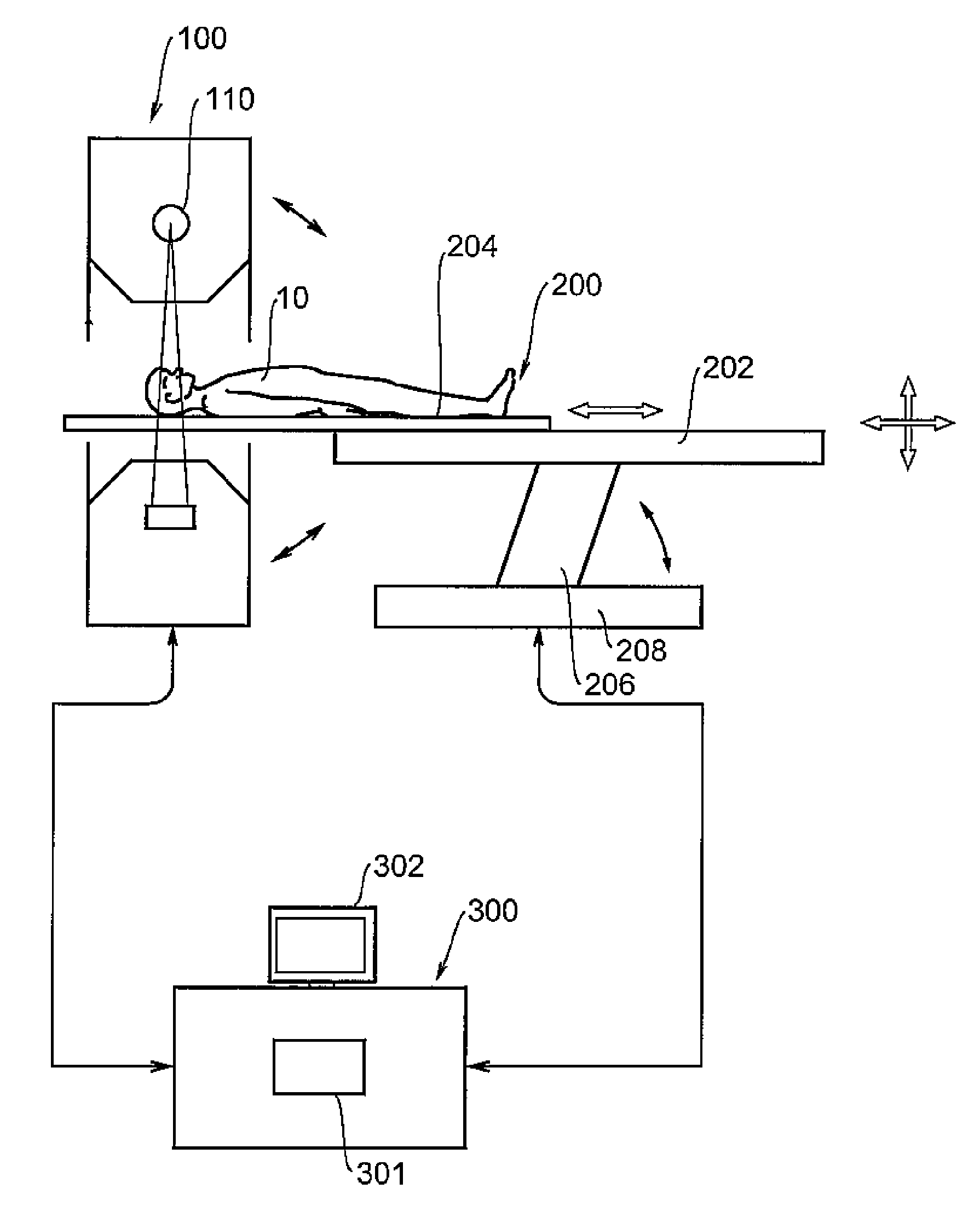
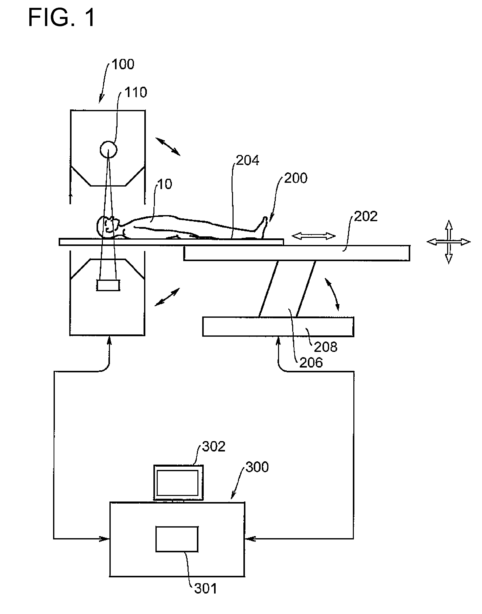
[0060]A first embodiment will explain an example for determining a tube current using a BMI. The tube current is given as a time product of currents and its unit is expressed in mAs. As a tube current determining criterion, an index related to image's noise is defined with respect to a reconstructed image. This index corresponds to an index specialized for cardiac imaging. This is hereafter called “Cardiac Image Noise Index: CINI”). It is also simply called “noise index CINI”. The noise index CINI is given by a standard deviation or the like of each pixel value, for example.

[0061]The tube current is determined so as to assume such a current that an image having noise coincident with the corresponding noise index CINI can be photographed. Image noise to be noted corresponds to image noise at a central portion of an ascending main artery. The tube current is determined in such a manner that the image noise of this portion coincides with the noise index CINI.

[0062]The determination of ...

second embodiment

[0070]A second embodiment will explain an example in which a tube current is determined based on the result of noise measurement at preliminary imaging. When coronary angiographic imaging is conducted, preliminary imaging using a small amount of contrast agent is carried out before actual imaging to examine timing or the like for contrast achievement. In such a case, the tube current may be determined using the result of preliminary imaging.

[0071]Determining the tube current utilizing the result of preliminary imaging is conducted using the following equation.

mAs(c)=k(pitch)*(Nb / Nc)̂2*(T(b) / T(c))*mAs(b)   (3)

[0072]wherein Nb: noise of image obtained by preliminary imaging, Tb: slice thickness at preliminary imaging, and mAs(b): tube current at preliminary imaging. Further, Nc indicates a desired noise index of an image obtained by the actual imaging, Tc indicates a slice thickness at the actual imaging, and mAs(c) indicates a tube current at the actual imaging. k(pitch) indicates a ...


PUM


Abstract

This invention is provided to determine a tube current at the imaging of the heart by an X-ray CT apparatus in such a manner that image noise becomes constant. Upon determining a tube current of an X-ray tube at the time that the heart is imaged by an X-ray CT apparatus, the tube current is determined based on an index related to image's noise and a BMI of a subject. The noise is at a central portion of an ascending main artery. Determining the tube current is conducted using a function with the BNI as a variable. The function is given in the form of a polynomial equation of the BMI. Each coefficient of the polynomial equation is a function of the index.

Description

CROSS REFERENCE TO RELATED APPLICATIONS[0001]This application claims the benefit of Chinese Patent Application No. 200710300497.9 filed Dec. 28, 2007, which is hereby incorporated by reference in its entirety.BACKGROUND OF THE INVENTION[0002]The embodiments described herein relate to an X-ray CT apparatus and an X-ray tube current determining method, and particularly to a technique for determining a tube current of an X-ray tube at the time that the heart is imaged or photographed by an X-ray CT (Computed Tomography) apparatus.[0003]Even of X-ray CT apparatuses, one called a VCT (Volume CT), an MDCT (Multi-row Detector CT) or a multi-slice CT or the like is configured so as to detect a cone beam X-ray radiated from an X-ray tube by means of a multi-row detector having 64 rows to 256 rows.[0004]This type of X-ray CT apparatus is used with emphasis on 3D-image photography of the heart or circulatory system by paying attention to the size of coverage in a body-axis direction and the le...

Claims


Application Information

Patent Timeline
IPC IPC(8): A61B6/00G01N23/083
CPCA61B6/032A61B6/488A61B6/027A61B6/542A61B6/544A61B6/504
Inventor JIANYING, LI
Owner GE MEDICAL SYST GLOBAL TECH CO LLC
Learn More