Implants and Methods for Repair, Replacement and Treatment of Disease

a technology for implants and joints, applied in the field of implants and methods for repair, replacement and treatment of diseases, can solve the problems of current treatments for repairing or ameliorating joint diseases, significant health care, economic and social costs, pain and disability in the adult population, etc., and achieve the effect of facilitating the positioning or physical stability of implants in patients

Inactive Publication Date: 2009-06-04
ZIMMER INC
View PDF99 Cites 56 Cited by
  • Summary
  • Abstract
  • Description
  • Claims
  • Application Information

AI Technical Summary

Benefits of technology

[0006]In some configurations, a region of a subchondral base comprising pores having a median pore diameter of about 100 microns to about 800 microns can provide a surface which can promote bone attachment and ingrowth, while a region comprising a cartilage-adherent surface can provide a surface which can promote chondrocyte and / or cartilage attachment and growth. In various configurations, a cartilage-adherent surface can be a region of a subchondral base comprising a plurality of pores having a median pore diameter of about 3 microns to about 20 microns, a cartilage adhesive, or a combination of a plurality of pores and a cartilage adhesive, and can comprise trabecular metal or a different material, such as a polymer. In certain configurations, an implant can further comprise a non-trabecular metal material which provides a surface for chondrocyte or cartilage attachment, such as, in non-limiting example, a chondrocyte-adherent ceramic or plastic. In addition, in some configurations, a trabecular metal component of an implant can have a geometry that promotes retention of chondrocytes, such as barbs, ridges or hooks.
[0017]In some configurations of these embodiments, a method can further include introducing a non-trabecular metal positioning structure to a recipient patient. Such a positioning structure can aid in the positioning or physical stability of an implant in the patient. In various configurations, a positioning structure can comprise a biocompatible metal or a biocompatible polymer. In configurations of these embodiments, a trabecular metal portion of an implant can be configured to attach to the positioning structure, and / or the positioning structure can be configured to attach to the subchondral base. In certain aspects, the positioning structure can be configured for engaging the bone, and can be, in non-limiting example, a screw, a cylinder, a plat, a rod, or a washer. In addition, in various aspects, a positioning structure can also comprise trabecular metal, and / or can comprise a material other than trabecular metal, such as, in non-limiting example, a biocompatible polymer, or a metal that is non-trabecular.

Problems solved by technology

Joint disease, defect, and injury are leading causes of pain and disability in the adult population.
The morbidity associated with joint disease, defect, and injury and their spectrum of associated disorders are responsible for significant health care, economic and social costs.
Current treatments for repairing or ameliorating a joint disease, defect, or injury, for example an osteochondral injury in which articular cartilage and underlying bone are damaged, can be expensive, inefficacious, painful, or lengthy.

Method used


Image

  • Implants and Methods for Repair, Replacement and Treatment of Disease
  • Implants and Methods for Repair, Replacement and Treatment of Disease
  • Implants and Methods for Repair, Replacement and Treatment of Disease

Examples


example 1

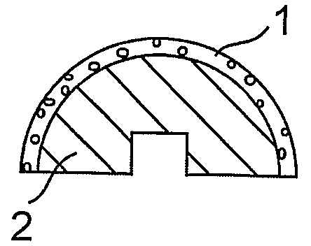
[0053]This example illustrates replacement of a femoral head in a patient suffering from a femoral head fracture with an implant.

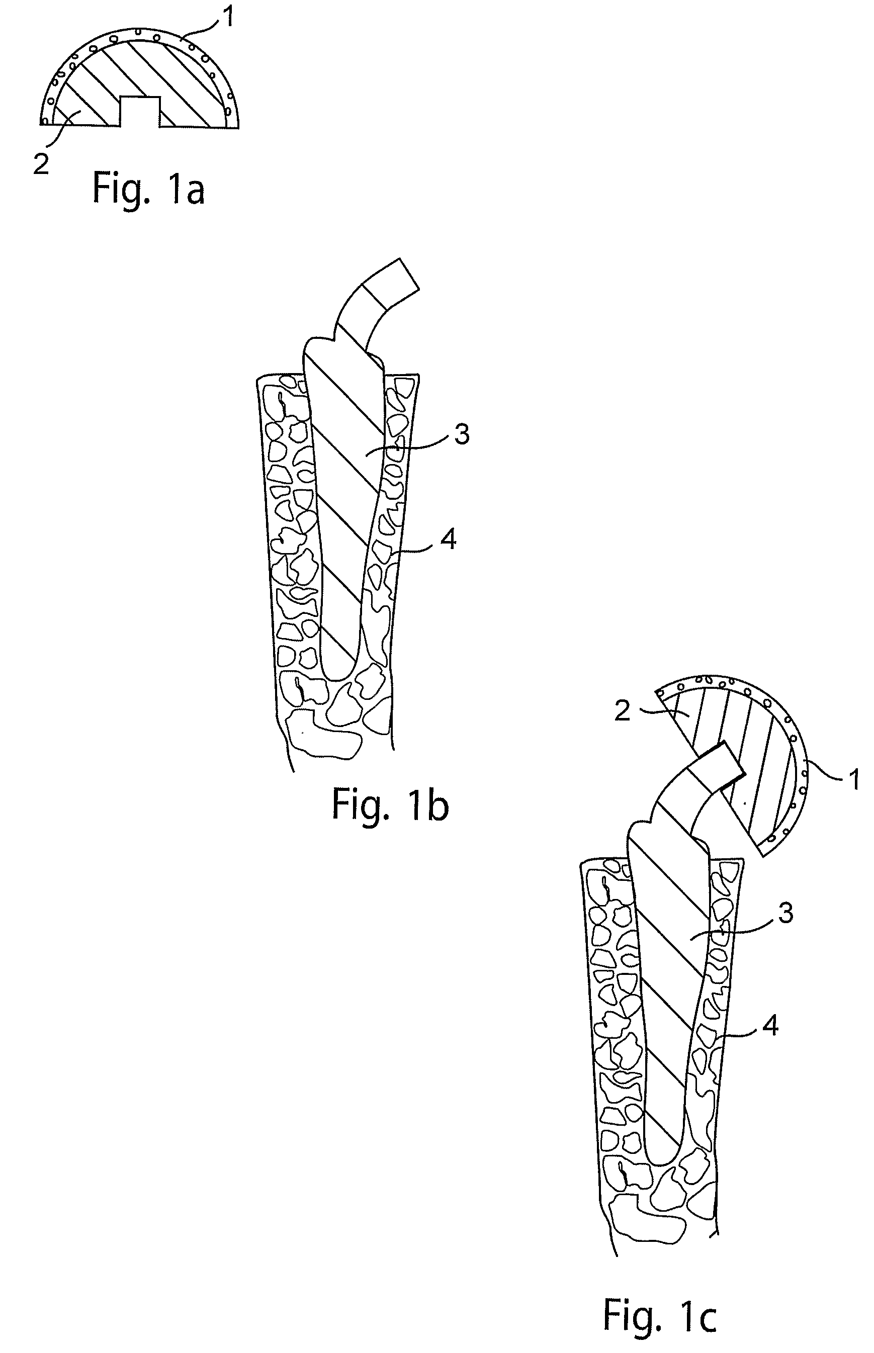
[0054]In this example, as shown in FIG. 1, a porous femoral head prosthesis (2) comprising a rounded surface comprising pores of median diameter of about 10 microns, is placed in an in vitro cell growth chamber. Juvenile chondrocytes are seeded onto the rounded surface, and cultured as described in Adkisson, H. D. et al., Clin. Orthop. 391S: S280-S294, 2001; and U.S. Pat. Nos. 6,235,316 and 6,645,316). The chondrocytes are permitted to grow until a layer of neocartilage (1) forms on the surface of the prosthesis (FIG. 1a). A surgeon then drives a spike-shaped subchondral base (3) comprising trabecular metal into the proximal femur (4) of the patient (FIG. 1b). The surgeon then attaches the femoral head prosthesis comprising neocartilage (1) and trabecular metal (2) to the subchondral base (3), thereby forming an implant comprising cartilage and a subchondr...

example 2

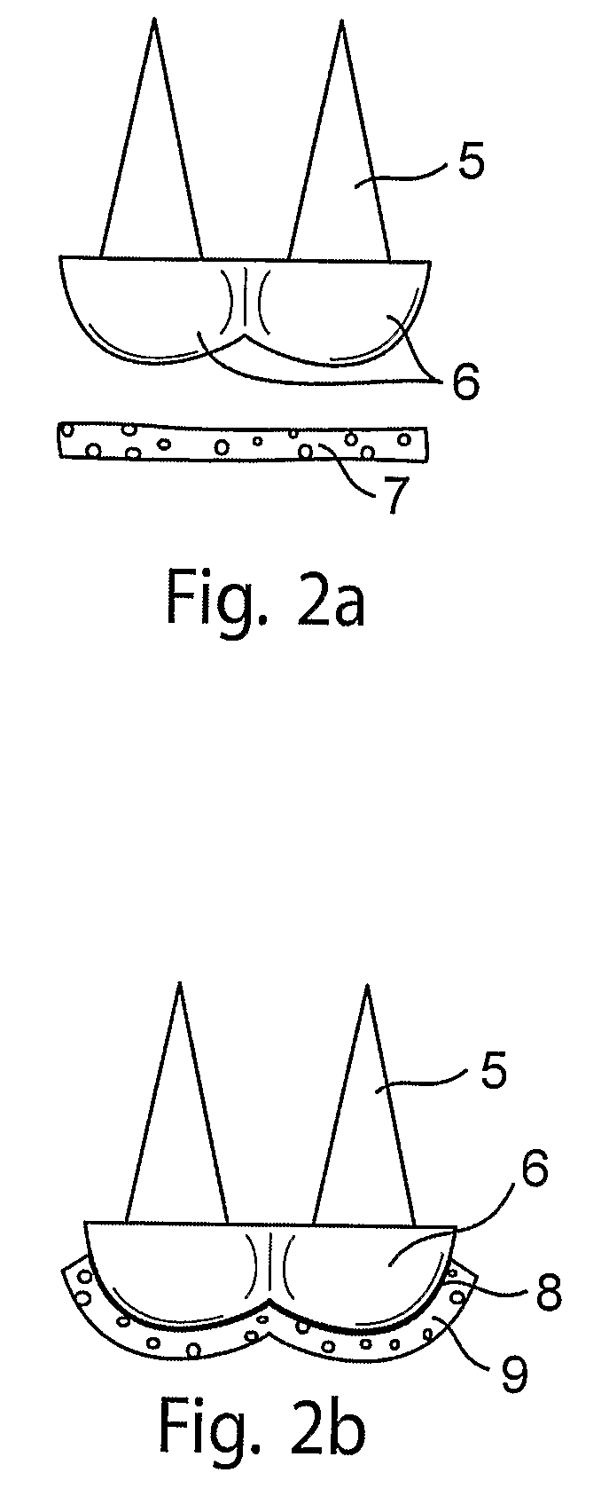
[0055]This example illustrates attachment of neocartilage to a subchondral base in vitro.

[0056]In this example, as shown in FIG. 2, a subchondral base (5) comprising trabecular metal and shaped to replace a condyle (6) is rendered adherent for cartilage by application of a fibrin adhesive to the rounded surfaces of the trabecular metal. The rounded surfaces of the base is then contacted with neocartilage grown in vitro (7) (FIG. 2a). The neocartilage (9) adheres to the adhesive layer (8), providing a layer of neocartilage following the contours of the surface of the base (9), thereby forming an implant which can replace a diseased or injured condyle of a patient (FIG. 2b).

example 3

[0057]This example illustrates use of a positioning structure to introduce an implant into a patient.

[0058]In this example, as shown in FIG. 3, a subchondral base (11) comprising trabecular metal and having an internal screw thread is seeded with chondrocytes which are cultured to form neocartilage (10), thereby providing an implant (FIG. 3a). A surgeon prepares a cylindrical-shape implant site which traverses the patient's cartilage and invades the bone in a condyle of the patient (13). The implant site has a depth and diameter corresponding to the implant. The surgeon then inserts a non-trabecular metal screw (14) into the site, leaving the screw partially exposed (FIG. 3b). The surgeon then lightly presses the implant into the site, thereby engaging the exposed screw (14) with the internal screw thread of the implant. The surgeon presses on the implant until the neocartilage (1) is flush with the patient's cartilage (FIG. 3c). The neocartilage can then heal smoothly with the pati...


PUM

PropertyMeasurementUnit
Pore sizeaaaaaaaaaa
Pore sizeaaaaaaaaaa
Pore sizeaaaaaaaaaa

Abstract

Implants comprising cartilage and trabecular metal, and methods of making the implants are disclosed. Further disclosed are therapeutic uses of the implants, which include methods of treatment or repair of an chondral or osteochondral defect, such as a chondral or osteochondral injury, lesion or disease. An implant comprises cartilage or chondrocytes and a subchondral base comprising trabecular metal. An implant can comprise a geometric shape such as a cylinder or an anatomical shape such as a condyle, and can be used in conjunction with a positioning structure.

Description

CROSS-REFERENCE TO RELATED APPLICATIONS[0001]This application claims priority from U.S. Provisional Application Ser. No. 60 / 712,004 filed on Aug. 26, 2005, which is incorporated herein by reference in its entirety.BACKGROUND[0002]Joint disease, defect, and injury are leading causes of pain and disability in the adult population. The morbidity associated with joint disease, defect, and injury and their spectrum of associated disorders are responsible for significant health care, economic and social costs. Current treatments for repairing or ameliorating a joint disease, defect, or injury, for example an osteochondral injury in which articular cartilage and underlying bone are damaged, can be expensive, inefficacious, painful, or lengthy. Alternative treatments are, therefore, needed.SUMMARY[0003]In view of these and related unmet needs, the present teachings disclose implants which can be used in the treatment, repair and / or partial or full replacement of a chondral or osteochondral ...

Claims


Application Information

Patent Timeline
IPC IPC(8): A61F2/02A61F2/28
CPCA61F2/28A61L27/56A61F2/30756A61F2/30767A61F2/34A61F2/36A61F2/3859A61F2002/2839A61F2002/30011A61F2002/30064A61F2002/3021A61F2002/30224A61F2002/30276A61F2002/30405A61F2002/30448A61F2002/30461A61F2002/30472A61F2002/30762A61F2002/30764A61F2002/30797A61F2002/30841A61F2002/30892A61F2002/30929A61F2002/30934A61F2002/30968A61F2002/3611A61F2002/365A61F2220/0008A61F2220/0025A61F2220/0041A61F2220/005A61F2220/0075A61F2230/0067A61F2230/0069A61F2230/0086A61F2250/0023A61F2310/00011A61F2310/00017A61F2310/00023A61F2310/00029A61F2310/00059A61F2310/00095A61F2310/00131A61F2310/00395A61F2310/00407A61F2310/00796A61F2310/00964A61F2310/00982A61F2310/00988A61L27/306A61L27/3612A61L27/3641A61L27/3645A61L27/3817A61L27/3843A61L27/446A61F2/30749A61F2002/30433
Inventor GAGE, GARYADKISSON, IV, H. DAVISBLANCHARD, CHERYL R.
Owner ZIMMER INC
Why Eureka
Try Eureka