
Vascular/Lymphatic Endothelial Cells

a technology of lymphatic and vascular endothelial cells and stem cells, which is applied in the field of nonembryonic stem cells, can solve the problems of inability to analyze the specific venous or arterial potential of different types of stem cells, and the signal involved in the fate of arterial, venous or lymphatic endothelium only starting to be unraveled, and achieves the effect of increasing expression

Inactive Publication Date: 2009-04-23
PROYECTO DE BIOMEDICINA CIMA +1
View PDF22 Cites 46 Cited by
  • Summary
  • Abstract
  • Description
  • Claims
  • Application Information

AI Technical Summary

Benefits of technology

[0013]One embodiment provides a method to differentiate cells comprising contacting an enriched population of stem cells that differentiate into more than one embryonic lineage with VEGF165, VEGF-C, VEGF-D or a combination thereof, so as to increase expression of one or more of prox-1, podoplanin or lyve-1 relative to the initial population.

Problems solved by technology

Although it has been shown that some of these pathways are well conserved from zebrafish to mouse, less information is available on whether they have a similar role in humans.
Furthermore, the signals involved in arterial, venous or lymphatic endothelium fate are only starting to be unraveled.
However, the specific venous or arterial potential of different types of stem cells has not been analyzed.
For example, ischemic disorders, such as atherosclerosis, are generally caused by obstruction of the arterial part of the vasculature, which results in oxygen deprivation and ultimately death of the target tissue.

Method used


Image

  • Vascular/Lymphatic Endothelial Cells
  • Vascular/Lymphatic Endothelial Cells
  • Vascular/Lymphatic Endothelial Cells

Examples


example 1

In vitro Analysis hMAPCs

[0266]Phenotypic and Functional Endothelial Potential of hAC133+ Cells and hMAPCs

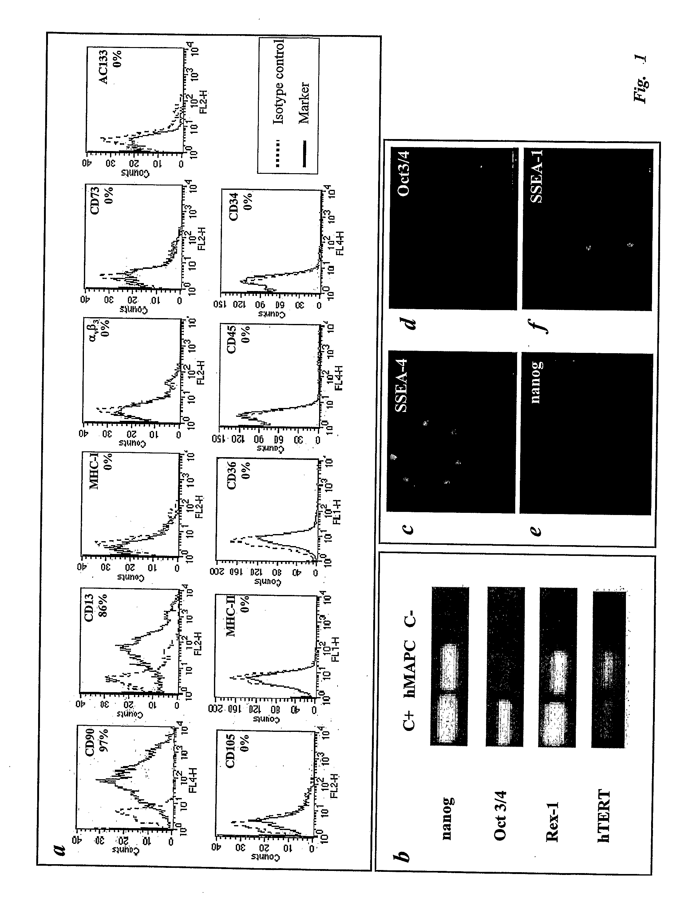
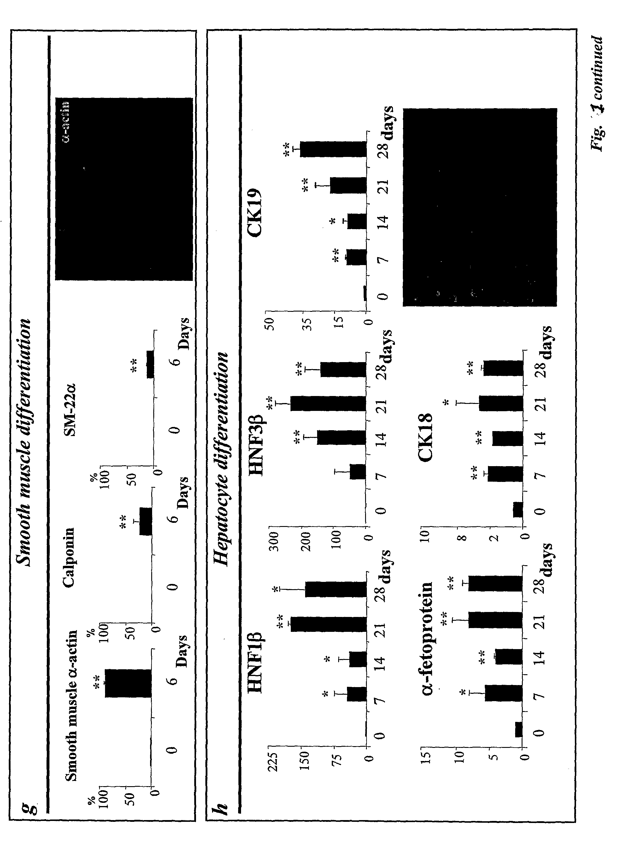
[0267]Having established and characterized hMAPCs capable of differentiating into mesoderm, endoderm and ectoderm derived tissues, the endothelial potential of hMAPCs was compared with that of hAC133+, a cell population previously shown to be enriched for endothelial, neuronal and hematopoietic progenitors (Gehling, 2000; Asahara, 1997). Culture of hAC133+ cells in the presence of VEGF165 induced down-regulation of hematopoietic markers (CD34 and CD45) and up-regulation of mature endothelial markers (CD36, CD105 and αvβ3) (FIG. 2a-b), as has been described Gehling et al. (2000). The majority of the cells in 21-day cultures (which are further designated as “hAC133-ECs) expressed VEGF receptors 1 (Flt-1) and 2 (KDR), angiopoietin receptors (Tie-1 and Tie-2; FIG. 2c-g) and were functional as demonstrated by uptake of acetylated-LDL (AcLDL; FIG. 2h) and vascular tube formation on mat...

example 2

In vivo Analysis of hMAPCs

[0277]Shh and Dll-4 Induce Arterial hMAPC-EC Differentiation and Arterial-Like Vessel Growth in vivo

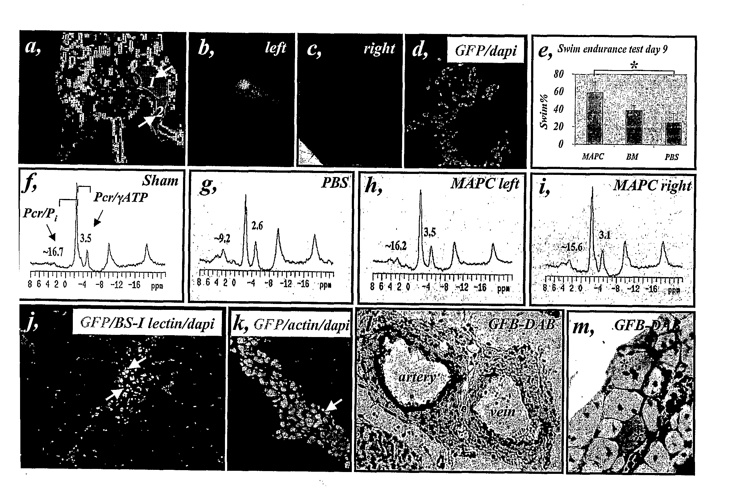
[0278]To determine whether the same factors could also induce hMAPC differentiation into arterial endothelium in vivo, 0.5×10⁶ undifferentiated hMAPCs in growth factor reduced matrigel containing either VEGF165 (“standard media”), or VEGF165+Shh+Dll4 (“arterial media”) was injected under the skin of nude mice (N=6 per group). To account for the effects of the admixed cytokines on host cells, the corresponding “cytokine-alone” groups were also included. In order to track the cells following implantation, hMAPCs were labeled with CFSE or iron particles (Resovist™; Arbab et al., 2003) before injection. Irrespective of the cytokine cocktail used, localized areas of CFSE-labeled cells (FIG. 9a) and single Resovist-labeled (Arbab et al., 2003) hMAPC-derived cells (FIG. 9b) persisted for at least 10 days in the matrigel plug as determined by in vivo live imaging and...


PUM


Abstract

The present invention provides methods to culture and use vascular endothelial cells, including lymphatic, venous and arterial endothelial cells.

Description

CROSS-REFERENCE TO RELATED APPLICATIONS[0001]This application claims priority under 35 U.S.C. 119(e) from U.S. Provisional Application Ser. No. 60 / 652,015 filed Feb. 10, 2005, which is herein incorporated by reference.FIELD OF THE INVENTION[0002]This invention relates to methods and compositions for differentiation of non-embryonic stem cells to cells of the endothelial lineage, particularly a vascular endothelial lineage with arterial, venous and / or lymphatic endothelial characteristics, culture conditions therefor and uses thereof.BACKGROUND OF THE INVENTION[0003]The vascular system is a bipolar complex network of arteries that transport oxygen-rich blood to all tissues, and veins that bring oxygen-deprived blood back to the heart (Carmeliet, 2004). The vascular system is the first system to develop in the embryo. Vasculogenesis, the in situ differentiation of primitive endothelial progenitors, termed angioblasts, into endothelial cells that aggregate into a primary capillary plex...

Claims


Application Information

Patent Timeline
IPC IPC(8): A61K35/12C12N5/06C12N5/071
CPCC12N5/069C12N5/0691C12N2501/11C12N2506/03C12N2501/165C12N2501/42C12N2501/13
Inventor PROSPER, FELIPEVERFAILLIE, CATHERINE M.LOPEZ-ARANGUREN, XABIERCLAVER, CARLOS CLAVELLUTTUN, AERNOUT
Owner PROYECTO DE BIOMEDICINA CIMA
Learn More