
High surface area anti-microbial coated endotracheal tube

a technology of anti-microbial coating and endotracheal tube, which is applied in the field of medical devices, can solve the problems of large volume of secretions and mucous at the junction of the tube and cuff, the previous method of preventing this buildup and the resultant infections have not been widely adopted in the respiratory field, and achieve the effect of reducing the density of pathogens

Inactive Publication Date: 2009-04-23
TELEFLEX MEDICAL INC
View PDF7 Cites 25 Cited by
  • Summary
  • Abstract
  • Description
  • Claims
  • Application Information

AI Technical Summary

Benefits of technology

The present invention provides an apparatus for reducing pathogens in the secretion buildup above the cuff of an endotracheal tube or other medical device. This is achieved by adding a high surface area structure attached to the cuff, which increases the surface area of silver or other antimicrobial coatings to neutralize pathogens. This invention effectively positions an antimicrobial agent proximate or into the secretion pool and not just around the perimeter of the device, potentially reducing the pathogen density within the secretion pool leading to a reduction in infections such as VAP.

Problems solved by technology

Infections, such as ventilator assisted pneumonia (VAP), can commonly result due to biofilm buildup around the device near or within the pool of secretions when using such a cuffed endotracheal or tracheostomy tube.
Previous methods of preventing this buildup and the resultant infections have not been widely embraced in medical devices in the respiratory field due to their lack of effectiveness.
However, the secretions and mucous pooling at the junction of the tube and cuff can be large in volume and often remote from the suction port.
Thus, these suction type devices are clinically limited in effectiveness to remove secretions as the suction port can be too remote from the main secretion buildup.

Method used


Image

  • High surface area anti-microbial coated endotracheal tube
  • High surface area anti-microbial coated endotracheal tube
  • High surface area anti-microbial coated endotracheal tube

Examples


first embodiment

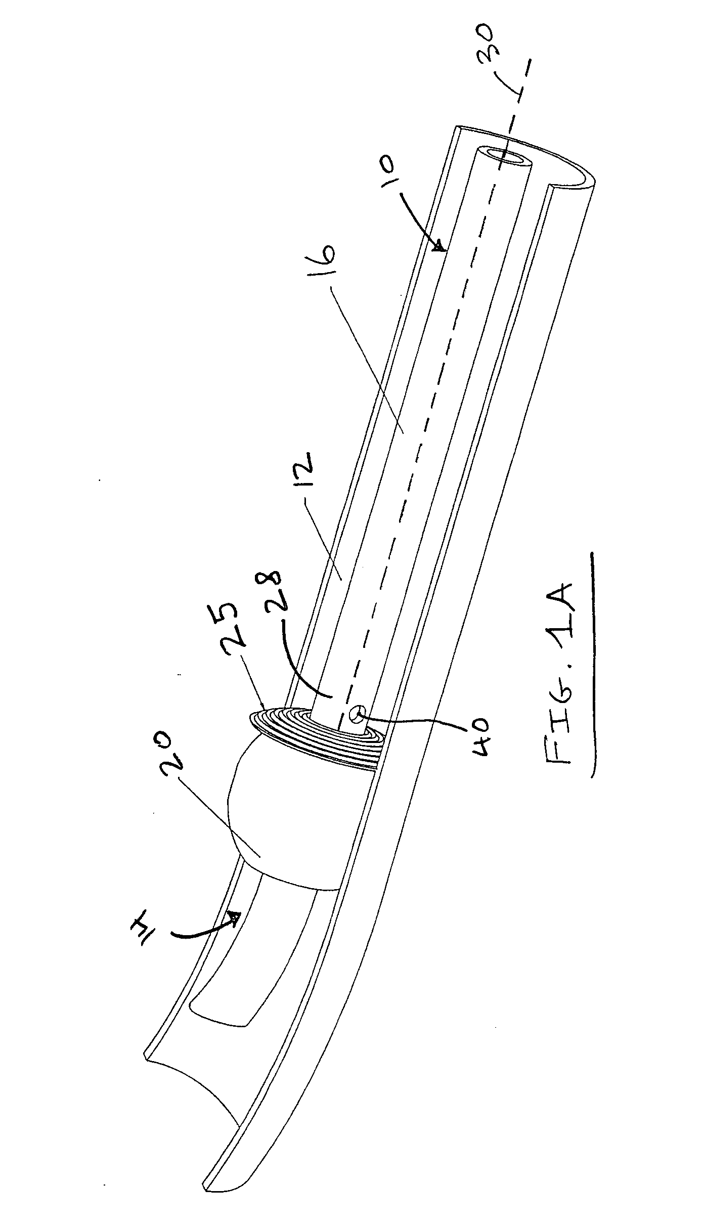
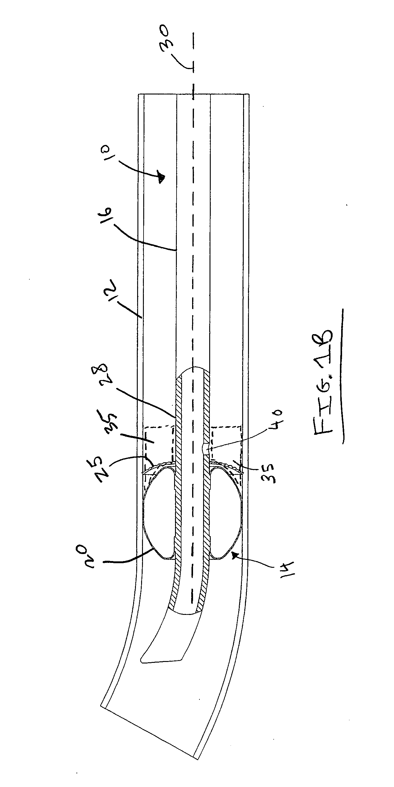
[0027]FIG. 1A is a schematic view illustrating an endotracheal or tracheostomy tube according to the invention. A tube 10 is positioned inside a patient's trachea 12. The tube 10 includes a distal end portion 14 and a proximal end portion (not shown) which generally extends out of the patient's oral airway opening when the tube 10 is inserted as shown. The tube 10 includes an elongated tubular member 16 which defines one or more lumens therein for providing an artificial airway as is well-known in the art. A distal cuff 20 is disposed at a point on or proximate the distal end portion 14 of the tube 10. The cuff 20 is an inflatable member which is used to position and secure the tube 10 when inserted into a patient's trachea, as is well known in the art.

[0028]On the proximal end or side of the cuff sub-assembly is a high surface area structure 25, having the general configuration or geometry shown in FIG. 1A. As used herein, the term “high surface area structure” shall mean any struc...

second embodiment

[0033]FIG. 3A is a schematic view illustrating an endotracheal tube or tracheostomy tube 50 according to the invention. Tube 50 includes the same tubular member 16 and cuff 20 as tube 10, but the distal end portion 54 includes a high surface area structure 55 which includes a soft disc shaped structure forming an annulus around the outer surface 58 of the tubular member 16 proximate the junction of the cuff 20 with tube 16. As shown in FIG. 3A, the disc of the high surface area structure 55 includes a plurality of slits 59 cut into the disc that form radially extending strips 57 to allow for more flexibility in the structure such that the tube 50 can be safely inserted into the trachea. This also allows the high surface area structure 55 to bend or collapse as the cuff is deflated or when the tube 50 is inserted. The high surface area structure 55 also includes an anti-microbial agent to allow for the tube device 50 to more effectively prevent the formation of biofilms and infection...

third embodiment

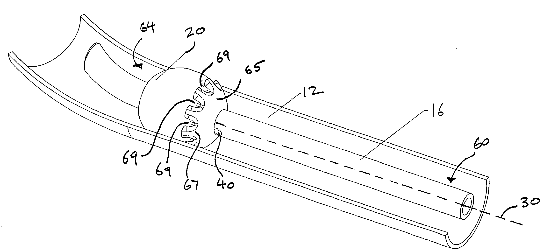
[0034]FIG. 4A is a schematic view illustrating an endotracheal or tracheostomy tube 60 according to the invention. Tube 60 includes the same tubular member 16 and cuff 20 as tubes 10 or 50, but the distal end portion 64 includes a high surface area structure 65 which includes an anti-microbial agent and is a dome-shaped element defining a plurality of recesses 69 within the outer surface 67 of the dome-shaped element. The dome-shaped element 65 overlies a proximal side of the inflatable cuff 20. FIG. 4B is a cross-sectional view of the endotracheal or tracheostomy tube shown in FIG. 4A. As shown in FIG. 4B, the dome shaped structure 65 extends radially away from central axis 30 and into region 35 to provide a greater contact area for secretions. The plurality of recesses 69 as well as the radial girth of the dome shaped element 65 both contribute to this increased surface area.


PUM


Abstract

The present invention provides a medical device such as an endotracheal tube or a tracheostomy tube having a distal inflation cuff and a high surface area structure disposed radially around the tube on a proximal side of the cuff. The high surface area structure includes an antimicrobial agent. The relatively higher surface area of the structure and radial extension thereof from the tube allows the antimicrobial agent to better extend into a secretion pool collected in the region around the intersection of the cuff and tube within a patient's trachea, so as to better prevent biofilms from forming and thereby prevent infections such as ventilator assisted pneumonia (VAP).

Description

FIELD OF THE INVENTION[0001]The present invention relates generally to medical devices. More particularly, the present invention relates to artificial airways for insertion into the trachea which are coated with an antimicrobial agent.BACKGROUND OF THE INVENTION[0002]This present invention addresses the problems incident with the insertion of medical devices such as endotracheal and tracheostomy tubes into a patient's airway. As is well known in the art, fluid secretions can pool around the distal cuff portion just proximal to the cuff around the tube when the device is dwelling inside a patient's trachea for an extended period of time. Infections, such as ventilator assisted pneumonia (VAP), can commonly result due to biofilm buildup around the device near or within the pool of secretions when using such a cuffed endotracheal or tracheostomy tube. Previous methods of preventing this buildup and the resultant infections have not been widely embraced in medical devices in the respira...

Claims


Application Information

Patent Timeline
Patent Type & Authority Applications(United States)
IPC IPC(8): A61M16/04
CPCA61M16/04A61M16/0434A61M16/0479A61M2202/203A61M16/0465
Inventor BURK, MARCDWYER, DANIEL PATRICK
Owner TELEFLEX MEDICAL INC
Learn More