[0013]In one aspect of the invention, the electrosurgical probe is designed to enhance the depth of current penetration into the tissue at a
voltage sufficiently low to minimize or completely avoid
vaporization,
necrosis or
ablation of the
tissue surface. In one embodiment, the return
electrode comprises an annular
electrode on the shaft spaced axially from the
active electrode about 5 to 50 mm, preferably about 10 to 30 mm. The return
electrode will generally have a larger exposed surface area than the
active electrode to minimize or eliminate
high current densities at the return electrode surface. Applicant has found that the depth of current penetration into the tissue may be increased (with a given
voltage) when the return electrode is spaced further away from the
active electrode on the shaft. Deeper current penetration allows a deeper, more uniform heating of the tissue (and thus more effective tissue contraction) without increasing the temperature of the surface tissue to a level that would result in irreversible damage, such as
vaporization,
necrosis or
ablation to the
tissue surface. Specifically, applicant has found that, at the voltages described herein (e.g., about 20 to 100 volts rms), the techniques of the present invention will allow sufficient current penetration into the tissue to contract collagen fibers at about 2.0 to 4.0 mm deep without causing
tissue necrosis at the
tissue surface.
[0014]According to the present invention, the dispersive return electrode is positioned relative to the active electrode such that the instrument effectively functions as a virtual unipolar
system in which the return electrode has substantially no effect on the electric fields surrounding the active electrode (similar to a true monopolar
system with a dispersive return pad). In this configuration, there is a lower impedance contact between the return electrode and the electrically conducting fluid surrounding return electrode. As the conductive volume becomes large, there is very little
potential difference around the return electrode so that the tissue surrounding the return electrode substantially behaves as a virtual return electrode. There is, therefore, almost no thermal heating around the return electrode. This configuration allows for deeper current penetration into the tissue, resulting in increased thermal heating and tissue contraction. In addition, this configuration still maintains the advantages of bipolar modalities; namely that the current path is substantially restricted to the region treated. Thus, if the treated region is the shoulder
capsule, the current will remain in this area, and will not flow through the heart or other sensitive organs, which minimizes the risk of arrythmias.
[0015]In a specific application of the invention, a method is provided for contracting or shrinking the collagen fibers within a joint capsular tissue, such as the shoulder, knee, hip, hand, foot,
elbow or the like, to eliminate capsular redundancy or to otherwise tighten the ligamous complex. In this method, an active electrode is percutaneously introduced through a portal in the joint and positioned adjacent to the joint capsular tissue. The cavity surrounding the joint is immersed in electrically conducting fluid, such as isotonic
saline, and a return electrode is positioned within the electrically conducting fluid. A
high frequency voltage difference is applied between the electrode terminal and the return electrode to induce contraction of the collagen fibers within the joint capsular tissue. In the representative embodiment, the active and return electrodes are both located on an electrosurgical probe, and the return electrode is configured (i.e., sized and spaced relative to the active electrode) to allow the
electric current to fully penetrate the
joint capsule, while maintaining the temperature of the surface tissue below the threshold of
cell vaporization or 100° C. Typically, the
joint capsule has a thickness in the range of about 2.0 to 4.0 mm. Accordingly, the return electrode is configured to allow the
electric current to penetrate about 2.0 to 4.0 mm of tissue, at a voltage level (e.g., about 40 to 90 volts rms) that is below the threshold for tissue vaporization at the tissue surface.
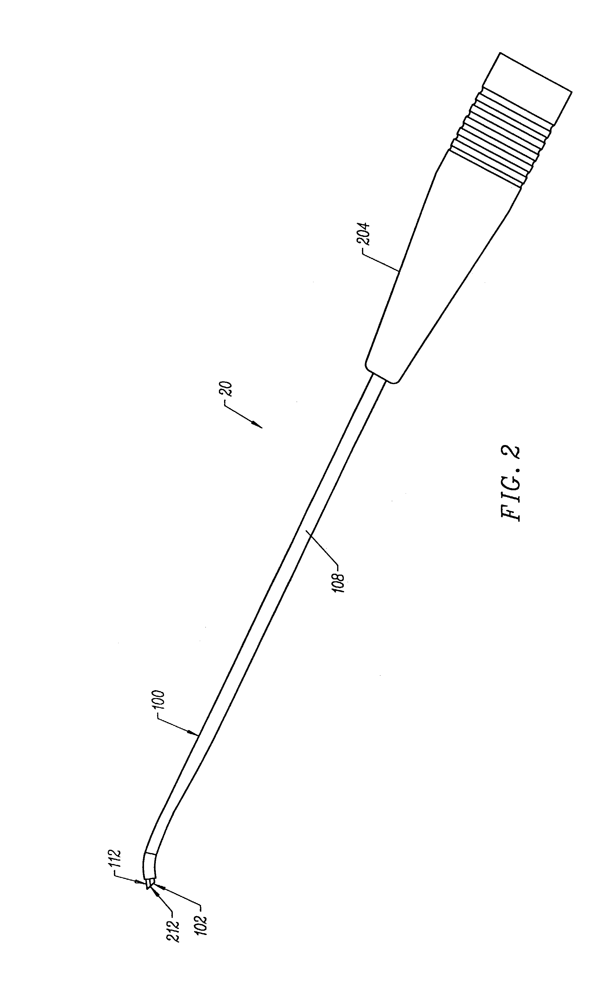
[0017]In one embodiment, the active electrode comprises a dome-shaped member having a plurality of holes mounted to an electrically insulating support member on the distal end portion of the instrument shaft. The instrument further comprises one or more electrical connectors extending through the instrument shaft, and at least partially through the holes in the dome-shaped active electrode to electrically couple the active electrode to a
high frequency power supply. The connectors each have distal ends that are slightly larger than the holes in active electrode so that these distal ends also serve to help fix the active electrode to the electrode support member. The dome-shaped active electrode has a substantially irregular surface with a plurality of holes or distal connector ends protruding therefrom. These irregularities (i.e., holes and protrusions) create
multiple edges on the surface of electrode that increase the current densities around electrode. This increased
current density enables the probe 400 to provide increased thermal penetration of RF energy for the same level of voltage to improve the contraction of collagen tissue. Thus, the present invention allows improved tissue contraction with relatively low power levels, and in a bipolar modality that minimizes current flow beyond the
target site into the patient's body.
[0019]The
system may optionally include a temperature controller coupled to one or more temperature sensors at or near the distal end of the probe. The controller adjusts the output voltage of the power supply in response to a temperature
set point and the measured temperature value. The temperature sensor may be, for example, a
thermocouple, located in the insulating support that measures a temperature at the distal end of the probe. In this embodiment, the temperature
set point will preferably be one that corresponds to a
tissue temperature that results in the contraction of the collagen tissue, i.e., about 60° C. to 70° C. Alternatively, the temperature sensor may directly measure the
tissue temperature (e.g.,
infrared sensor). This embodiment is advantageous in situations when the surgeon is moving the probe transversely across the tissue.
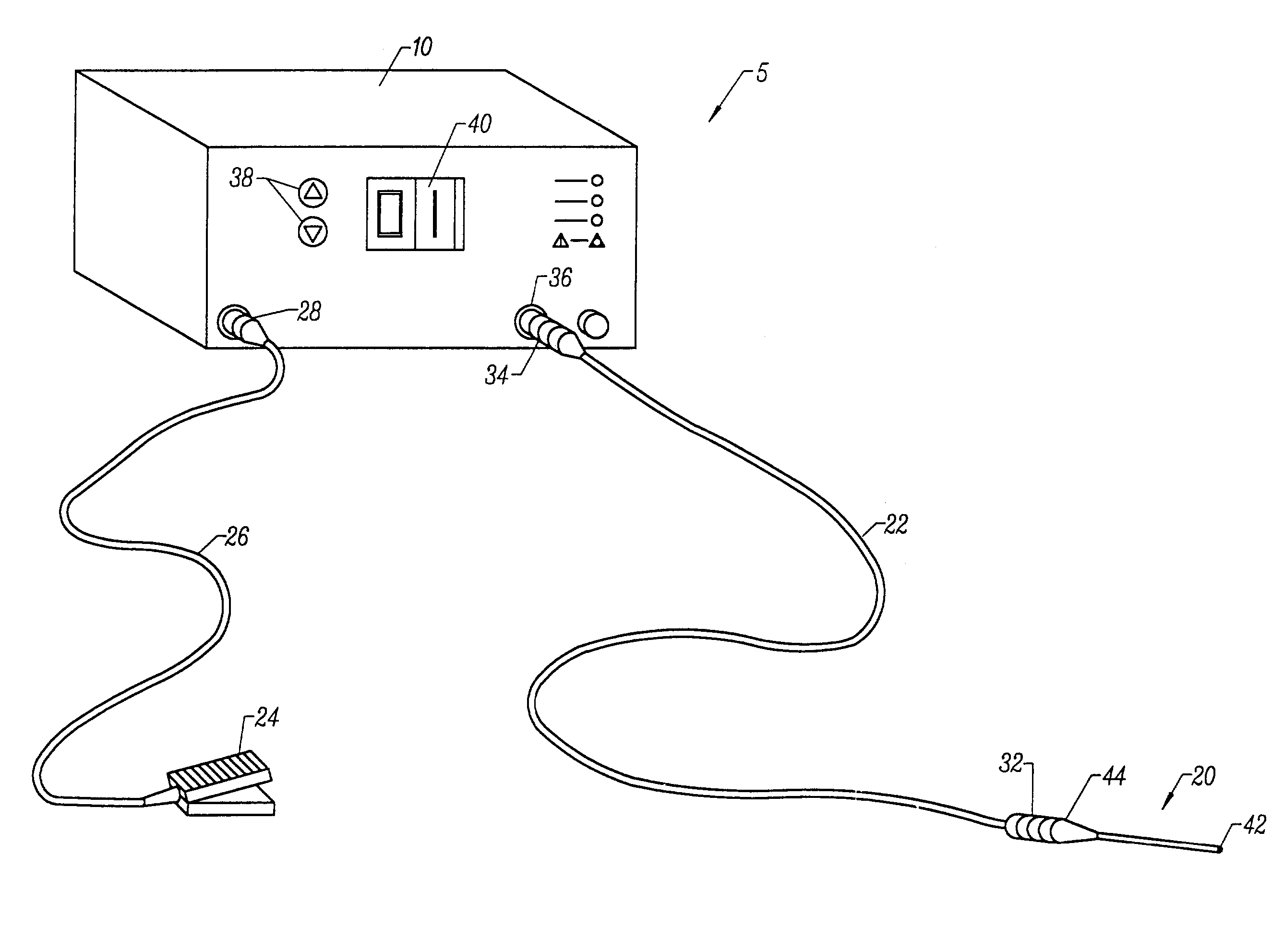
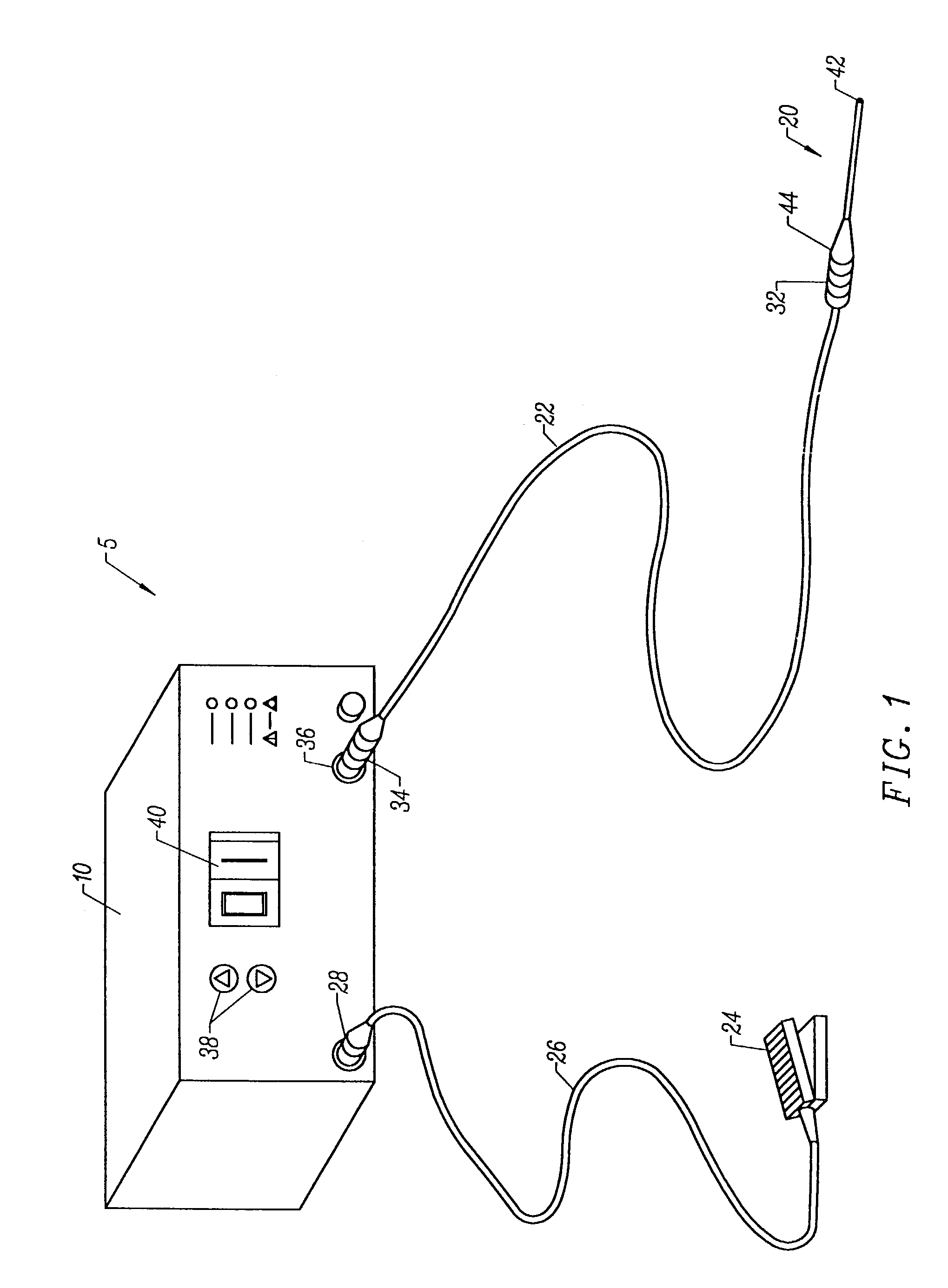
[0020]In another aspect of the invention, an electrosurgical system comprises a
high frequency power supply and a
surgical instrument capable of both bipolar and monopolar electrosurgical techniques without having to disconnect the instrument from the power supply. In this embodiment, a switch is included in the system to switch the return terminal on the power supply between a dispersive return pad coupled to the
surgical instrument (monopolar mode) and a return electrode on the
surgical instrument spaced proximally from the active electrode (bipolar mode).
