
Devices and methods for repairing cardiac valves

a technology for cardiac valves and valves, applied in the field of devices and methods for repairing cardiac valves, can solve the problems of increasing the workload on the heart, relative obstruction of blood flow, and affecting the proper functioning of one or more of these valves, so as to facilitate proper leaflet coaptation, improve the repair process, and improve the effect of cardiac valve function

Inactive Publication Date: 2008-12-25
MT SINAI SCHOOL OF MEDICINE
View PDF30 Cites 48 Cited by
  • Summary
  • Abstract
  • Description
  • Claims
  • Application Information

AI Technical Summary

Benefits of technology

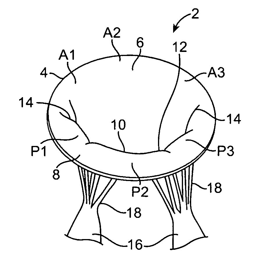
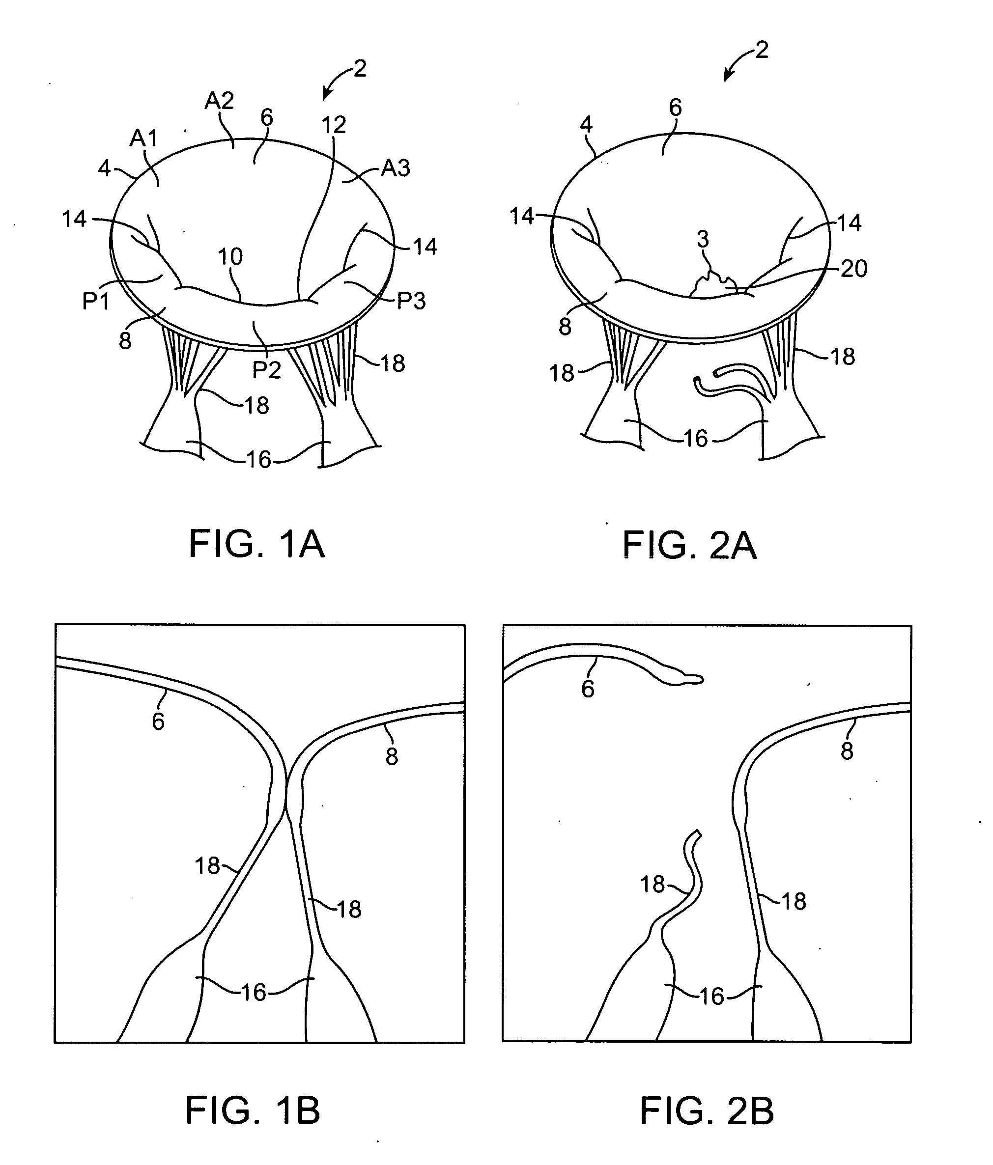
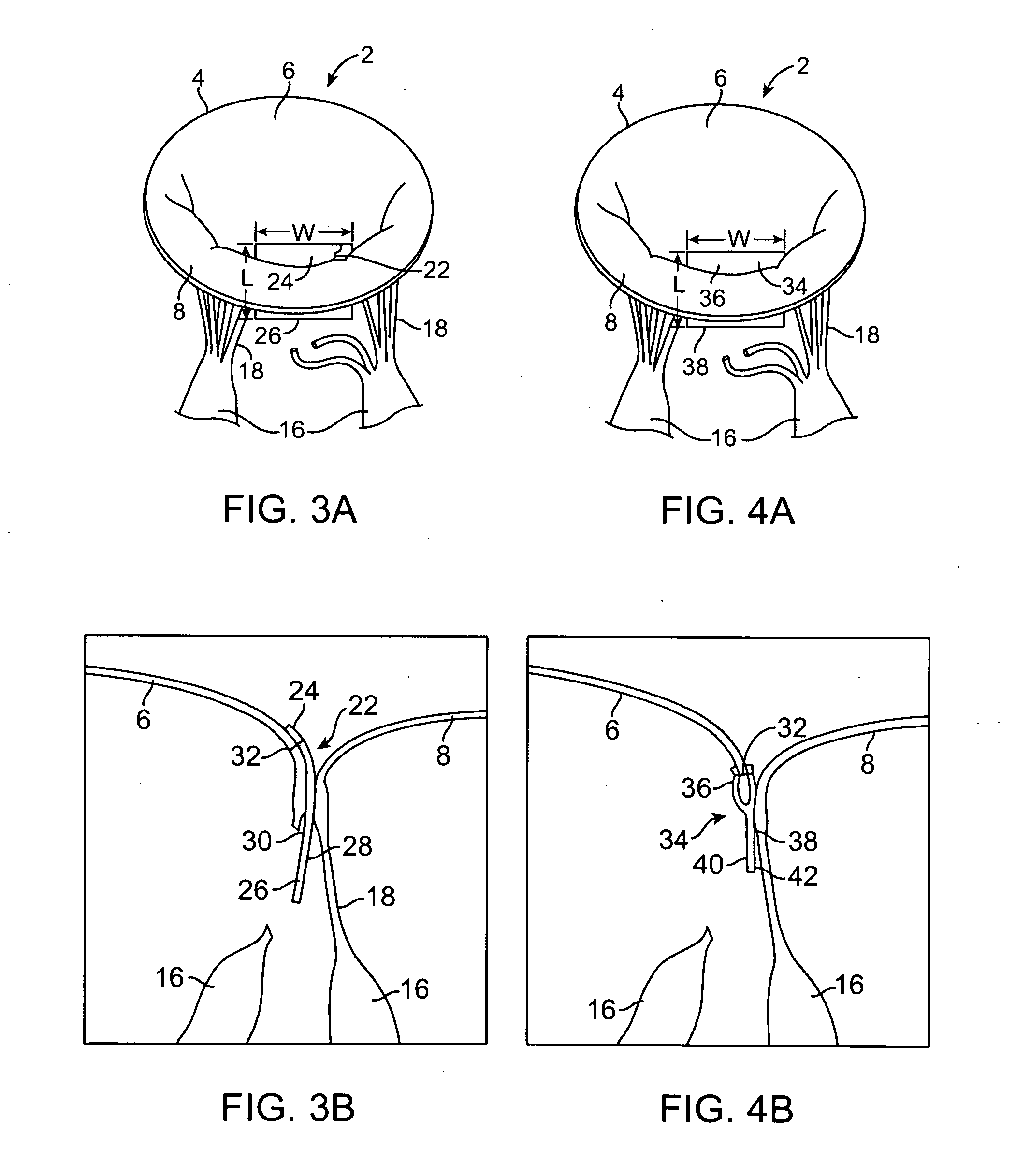
[0023]A feature of the present invention is the provision of an implantable device for facilitating proper leaflet coaptation without affecting the mobility of the leaflet and without reducing the effective valve area. The device is affixed to the affected leaflet at, over or under at least a portion of its prolapsing segment and provides a normalized coaptation surface area against which the opposing leaflet(s) may coapt. In certain embodiments, the device immobilizes or restrains the prolapsing portion or segment of the affected leaflet in order to permit leaflet coaptation during systole. By restraining the prolapsing segment and / or by providing an improved coaptation plane or surface, the devices facilitate coaptation of the leaflets(s) thereby eliminating the regurgitation.

Problems solved by technology

Various disease processes can impair the proper functioning of one or more of these valves.
In addition, damage to the ventricle from prior heart attacks (i.e., myocardial infarction secondary to coronary artery disease) or other heart diseases (e.g., cardiomyopathy) can distort the valve's geometry causing it to dysfunction.
Valve stenosis is present when the valve does not open completely causing a relative obstruction to blood flow.
Both of these conditions increase the workload on the heart and are very serious conditions.
If left untreated, they can lead to debilitating symptoms including congestive heart failure, permanent heart damage and ultimately death.
Dysfunction of the left-sided valves—the aortic and mitral valves—is typically more serious since the left ventricle is the primary pumping chamber of the heart.
Many dysfunctional valves, however, are diseased beyond the point of repair.
In addition, valve repair is usually more technically demanding and only a minority of heart surgeons are capable of performing complex valve repairs.
The aortic valve is more prone to stenosis, which typically results from buildup of calcified material on the valve leaflets and usually requires aortic valve replacement.
Although mitral stenosis, which usually results from inflammation and fusion of the valve leaflets, can often be repaired by peeling the leaflets apart from each other (i.e., a commissurotomy), as with aortic stenosis, the valve is often heavily damaged and may require replacement.
Lesions in any of these components can cause the valve to dysfunction, leading to mitral regurgitation—the regurgitation of blood from the left ventricle to the left atrium during systole.
Physiologically, mitral regurgitation results in increased cardiac work since the energy consumed to pump some of the stroke volume of blood back into the left atrium is wasted.
Overtime, the volume overload on the heart leads to myocardial remodeling in the form of left ventricular dilation and / or hypertophy.
It also leads to increased pressures in the left atrium which results in the back up of fluid in the lungs and shortness of breath—a condition known as congestive heart failure.
Annular dilatation or distortion results in separation of the free margins of the two leaflets.
The increased pressures in the right heart can lead to dilatation of the chambers and concomitant tricuspid annular dilatation.
The most common cause of insufficiency of the mitral valves in western countries is due to Type II dysfunction (leaflet prolapse).
Most surgeons, outside of specialized centers, rarely tackle these complex repairs and these patients usually receive a valve replacement.
Initial studies showed a high rate of failure of the edge-to-edge repair particularly in patients with mitral regurgitation resulting from rheumatic fever and that a concomitant annuloplasty should be performed in every patient.
However, it has been found that the edge-to edge repair, particularly the double orifice technique, results in a significant decrease in mitral valve area which may result in mitral stenosis.
Even without physiologic mitral stenosis, the decrease in orifice area increases flow velocities and turbulence, which can lead to fibrosis and calcification of the functioning valve segments.
This will likely impact the long-term durability of this repair.
Another factor, which may impact the long-term durability of the edge-to-edge technique, is the increased stress on the subvalvular apparatus of all segments.
In sum, current clinical data does not support the routine use of the edge-to-edge technique for the treatment of Type II mitral regurgitation.
Although most patients tolerate limited periods of cardiopulmonary bypass and cardiac arrest well, these maneuvers are known to adversely affect all organ systems.
If severe, these complications can lead to permanent disability or death.
The risk of these complications is directly related to the amount of time the patient is on the heart-lung machine (“pump time”) and the amount of time the heart is stopped (“cross-clamp time”).
Complex valve repairs can push these time limits even in the most experienced hands.
Even if he or she is fairly well versed in the principles of mitral valve repair, a less experienced surgeon is often reluctant to spend 3 hours trying to repair a valve since, if the repair is unsuccessful, he or she will have to spend up to an additional hour replacing the valve.
However the use of these minimally invasive procedures has been limited to a handful of surgeons at specialized centers in a very selected group of patients.
Even in their hands, the most complex valve repairs cannot be performed since dexterity is limited and the whole procedure moves more slowly.

Method used


Image

  • Devices and methods for repairing cardiac valves
  • Devices and methods for repairing cardiac valves
  • Devices and methods for repairing cardiac valves

Examples


Embodiment Construction

[0033]Before the present invention is described, it is to be understood that this invention is not limited to particular embodiments described, as such may, of course, vary. It is also to be understood that the terminology used herein is for the purpose of describing particular embodiments only, and is not intended to be limiting, since the scope of the present invention will be limited only by the appended claims.

[0034]Where a range of values is provided, it is understood that each intervening value, to the tenth of the unit of the lower limit unless the context clearly dictates otherwise, between the upper and lower limit of that range and any other stated or intervening value in that stated range is encompassed within the invention. The upper and lower limits of these smaller ranges may independently be included in the smaller ranges is also encompassed within the invention, subject to any specifically excluded limit in the stated range. Where the stated range includes one or bot...


PUM


Abstract

Devices and methods for the repair of a defective cardiac valve are provided. The implantable devices provide a leaflet coaptation surface and correct for one or more prolapsing segments of a valve leaflet. The methods involve implanting one or more devices within the defective cardiac valve. In certain embodiments, the devices and methods correct for billowing leaflets and / or a dilated valve annulus.

Description

FIELD OF THE INVENTION[0001]The invention relates to devices and methods for facilitating and simplifying the repair of cardiac valves.BACKGROUND OF THE INVENTION[0002]The human heart has four valves that control the direction of blood flow in the circulation. The aortic and mitral valves are part of the “left” heart and control the flow of oxygen-rich blood from the lungs to the body, while the pulmonic and tricuspid valves are part of the “right” heart and control the flow of oxygen-depleted blood from the body to the lungs. The aortic and pulmonic valves lie between a pumping chamber (ventricle) and major artery, preventing blood from leaking back into the ventricle after it has been ejected into the circulation. The mitral and tricuspid valves lie between a receiving chamber (atrium) and a ventricle preventing blood from leaking back into the atrium during ejection.[0003]Various disease processes can impair the proper functioning of one or more of these valves. These include deg...

Claims


Application Information

Patent Timeline
Patent Type & Authority Applications(United States)
IPC IPC(8): A61F2/24
CPCA61F2/2463
Inventor FILSOUFI, FARZAN
Owner MT SINAI SCHOOL OF MEDICINE
Learn More