
Intraocular pressure-regulated early genes and uses thereof

a technology of intraocular pressure and genes, applied in the field of intraocular pressure-regulated early genes, can solve the problems of glaucoma that is difficult to treat, affects the vision of millions of people, and the annual cost of glaucoma has reached billions of dollars, so as to prevent rgc death, inhibit expression or activity, and increase expression or activity

Active Publication Date: 2008-12-11
MCGILL UNIV
View PDF5 Cites 6 Cited by
  • Summary
  • Abstract
  • Description
  • Claims
  • Application Information

AI Technical Summary

Benefits of technology

[0019]The present invention also relates to pharmaceutical compositions used to treat glaucoma, chronic ocular degeneration or RGC death. Thus, the present invention relates to a pharmaceutical composition comprising an effective amount of at least one substance that inhibits the expression or activity of up-regulated IPREGs and a pharmaceutically acceptable carrier, wherein the composition is administered to treat glaucoma. In another embodiment, the pharmaceutical composition is administered to treat chronic ocular degeneration or RGC death. In addition, the invention also relates to a pharmaceutical composition comprising an effective amount of at least one substance that increases the expression or activity of down-regulated IPREGs and a pharmaceutically acceptable carrier wherein the composition is administered to treat glaucoma or, in a further embodiment, chronic ocular degeneration or RGC death. The invention further relates to a pharmaceutical composition that comprises an effective amount of at least one substance that inhibits the expression or activity of up-regulated IPREGs and at least one substance that increases the expression or activity of down-regulated IPREGs and an acceptable pharmaceutical carrier, wherein the composition is administered to treat glaucoma, chronic ocular degeneration or RGC death. The IPREGs having altered expression include those having altered expression induced by high IOP, those having altered expression induced by administration of α2 macroglobulin protein and those having altered expression induced by both.

Problems solved by technology

Glaucoma impairs the vision of millions people worldwide and is one of the leading causes of blindness.
In fact, the annual cost of glaucoma has reached billions of dollars in the United States alone.
However, glaucoma is difficult to treat because the exact onset of high IOP is unpredictable and generally unapparent until peripheral vision loss occurs, at which point irreversible RGC loss is often advanced.
However, despite the normalization of IOP, sustained RGC death and clinical evolution towards glaucoma often continue, which suggests that high IOP may not be the direct cause of RGC apoptosis.
However, these mechanisms alone can not explain why only RGCs should be susceptible to apoptosis instead of all cells in the inner retinal layer that are exposed to the potentially deleterious effects of altered glutamate / nitric oxide / Ca++ and to mechanical stress.

Method used


Image

  • Intraocular pressure-regulated early genes and uses thereof
  • Intraocular pressure-regulated early genes and uses thereof
  • Intraocular pressure-regulated early genes and uses thereof

Examples


example 1

Intraocular Pressure Regulated Genes

Induction of Intraocular Pressure

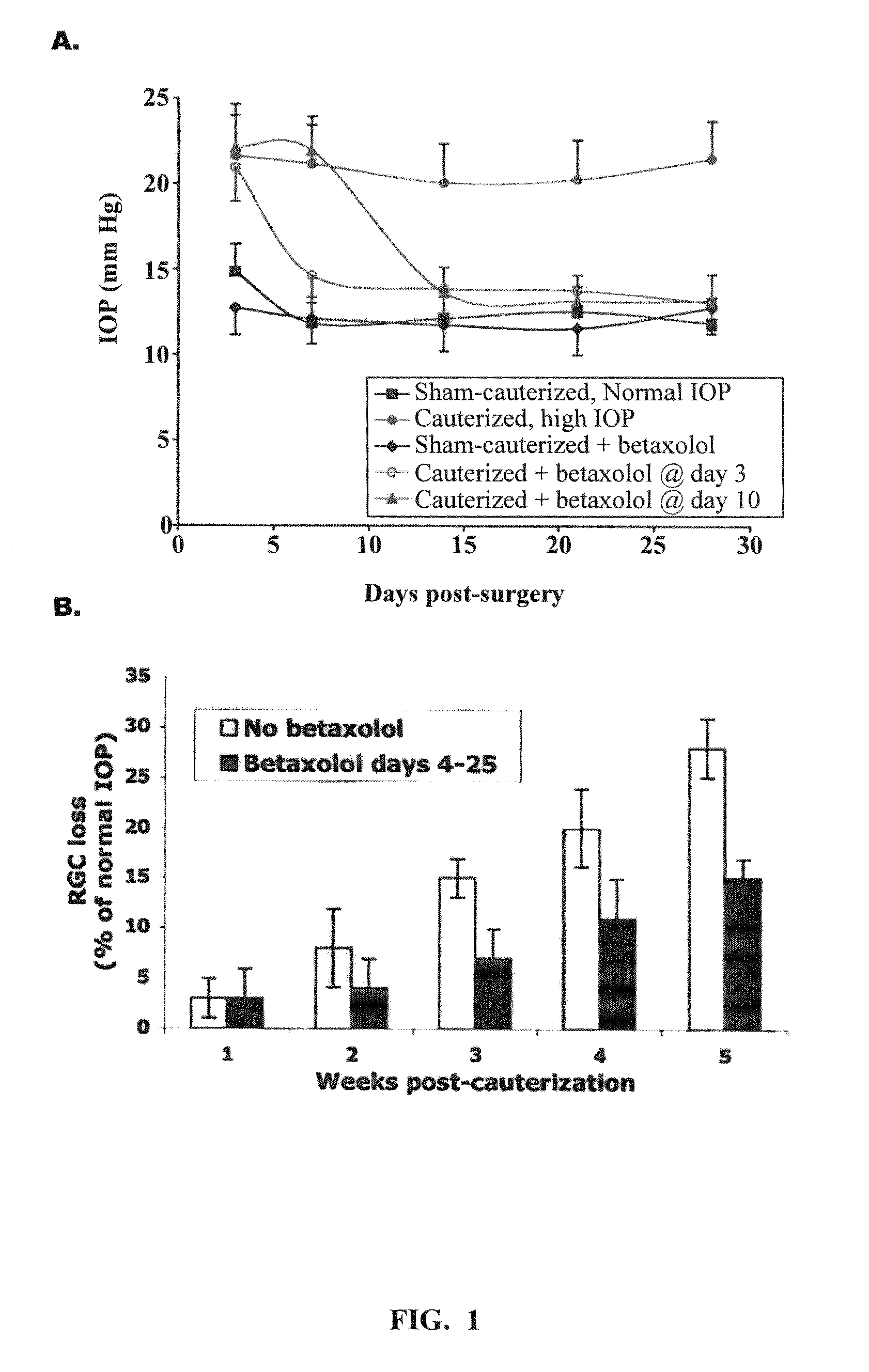
[0072]High IOP. Episcleral cauterization of rat eyes was performed under anesthesia. After a conjunctival incision, extra-ocular muscles were isolated and the major limbal draining veins were identified based on location, relative immobility, larger caliber and branching pattern. Cauterization of three episcleral vessels in the right eye was done with a 30″ cautery tip. The left eye in each animal was used as normal IOP control after sham-surgery (conjunctival incisions with no cauterization).

[0073]IOP measurements. IOP was gauged using a Tonopen XL tonometer under light anesthesia (intramuscular injection of ketamine, 4 mg kg; xylazine, 0.32 mg / kg; and acepromazine, 0.4 mg / kg). The accuracy of the readings of the Tonopen compared with other instruments, even under anesthesia, had already been determined. The average normal IOP of rats under anesthesia was 12 mm Hg (range 10-14 mm Hg), and in cauterized eyes it was...

example 2

Validation of a Role for α2 Macroglobulin in Glaucoma

Localization of α2 Macroglobulin and its Receptor in Retina

[0095]Retrograde RGC labeling RGCs were labeled with 3% DII (1,1-dioctadecyl-3,3,3,3-tetramethylindocarbocyanine perchlorate) or with 3% fluorogold. Briefly, Wistar rats were anesthetized and their heads were mounted in a stereotactic apparatus. Both superior colliculi (SC) were exposed and the dye was injected in two adjacent sites at the SC of each hemisphere (5.8 mm behind Bregma, 1.0 mm lateral, and depth 4.5 mm for the first release of dye solution and 3.5 mm for the second release).

[0096]Flat mounted retinas and RGC counting 7 days after dye injection, the vasculature of the rats were perfused-fixed (transcardiac perfusion in phosphate-buffer (PB), followed by 4% PFA in PB) and the eyes were enucleated. After post-fixing for 1 hour cuts were made through the sclera to form a Maltese cross pattern and retinas detached from the eyecup at the optic nerve head. The retin...

example 3

Changes in Intraocular Pressure Regulated Genes after Intraocular α2 Macroglobulin Protein Injection

[0121]The following experiment was conducted in order to assess changes in gene expression induced in the retina after intraocular injection of α2 macroglobulin protein. 2 μg of α2 macroglobulin protein was intraocularly injected into the right eye of rats (n=3) and the left eye was used as a control. One rat was tested at Day 3, one rat was tested at Day 7 and one rat was tested at Day 14. Retinas were isolated and mRNA was collected. Samples from each of the rats were studied by gene microassays. Retinas were carefully dissected out to insure that only retinal mRNAs were prepared for gene microarray studies. Samples were obtained from control eyes.

[0122]RNA preparation. Total RNA was isolated from retinal tissue using TRIZOL® (Life Technologies). RNA was then further purified using the RNAEASY® (QIAGEN®). The integrity of the RNA samples was assessed by running aliquots on RNA 6000 ...


PUM

PropertyMeasurementUnit
Timeaaaaaaaaaa
Pressureaaaaaaaaaa
Stress optical coefficientaaaaaaaaaa

Abstract

The present invention relates to methods to treat glaucoma and glaucoma-related conditions through the regulation of changes in gene expression that are mediated by high intraocular pressure or α2 macgroglobulin administration. Glaucoma, retinal ganglion cell (RGC) death and chronic ocular hypertension are treated using pharmaceutical compositions which comprise substances that inhibit the expression or activity of intraocular pressure-regulated early genes (IPREGs) or their gene products that are up-regulated by high intraocular pressure or α2 macgroglobulin administration and / or which increase the expression or activity of IPREGs or their gene products that are down-regulated by high intraocular pressure or α2 macgroglobulin administration. The invention also relates to methods of identifying an IPREG and methods to test for chronic ocular degeneration and the onset of RGC stress in an individual by measuring the expression level of IPREG proteins.

Description

RELATED APPLICATIONS[0001]This application is a continuation-in-part of International Application No. PCT / US2006 / 045169, which designated the United States and was filed on Nov. 22, 2006, published in English, which claims the benefit of U.S. Provisional Application No. 60 / 739,570, filed on Nov. 22, 2005. The entire teachings of the above applications are incorporated herein by reference.BACKGROUND OF THE INVENTION[0002]Glaucoma impairs the vision of millions people worldwide and is one of the leading causes of blindness. Accounting for a significant number of patient visits to ophthalmologists' offices in North America, hundreds of thousands of new cases of glaucoma are diagnosed in the United States each year with many of those cases afflicting those of the older population. In fact, the annual cost of glaucoma has reached billions of dollars in the United States alone.[0003]In open angle glaucoma, the most frequent form of glaucoma, visual field loss is caused by progressive opti...

Claims


Application Information

Patent Timeline
IPC IPC(8): A61K39/395A61K38/16G01N33/68C12Q1/68C07H21/04A61P27/06A61K39/00
CPCA61K2039/505C07K14/47C07K16/18C07K16/30C07K2316/96G01N33/5308G01N2800/168A61P27/06C07K2317/76
Inventor SARAGOVI, H. URI
Owner MCGILL UNIV
Learn More