Detecting and Quantifying Host Cell Proteins in Recombinant Protein Products

Inactive Publication Date: 2008-10-23
GENENTECH INC
View PDF0 Cites 18 Cited by
  • Summary
  • Abstract
  • Description
  • Claims
  • Application Information

AI Technical Summary

Problems solved by technology

Although there are commercial assays and reagents available to detect immunoglobulins, DNA, endotoxins, viruses, etc., there are currently no commercial reagents or analytical methods available for the detection and quantification of process-specific HCPs.
Failure to remove these contaminants early in the drug development process can result in reduced efficiency and delay in approval of a given biopharmaceutical drug.
Under this model, a null cell (a cell similar to or identical to the cell used for production of the biopharmaceutical) is used to generate host cell proteins or other potential contaminants, but is incapable of producing the biopharmaceutical product itself.
Many small potential HCPs, however, are weak immunogens that do not elicit an adequate antibody response in the host animal.
It offers good resolution and sensitivity, but suffers from subjective interpretation of band comparison and is technique dependent.
HPLC can be quantitative with high resolution, but offers only low sensitivity, as well as being subjective and costly.
A western blot offers reasonable sensitivity, but it is only qualitative and the antibodies used may fail to detect some contaminants.
Immunoassays offer very high sensitivity and can offer an objective endpoint, but there is no resolution of individual components and again the antibodies used may fail to detect some contaminants.
Moreover, typical immunoassays require a two-step process otherwise there can be a significant number of “false negatives” because the assay failed to detect bound antibody.
False negatives result when the HCP detection assay fails to detect an HCP present in the biopharmaceutical product preparation.
In other words, some HCPs may not be able to form a complex with the capture and detection antibodies, thus remaining undetectable in the assay, and resulting in a false negative measurement.

Method used


Image

  • Detecting and Quantifying Host Cell Proteins in Recombinant Protein Products
  • Detecting and Quantifying Host Cell Proteins in Recombinant Protein Products
  • Detecting and Quantifying Host Cell Proteins in Recombinant Protein Products

Examples


example 1

HCP Pool (30) Production from Parental Cells (10)

[0039]Frozen parental cells were thawed, in a 37° C. water bath for an appropriate amount of time. The thawed cells were mixed, and an aliquot (i.e. 1 ml) of the cells taken and mixed with a pre-warmed growth medium (at about 37° C.). The ratio of the growth medium to the cell concentration was about 9:1. The cell culture was then centrifuged to form a cell pellet to better separate the cells from impurities. The cell pellet was re-suspended in a similar amount of growth medium. The cell culture was incubated at about 37° C. (i.e., about 90% humidity and about 5% CO2). The cell culture was transferred to a spinner flask, and after about 7 days to a bioreactor. Once the cells were ready to harvest, the cell culture was centrifuged and filtered using a 0.2 μm filter.

[0040]A BCA protein assay was performed to determine the total protein concentration. A well known BCA technique and kit uses a combination of working reagents, which are th...

example 2

Immunization (40)

[0051]The HCP pools (30) are then utilized for custom polyclonal antisera production. An example immunization treatment as discussed above and as produced in Example 2 is based on a seven-day routine.

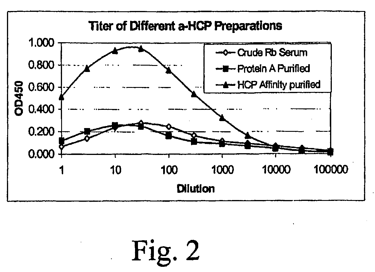
[0052]The two HCP preparations, pools I and II in 10 ml aliquots, were subjected to custom polyclonal antisera production procedures. Two New Zealand White rabbits for each immunogen were used with a standard immunization protocol based on a seven-day routine. Animals were pre-bled on day 0. The initial injection (1 mg each) was administered subcutaneously in complete Freund's adjuvant (CFA) and all subsequent injections (every two weeks, 1 mg each) were in incomplete Freund's adjuvant (IFA). About 25 ml of sera were collected from each rabbit seven days after each boost. The titer from the first bleed was assessed using EIA. All of the rabbits exhibited good binding titers to the HCP immunogens (titer is defined as the antiserum dilution that produces a net OD of 1.0 i...

example 3

HCP Affinity Chromatography (75)

[0054]As mentioned above, the crude polyclonal anti-HCP antibodies produced from the sera are purified using HCP affinity chromatography. The affinity media includes HCPs from the initial HCP pool (30) that were coupled to beads. The HCP-coupled affinity beads were packed into a column. Crude diluted rabbit serum (50) was loaded onto the column. The buffered serum was washed, eluted, and then followed by an acid strip. The loading flowthrough was also collected and subjected to recapture for any unbound HCPs. The elution, acid strip, and the flowthrough fractions were evaluated using EIA and WB for titers and specificity.

[0055]Five grams of CNBr activated Sepharose 4 Fast Flow beads (Amersham Biosciences, Uppsula, SE) was hydrated in PBS, pH 7 buffer in a 50 ml centrifuge tube, yielding about 20 ml of drained gel. 12 ml of concentrated HCP preparation in PBS (5.14 mg / ml) via stirred cell with YM3 membrane was added to the drained gel to form a slurry....


PUM


Abstract

A single-step immunoassay method, kit, and reagents for detecting and quantifying contaminant host cell proteins in a recombinant protein sample are described. The method includes the step of adding to immobilized medium comprising a capture reagent including anti-host cell protein antibodies, both the recombinant protein sample and a detection reagent comprising anti-host cell protein antibodies and a detectable moeity. The recombinant protein sample and two reagents are added simultaneously. This single-step format provides greater interaction between the capture antibody, the contaminant host cell proteins that may be present in the recombinant protein sample, and the detection antibody. By providing the opportunity for both antibodies and HCPs to interact at the same time, the one step format allows the formation of the “capture antibody-HCP-detection antibody” complex with all possible HCPs present. Thus, the HCP assay sensitivity is significantly improved, and the possibility of a false negative is significantly reduced. In addition, the one step format shortens the assay turnaround time and provides a convenient tool for measuring the HCP in the product during the course of the entire purification procedure. Quality control of the process is increased and the protein purification efficiency during process development can be measured.

Description

BACKGROUND OF INVENTION[0001]For recombinant biopharmaceutical proteins to be acceptable for administration to human patients, it is important that residual contaminants resulting from the manufacture and purification process are removed from the final biological product. These process contaminants include culture medium proteins, immunoglobulin affinity ligands, viruses, endotoxin, DNA, and host cell proteins. These host cell contaminants include process-specific host cell proteins (HCPs), which are process-related impurities / contaminants in the biologics derived from recombinant DNA technology. U.S. and foreign regulations often require removal of such contaminants. For example, the U.S. Food and Drug Administration (FDA) requires that biopharmaceuticals intended for in vivo human use should be as free as possible of extraneous immunoglobulin and non-immunoglobulin contaminants, and requires tests for detection and quantitation of potential contaminants, such as HCPs. As well, the...

Claims


Application Information

Patent Timeline
IPC IPC(8): G01N33/53G01N33/566G01N33/543G01N33/551G01N33/68
CPCG01N33/54306G01N33/6854
Inventor WANG, YAJUN "JENNIFER"LIU, MENG-YUAN "PATRICK"
Owner GENENTECH INC
Why Eureka
Try Eureka