
Method For Providing Dna Fragments Derived From An Archived Sample

Active Publication Date: 2008-09-11
EPIGENOMICS AG
View PDF8 Cites 25 Cited by
  • Summary
  • Abstract
  • Description
  • Claims
  • Application Information

AI Technical Summary

Benefits of technology

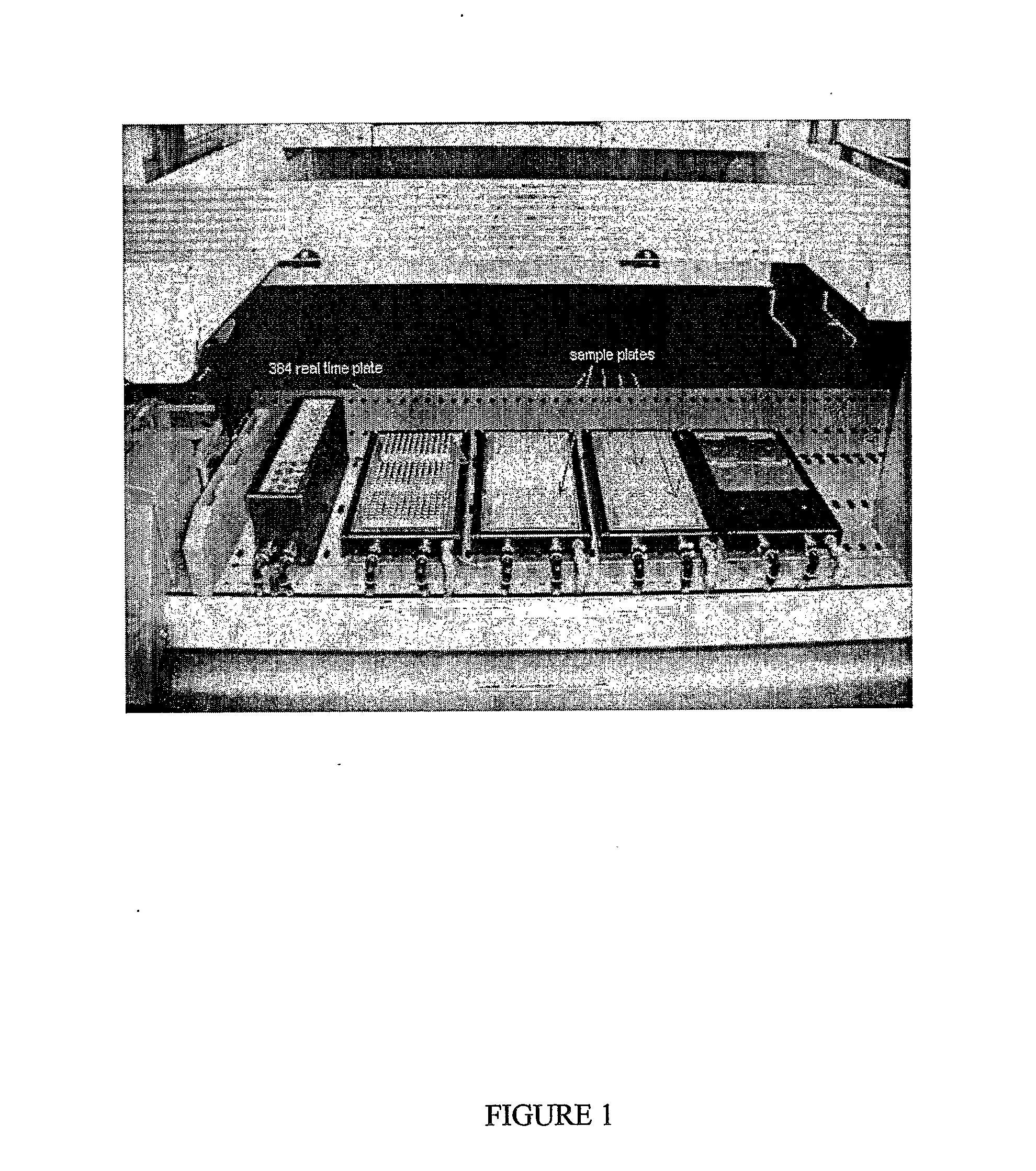
[0053] In particular aspects, the exemplary inventive method has the following advantages: It has a low handling effort, because it includes only a very limited amount of steps which has to be carried out. Additionally, it is possible to carry out the method of the invention in a plate scale. Moreover the different steps can also be automated and therefore robotics can also be used.
[0054] The execution in plate scale and the suitability of the method for automatisation also lead to a reduction in costs. In addition the costs are further reduced by the use of devices and solutions which are available at low expenses.
[0055] Another advantage of the method of the invention is that every step can easily be performed because only standard laboratory equipment is necessary for its execution.
[0056] Because of its simpleness, its suitability for automatisation, its low handling effort as well as its easy handling, the method o

Problems solved by technology

But unfortunately, this resource is only minimally accessible by molecular biological means, in particular by the most promising and modern methods such as those for the analysis of the methylation pattern.
This is because of difficulties in obtaining sufficiently large amounts of high quality genomic DNA at low costs and with minimal handling effort.
These difficulties are based on the degradation of DNA and RNA due to fixation and storage conditions of the sample or biopsy, and on the insufficient methods for the preparation of DNA.
This has the disadvantage that a lot of proteins are covalently linked to the genomic DNA and also the genomic DNA becomes cross-linked.
Consequently, genomic DNA tends to be of small fragment size, has a low integrity, and is contaminated by proteins, peptides and / or amino acids which are cross linked with the DNA and which also interfere with further analysis.
As it is well known, such a procedure leads only to comparably low yields of genomic DNA, the DNA having only a small fragment length.
Furthermore, the DNA is also not suitable for more sophisticated analysis methods, because still a lot of interfering proteins, peptides and / or amino acids are linked to the DNA.
Such methods usually lead to larger amounts of genomic DNA with a lot of contaminating RNA and protein.
In addition, the contaminants cannot be easily removed because they are cross-linked to the genomic DNA by the fixative.
This procedure leads only to very low yields of DNA.
This solution has the disadvantage of a low reproducibility and is not applicable when DNA is going to be analysed for methlyation as amplification erases methylation signals.
Consequently, genomic DNA isolated according to prior art methods is not suitable for analysis of the methylation pattern as explained in detail below.
On the other hand, hypermethylation frequently leads to a suppression of the expression.
This leads to an alteration of the expression of the gene.
This results in a deacetylation of the histones, which itself leads to a tighter package of the DNA.
Additionally, because in general the amount of the tissue sample or biopsy is very small, it is necessary that the methods for DNA preparation result in high yields of DNA.
Because of all these requirements, and given the prior art methods, archived samples, despite being a major resource in medical science, can only be minimally used for the efficient analysis of the DNA methylation.
However this method is conflicting with regard to the aim to provide as long as possible DNA fragments.
As is well known in the relevant art, such treatment reduces the integrity of DNA resulting in a random breakage of the DNA strand and therefore the length of the DNA fragments.

Method used


Image

  • Method For Providing Dna Fragments Derived From An Archived Sample
  • Method For Providing Dna Fragments Derived From An Archived Sample
  • Method For Providing Dna Fragments Derived From An Archived Sample

Examples


example 1

a) Paraffin Removal Step

Chemical Needed:

Limonenel45, Fluka Chemika, Art Nr. 89188

Procedure:

[0432] 1. Spin safe look or screw cap reaction tubes (containing 1-5 sections of a paraffin-embedded formalin-fixed tissue) for 1 min at 5,000×g [0433] 2. Add 1 ml limonene to each tube. Push all pieces into the liquid. In some cases sample material is already very brittle with small pieces of paraffin / tissue in tube—make sure that material sticking to the tube cap goes back into the tube. [0434] 3. 1 h incubation at room temperature at 1,000 rpm in thermomixer, vortex vigorously at least 3 times during incubation. [0435] 4. Place tubes into centrifuge and spin at 16,000 rcf (=16,000×g) for 5 min, the tissue will pellet at the tube bottom. [0436] 5. Use a 1 ml pipet to suck up the limonene from each sample. Great care should be taken not to disturb the pellet! Place pipet tip opposite the pellet onto the tube and gently allow limonene to enter the pipet tip; for removal of last droplets...

example 2

[0541] All steps will be done in the tubes that the samples are provided in, i.e. the tubes provided by the supplier of the samples, these can be both 1.5 and 2.0 ml (preferred) tubes. Please double check that tube formats fit into the centrifuge.

[0542] The solvents and buffers can be delivered with either single channel pipettes or multipettes

a) Removal of Paraffin

Chemical Needed:

Limonenel45, Fluka Chemika, Art Nr. 89188

Procedure:

[0543] 1. Add 1 ml limonene to each tube (containing 1 to 5 slides of paraffin-embedded formalin-fixed surgery sample). Push all pieces into the liquid. In some cases sample material is already very brittle with small pieces of paraffin / tissue in tube—make sure that material sticking to the tube cap goes back into the tube. [0544] 2. 1 h incubation at room temperature at 1,000 rpm, vortex vigorously at least 3 times during incubation. [0545] 3. Place tubes into centrifuge and spin at 16,000 rcf (=16,000×g) for 5 min, the tissue will pellet at the...

example 3

[0649] All steps will be done in the tubes that the samples are provided in. The tubes can be both 1.5 and 2.0 ml tubes. Please double check that tube formats fit into the centrifuge.

[0650] The solvents and buffers can be delivered with either single channel pipettes or multipettes

a) Removal of Paraffin

Chemical Needed:

Limonenel45, Fluka Chemika, Art Nr. 89188

Procedure:

[0651] 1. Add 1 ml limonene to each tube (containing 1 to 5 sections of a paraffin-embedded formalin-fixed tissue). Push all pieces into the liquid. In some cases sample material is already very brittle with small pieces of paraffin / tissue in tube. Make sure that material sticking to the tube cap goes back into the tube. [0652] 2. 1 h incubation at RT at 1,000 rpm, vortex vigorously at least 3 times during incubation. [0653] 3. Place tubes into centrifuge and spin at 16,000 rcf (=16,000×g) for 5 min, the tissue will pellet at the tube bottom. [0654] 4. Use a 1 ml pipet to suck up the limonene from each sample...


PUM

PropertyMeasurementUnit
Temperatureaaaaaaaaaa

Abstract

Aspects of the present invention relate to compositions and methods for providing DNA fragments from an archived sample (e.g., paraffin-embedded and / or fixed-tissue biopsies, etc). Particular aspects provide methods whereby high yields of DNA are isolated as well as a substantial portion of the DNA consists of long DNA fragments, and where the isolated genomic DNA is free of associated or cross-linked contaminants like proteins, peptides, amino acids or RNA. The methods are facile, cost-effective, an are characterized by high reproducibility and reliability. Particular aspects provide methods for providing DNA fragments derived from an archived sample, wherein the yield of DNA before, for example, an amplification step is at least 20%, and amplicons up to a length of about 1,000 base pairs are amplifiable.

Description

CROSS-REFERENCE TO RELATED APPLICATIONS [0001] This application claims the benefit of priority to U.S. Provisional Patent Application Ser. No. 60 / 614,697, filed 30 Sep. 2004, and entitled “Work Flow,” and which is incorporated herein by reference in its entirety.FIELD OF THE INVENTION [0002] The invention relates generally to novel and substantially improved compositions and methods for providing DNA fragments derived from an archived sample (e.g., paraffin-embedded and / or fixed-tissue biopsies, etc), and for analyses of same. BACKGROUND OF ASPECTS OF THE INVENTION [0003] Samples or biopsies are archived many times by default in diagnostic routines. This is done in order to conserve the tissue and to prepare it for subsequent histological examinations. Such conservation is necessary to ensure that the biopsy has not changed after the removal, the observed findings correspond to the situation of the patient, and to prevent degradation of cell structures. Accordingly, the tissue sampl...

Claims


Application Information

Patent Timeline
IPC IPC(8): C12Q1/68C07H1/00C12P19/34
CPCC12N15/1003C12Q1/6806C12Q2527/127C12Q2523/125C12Q2521/531C12Q2547/107C12Q1/6858C12Q1/686C12Q2521/101C12Q2527/137C12Q2527/149
Inventor BALLHAUSE, MATTHIASBERLIN, KURTDIETRICH, DIMOLUKAS, ANTJE KLUTHSCHUSTER, MATTHIASWAGNER, UTEWASSERKORT, REINHOLDZIEBARTH, HEIKE
Owner EPIGENOMICS AG
Learn More